Click image to see more details
-

-
-
-
-
+1
Product Info Summary
| SKU: | A05175-1 |
|---|---|
| Size: | 100 μl/vial |
| Reactive Species: | Human, Mouse, Rat |
| Host: | Rabbit |
| Application: | ELISA, IP, IF, IHC, ICC, WB |
Customers Who Bought This Also Bought

Product info
Product Name
Anti-Ribosomal protein L4/RPL4 Antibody
SKU/Catalog Number
A05175-1
Size
100 μl/vial
Form
Liquid
Description
Boster Bio Anti-Ribosomal protein L4/RPL4 Antibody catalog # A05175-1. Tested in WB, IHC, IF, ICC, IF, IP, ELISA applications. This antibody reacts with Human, Mouse, Rat.
Storage & Handling
12 months from date of receipt,-20℃ as supplied. 6 months 2 to 8℃ after reconstitution. Avoid repeated freezing and thawing.
Cite This Product
Anti-Ribosomal protein L4/RPL4 Antibody (Boster Biological Technology, Pleasanton CA, USA, Catalog # A05175-1)
Host
Rabbit
Contents
500 μg/ml antibody with PBS, 0.02% NaN3, 1 mg stabilizing protein and 50% glycerol
*This antibody is supplied in a stabilized formulation.
Compatibility with conjugation reactions depends on the chemistry of the conjugation method used.
For conjugation methods that are not compatible with the stabilizing components present in this formulation, a carrier-free antibody format is required.
Clonality
Polyclonal
Immunogen
E.coli-derived human Ribosomal protein L4/RPL4 recombinant protein (Position: M1-K424).
Reactive Species
A05175-1 is reactive to RPL4 in Human, Mouse, Rat
Calculated molecular weight
47.7 kDa
Background of RPL4
Ribosomes, the organelles that catalyze protein synthesis, consist of a small 40S subunit and a large 60S subunit. Together these subunits are composed of 4 RNA species and approximately 80 structurally distinct proteins. This gene encodes a ribosomal protein that is a component of the 60S subunit. The protein belongs to the L4E family of ribosomal proteins. It is located in the cytoplasm. As is typical for genes encoding ribosomal proteins, there are multiple processed pseudogenes of this gene dispersed through the genome.
Antibody Validation
Boster validates all antibodies on WB, IHC, ICC, Immunofluorescence, and ELISA with known positive control and negative samples to ensure specificity and high affinity, including thorough antibody incubations.
Application & Images
Applications
A05175-1 is guaranteed for ELISA, IP, IF, IHC, ICC, WB Boster Guarantee
Assay Dilutions Recommendation
The recommendations below provide a starting point for assay optimization. The actual working concentration varies and should be decided by the user.
Western blot, 1:1000-5000
Immunohistochemistry, 1:50-400
Immunofluorescence, 1:50-400
Immunocytochemistry/Immunofluorescence, 1:50-400
Immunoprecipitation, 1:50
ELISA, 1:100-1000
Validation Images & Assay Conditions
Click image to see more details
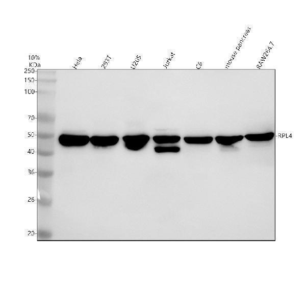
Western blot analysis of Ribosomal protein L4/RPL4 using anti-Ribosomal protein L4/RPL4 antibody (A05175-1).
Electrophoresis was performed on a 10% SDS-PAGE gel at 80V (Stacking gel) / 120V (Resolving gel) for 2 hours. The sample well of each lane was loaded with 30 ug of sample under reducing conditions.
Lane 1: human Hela whole cell lysates,
Lane 2: human 293T whole cell lysates,
Lane 3: human U2OS whole cell lysates,
Lane 4: human Jurkat whole cell lysates,
Lane 5: rat C6 whole cell lysates,
Lane 6: mouse pancreas tissue lysates,
Lane 7: mouse RAW264.7 whole cell lysates.
After electrophoresis, proteins were transferred to a nitrocellulose membrane at 150 mA for 50-90 minutes. Blocked the membrane with 5% non-fat milk/TBS for 1.5 hour at RT. The membrane was incubated with rabbit anti-Ribosomal protein L4/RPL4 antigen affinity purified polyclonal antibody (A05175-1) at 1:1000 overnight at 4°C, then washed with TBS-0.1%Tween 3 times with 5 minutes each and probed with a goat anti-rabbit IgG-HRP secondary antibody at a dilution of 1:5000 for 1.5 hour at RT. The signal is developed using an ECL Plus Western Blotting Substrate (Catalog # AR1196-200) with Tanon 5200 system. A specific band was detected for Ribosomal protein L4/RPL4 at approximately 48 kDa. The expected band size for Ribosomal protein L4/RPL4 is at 48 kDa.

Click image to see more details
IHC analysis of Ribosomal protein L4/RPL4 using anti-Ribosomal protein L4/RPL4 antibody (A05175-1).
Ribosomal protein L4/RPL4 was detected in a paraffin-embedded section of human colon cancer tissue. Heat mediated antigen retrieval was performed in EDTA buffer (pH 8.0, epitope retrieval solution). The tissue section was blocked with 10% goat serum. The tissue section was then incubated with 1:100 rabbit anti-Ribosomal protein L4/RPL4 Antibody (A05175-1) overnight at 4°C. Peroxidase Conjugated Goat Anti-rabbit IgG was used as secondary antibody and incubated for 30 minutes at 37°C. The tissue section was developed using HRP Conjugated Rabbit IgG Super Vision Assay Kit (Catalog # SV0002) with DAB as the chromogen.

Click image to see more details
IHC analysis of Ribosomal protein L4/RPL4 using anti-Ribosomal protein L4/RPL4 antibody (A05175-1).
Ribosomal protein L4/RPL4 was detected in a paraffin-embedded section of mouse bladder tissue. Heat mediated antigen retrieval was performed in EDTA buffer (pH 8.0, epitope retrieval solution). The tissue section was blocked with 10% goat serum. The tissue section was then incubated with 1:100 rabbit anti-Ribosomal protein L4/RPL4 Antibody (A05175-1) overnight at 4°C. Peroxidase Conjugated Goat Anti-rabbit IgG was used as secondary antibody and incubated for 30 minutes at 37°C. The tissue section was developed using HRP Conjugated Rabbit IgG Super Vision Assay Kit (Catalog # SV0002) with DAB as the chromogen.

Click image to see more details
IHC analysis of Ribosomal protein L4/RPL4 using anti-Ribosomal protein L4/RPL4 antibody (A05175-1).
Ribosomal protein L4/RPL4 was detected in a paraffin-embedded section of rat bladder tissue. Heat mediated antigen retrieval was performed in EDTA buffer (pH 8.0, epitope retrieval solution). The tissue section was blocked with 10% goat serum. The tissue section was then incubated with 1:100 rabbit anti-Ribosomal protein L4/RPL4 Antibody (A05175-1) overnight at 4°C. Peroxidase Conjugated Goat Anti-rabbit IgG was used as secondary antibody and incubated for 30 minutes at 37°C. The tissue section was developed using HRP Conjugated Rabbit IgG Super Vision Assay Kit (Catalog # SV0002) with DAB as the chromogen.

Click image to see more details
IF analysis of Ribosomal protein L4/RPL4 using anti-Ribosomal protein L4/RPL4 antibody (A05175-1).
Ribosomal protein L4/RPL4 was detected in an immunocytochemical section of U2OS cells. Enzyme antigen retrieval was performed using IHC enzyme antigen retrieval reagent (AR0022) for 15 mins. The cells were blocked with 10% goat serum. And then incubated with 1:100 rabbit anti-Ribosomal protein L4/RPL4 Antibody (A05175-1) overnight at 4°C. DyLight®488 Conjugated Goat Anti-Rabbit IgG (BA1127) was used as secondary antibody at 1:500 dilution and incubated for 30 minutes at 37°C. The section was counterstained with DAPI. Visualize using a fluorescence microscope and filter sets appropriate for the label used.
Specific Publications For Anti-Ribosomal protein L4/RPL4 Antibody (A05175-1)
Loading publications
Recommended Resources
Here are featured tools and databases that you might find useful.
- Boster's Pathways Library
- Protein Databases
- Bioscience Research Protocol Resources
- Data Processing & Analysis Software
- Photo Editing Software
- Scientific Literature Resources
- Research Paper Management Tools
- Molecular Biology Software
- Primer Design Tools
- Bioinformatics Tools
- Phylogenetic Tree Analysis
Customer Reviews
Have you used Anti-Ribosomal protein L4/RPL4 Antibody?
Share your experimental results or join a short interview to earn up to $1,000 in product credits or other rewards.
0 Reviews For Anti-Ribosomal protein L4/RPL4 Antibody
Customer Q&As
Have a question?
Find answers in Q&As, reviews.
Can't find your answer?
Submit your question
