This website uses cookies to ensure you get the best experience on our website.


- Table of Contents
RPS7 shows general cytoplasmic staining in tissue IHC (HPA tissue IHC). This guide uses the catalog antibody’s illustrated human breast staining at 1:100 as a starting point for paraffin-section planning (datasheet: A04736).

Expected localisation, validated protocols, controls and antibodies — the at-a-glance facts below, then the full design guide.
| Expected localisation | General cytoplasmic tissue staining (HPA tissue IHC) | |
| Staining pattern | Cytoplasmic staining in glandular and epithelial cells (HPA tissue IHC) | |
| Antigen retrieval | Tris-EDTA pH 9.0 HIER, 95–98 °C, 20 min (rule: nuclear antigen) | |
| Positive control | Appendix+4 more · see all | |
| Negative control | Adipose tissue+1 more · see all |
| Fixation | Keep paraffin-section fixation consistent. (standard IHC practice; not target-specific) | |
| Caveat | Staining and RNA show only medium consistency (HPA tissue IHC) | |
| Regulation | No stimulus-linked regulation annotated (UniProt) | |
| Isoform / epitope | No annotated isoforms; one 1–194 chain (UniProt) |
The catalog antibody protocol is paired with published RPS7 IHC methods for colorectal cancer tissue microarrays and paraffin sections (PMC4868722; PMC9584673).
| Sample | Paraffin-embedded rat brain tissue; fixative not specified (datasheet A04736) |
| Fixation | Image fixative and duration unreported (datasheet A04736); verify before use. |
| Sectioning | 4–5 µm sections on charged slides (standard) |
| Deparaffinisation | Xylene, graded ethanol series to water (standard) |
| Antigen retrieval | Heat-induced epitope retrieval in Tris-EDTA buffer, pH 9.0, 20 min at 95–98 °C (standard rule: nuclear antigen) |
| Peroxidase block | 3% H2O2, 10 min, room temperature (standard) |
| Blocking | 10% normal serum of the secondary host, 30 min, room temperature (standard) |
| Primary antibody | Rabbit anti-RPS7, 1:50-1:200 (datasheet A04736) |
| Primary incubation | Overnight at 4 °C (standard) |
| Detection | HRP-polymer secondary, DAB chromogen 5–10 min (standard) |
| Counterstain | Hematoxylin, blue, dehydrate and mount (standard) |
| Expected result | RPS7-positive staining in glandular cells of appendix (HPA tissue IHC: High). HPA tissue profile: General cytoplasmic expression. No signal in the no-primary control. |
RPS7 is a small ribosomal subunit protein found in the cytoplasm and nucleolus, with centrosomal localization also reported; it has no transmembrane segment (UniProt P62081). In paraffin tissue sections, expect predominantly cytoplasmic staining across many cell types (HPA: general cytoplasmic expression; low tissue specificity). HPA rates its tissue IHC evidence Approved, with medium consistency between antibody staining and RNA expression (HPA: tissue IHC).
| Cytoplasmic staining is strong in appendix glandular cells and bone marrow hematopoietic cells (HPA: High in both). | This fits the reported IHC pattern and gives useful positive tissue checks (HPA: general cytoplasmic expression; High in both). Compare cell types within each section, since HPA reports tissue staining with medium consistency against RNA expression (HPA: tissue IHC). |
| Signal is confined to cell borders or another sharply membrane-like pattern. | Treat a predominantly membrane-bound result as suspect: RPS7 has no transmembrane segment, while HPA describes general cytoplasmic IHC staining (UniProt P62081 topology; HPA: tissue IHC). Nuclear or nucleolar staining alone is less decisive because UniProt also places RPS7 in the nucleolus (UniProt P62081). |
| Adipocytes or smooth muscle cells stain strongly and reproducibly. | These are discordant with the supplied HPA examples, where both cell types are Not detected (HPA: adipocytes and smooth muscle cells). Check for antibody cross-reactivity or endogenous chromogenic activity before calling them RPS7-positive (general IHC practice). Do not treat either example as a universal negative control (HPA: low tissue specificity; medium consistency). |
| Brown signal covers tissue, extracellular spaces, or the negative control without clear cellular boundaries. | A widespread deposit is background rather than a readable RPS7 localization pattern (general IHC practice; HPA: general cytoplasmic expression). Review blocking, washes, detection reagent, and chromogen development alongside a control that omits primary antibody (general IHC practice). |
| No staining appears in appendix glandular cells or bone marrow hematopoietic cells (HPA: High in both). | An absent signal in these reported high-staining cells makes the run difficult to interpret (HPA: High in both). First check section integrity, positive control, antibody and detection steps, and counterstain visibility (general IHC practice). HPA's Approved rating does not establish fixation sensitivity or guarantee every specimen will stain (HPA: tissue IHC). |
| Compartment and topology | RPS7 is cytoplasmic and nucleolar, with centrosomal localization reported and no transmembrane segment (UniProt P62081). HPA's tissue IHC profile is generally cytoplasmic (HPA: tissue IHC). Assess the dominant cellular pattern rather than rejecting every nuclear focus (UniProt P62081; HPA: tissue IHC). |
| Cell type and tissue context | HPA reports High staining in several epithelial, hematopoietic, neural, and glial cell groups; prostate glandular cells and skeletal muscle myocytes are Low, while adipocytes and smooth muscle cells are Not detected in the supplied examples (HPA: tissue IHC). Choose comparisons by cell type, not tissue name alone (general IHC practice). |
| Strength of validation | The listed antibody HPA056586 is Approved for IHC, and the tissue profile has medium consistency with RNA expression (HPA: antibody validation; tissue IHC). An IHC Enhanced designation is not supplied (HPA: antibody validation). Interpret an unexpected compartment or cell type with controls rather than assuming the pattern is confirmed independently (general IHC practice). |
| Epitope and fixation sensitivity | The supplied record gives no antibody epitope or target-specific fixation effect (UniProt P62081; HPA: supplied records). Antigen retrieval can be checked against the catalog antibody's IHC instructions as a general paraffin IHC workflow step; do not infer an RPS7-specific retrieval requirement from HPA staining levels (general IHC practice; HPA: tissue IHC). |
| Situation | Likely cause | Next action |
|---|---|---|
| Known high-staining cells show no chromogenic signal (HPA: High in appendix glandular cells and bone marrow hematopoietic cells). | The run may have a detection, reagent, retrieval, or section problem (general IHC practice); the supplied sources identify no RPS7-specific fixation failure mode (UniProt P62081; HPA: supplied records). | Inspect a positive control and the catalog antibody's IHC instructions; verify reagent sequence, antigen retrieval, and chromogen activity (general IHC practice). |
| The entire section is brown, including spaces outside cells. | Nonspecific reagent binding or excess chromogen development can obscure cellular localization (general IHC practice). | Check the primary-omission control, blocking and washes, and development time; score RPS7 only where cells show interpretable staining (general IHC practice; HPA: general cytoplasmic expression). |
| Signal appears in adipocytes or smooth muscle cells despite the supplied HPA Not detected examples (HPA: tissue IHC). | Cross-reactivity or endogenous detection activity is possible (general IHC practice); HPA's medium RNA-staining consistency also limits a categorical biological conclusion (HPA: tissue IHC). | Compare primary-omission and detection controls, then assess the cell boundaries and cytoplasmic pattern before assigning positivity (general IHC practice; HPA: general cytoplasmic expression). |
| Staining is restricted to membranes or has no recognizable cytoplasmic component. | That distribution conflicts with HPA's general cytoplasmic IHC profile and RPS7's lack of a transmembrane segment (HPA: tissue IHC; UniProt P62081 topology). | Recheck the negative control and tissue morphology; repeat with the IHC-validated antibody's documented conditions if the localization remains discordant (general IHC practice; HPA: antibody IHC Approved). |
| A section shows mixed cytoplasmic and nuclear or nucleolar signal. | RPS7 has reported cytoplasmic and nucleolar locations, so a nuclear component is biologically plausible (UniProt P62081); HPA summarizes tissue IHC as generally cytoplasmic (HPA: tissue IHC). | Score cytoplasmic and nuclear components separately, verify cellular morphology, and use controls before treating an unusually dominant nuclear pattern as specific (general IHC practice). |
| What should an IF/ICC comparison show? | HPA reports endoplasmic reticulum localization as Approved and cytosol localization as Supported in ICC-IF; these are modality-specific observations (HPA: subcellular ICC-IF). | For an IF/ICC comparison, look for cellular signal compatible with those compartments; HPA lists images from A-431, U-251MG, and U2OS cells (HPA: subcellular ICC-IF). |
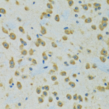
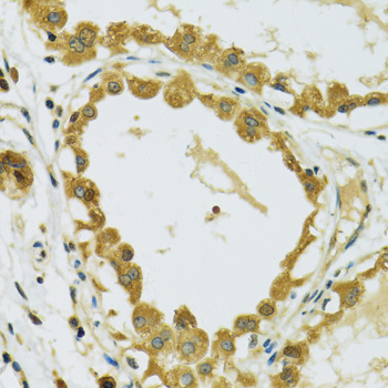

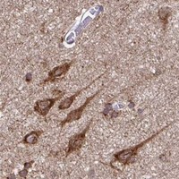


Comprehensive Human Protein Atlas IHC scoring per tissue (reliability: Approved — Medium consistency between antibody staining and RNA expression data.). Rows are taken directly from the HPA tissue chart — click any row's HPA link to view the source.
| Tissue | Cell type | Level | Evidence | Source |
|---|---|---|---|---|
| Appendix | Glandular cells | High | Protein (IHC) | HPA → |
| Bone marrow | Hematopoietic cells | High | Protein (IHC) | HPA → |
| Breast | Glandular cells | High | Protein (IHC) | HPA → |
| Bronchus | Respiratory epithelial cells | High | Protein (IHC) | HPA → |
| Caudate | Neuronal cells | High | Protein (IHC) | HPA → |
Troubleshoot RPS7 chromogenic IHC by checking retrieval, compartment pattern, controls and cell level scoring before interpreting changes in staining.
Validated anti-RPS7 data cover paraffin-section IHC in rat brain, human breast, and mouse spinal cord (A04736 image captions), and IF in human HeLa cells (M04736 image caption).
A04736 lists IHC and ICC/IF for human, mouse, and rat (A04736 applications and reactivity); its IHC images show paraffin-embedded rat brain, human breast, and mouse spinal cord (A04736 image captions). M04736 lists IF for human, mouse, and rat (M04736 applications and reactivity), with an IF image of paraformaldehyde-fixed, permeabilized HeLa cells (M04736 image caption).
Which to pick: Choose A04736 for paraffin-section tissue IHC (A04736 applications and image captions); its IHC captions do not report the fixative (A04736 image captions). Choose M04736 for cell IF (M04736 application and image caption), or A04736 when ICC is required (A04736 applications). Both list human, mouse, and rat reactivity, but only A04736 lists IHC (A04736 and M04736 applications and reactivity).