This website uses cookies to ensure you get the best experience on our website.


- Table of Contents
Plan RPS9 chromogenic IHC in paraffin sections using the cytoplasmic tissue pattern reported by HPA (HPA tissue IHC). This guide covers fixation, staining interpretation and controls, including marrow peroxidase background (standard IHC practice).

Expected localisation, validated protocols, controls and antibodies — the at-a-glance facts below, then the full design guide.
| Expected localisation | Cytoplasmic tissue staining (HPA tissue IHC) | |
| Staining pattern | Cytoplasmic in glandular and hematopoietic cells (HPA tissue IHC) | |
| Antigen retrieval | Tris-EDTA pH 8.0 HIER, heat-mediated (datasheet A05633-1) | |
| Positive control | Bone marrow+4 more · see all | |
| Negative control | Adipose tissue+4 more · see all |
| Fixation | Keep fixation consistent across paraffin sections (standard IHC practice; not target-specific) | |
| Caveat | Endogenous peroxidase in marrow may raise background (standard IHC practice) | |
| Regulation | Expression regulation unannotated (UniProt) | |
| Isoform / epitope | No annotated isoforms; chain spans residues 2–194 (UniProt) |
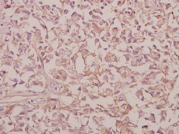
The catalog antibody uses heat-mediated Tris-EDTA retrieval at pH 8.0 (datasheet A05633-1). One published RPS9 IHC protocol follows (PMC5604204).
| Sample | Paraffin-embedded Human brain tissue; fixative not specified (datasheet A05633-1) |
| Fixation | Image fixative and duration unreported (datasheet A05633-1); verify before use. |
| Sectioning | 4–5 µm sections on charged slides (standard) |
| Deparaffinisation | Xylene, graded ethanol series to water (standard) |
| Antigen retrieval | Heat retrieval: Tris-EDTA pH 8.0 (datasheet A05633-1); 20 min, 95–100 °C (standard) |
| Peroxidase block | 3% H2O2, 10 min, room temperature (standard) |
| Blocking | 10% normal serum of the secondary host, 30 min, room temperature (standard) |
| Primary antibody | Rabbit anti-RPS9, 1:100 - 1:300 (datasheet A05633-1) |
| Primary incubation | Overnight at 4 °C (standard) |
| Detection | HRP-polymer secondary, DAB chromogen 5–10 min (standard) |
| Counterstain | Hematoxylin, blue, dehydrate and mount (standard) |
| Expected result | RPS9-positive staining in hematopoietic cells of bone marrow (HPA tissue IHC: High). HPA tissue profile: Cytoplasmic expression in several tissues. No signal in the no-primary control. |
RPS9 is a small ribosomal subunit protein reported in the cytoplasm and nucleolus, with no transmembrane segment (UniProt P46781: function, location, topology). For paraffin section IHC, expect predominantly cytoplasmic staining in several tissues, including strong staining in selected glandular and hematopoietic cells (HPA: tissue IHC). HPA rates its tissue IHC profile Approved, with medium consistency between antibody staining and RNA expression (HPA: tissue IHC reliability).
| Clear cytoplasmic staining in pancreatic exocrine glandular cells or bone marrow hematopoietic cells. | These are suitable positive patterns: both cell populations are rated High by HPA (HPA: pancreas and bone marrow tissue IHC). Judge the signal in the named cells, since a tissue section contains multiple cell types (standard IHC practice). |
| A nucleolar component accompanies otherwise plausible cytoplasmic staining. | Nucleolar localization is compatible with RPS9 biology (UniProt P46781: subcellular location). HPA describes the tissue IHC profile as cytoplasmic, so a prominent nuclear pattern alone needs independent confirmation before being called RPS9 specific (HPA: tissue IHC profile; standard IHC practice). |
| Staining is confined to cell borders or extracellular material, with little cytoplasmic signal. | That distribution conflicts with the reported cytoplasmic and nucleolar locations and absence of a transmembrane segment (UniProt P46781: location, topology). Treat it as a suspect IHC pattern and check the detection controls before assigning it to RPS9 (standard IHC practice). |
| Strong staining appears in cardiomyocytes or adipocytes, while expected positive cells are weak. | HPA reports RPS9 as Not detected in cardiomyocytes and adipocytes (HPA: heart muscle and adipose tissue IHC). The mismatch can suggest cross-reactivity or endogenous detection activity; it does not establish either cause from one slide (standard IHC practice). |
| Diffuse color covers many compartments, or no signal appears in a known positive cell population. | Diffuse color prevents a confident compartment call (standard IHC practice). Absent staining in a High HPA population, such as duodenal glandular cells, leaves the run uninterpretable until controls and tissue preservation are reviewed (HPA: duodenum tissue IHC; standard IHC practice). |
| Cell and tissue choice | Use a named High population, such as small intestinal glandular cells, to assess the run (HPA: small intestine tissue IHC). HPA reports low tissue RNA specificity, so an unlisted tissue is not automatically negative (HPA: tissue RNA specificity). |
| Intracellular location | RPS9 is reported in cytoplasm and nucleolus, including cytoplasmic mRNP granules (UniProt P46781: subcellular location). HPA's observed tissue IHC profile is cytoplasmic; score what the stained cells show, without requiring visible granules (HPA: tissue IHC profile; standard IHC practice). |
| Antibody validation | The listed antibody, HPA048746, has Approved IHC status; no ICC status is supplied (HPA: antibody validation). HPA's tissue reliability description is medium consistency with RNA expression, so validate an unexpected pattern independently (HPA: tissue IHC reliability; standard IHC practice). |
| Sequence and epitope interpretation | UniProt reports one processed chain spanning residues 2–194, no signal peptide or propeptide, and no listed isoforms (UniProt P46781: processing, isoforms). It lists modified residues, but the supplied sources do not locate this antibody's epitope or establish a staining effect (UniProt P46781: modified residues; HPA: antibody record). |
| IF/ICC Q&A | What pattern should IF/ICC show? Cytoplasmic or nucleolar signal is biologically plausible (UniProt P46781: location), but HPA supplies no main ICC location, image bearing cell line, or ICC validation for the listed antibody; treat an IF result as unverified here (HPA: subcellular and antibody records). |
| Situation | Likely cause | Next action |
|---|---|---|
| High background obscures cell boundaries. | Nonspecific reagent binding, endogenous detection activity, or excess detection signal are possible general IHC causes (standard IHC practice). | Inspect a no primary control, adjust blocking or detection conditions, and compare with a named HPA High population before scoring RPS9 (standard IHC practice; HPA: tissue IHC). |
| No staining in pancreatic exocrine glandular cells. | A failed run, unsuitable reagent conditions, or poor tissue preservation are possibilities; HPA rates these cells High (standard IHC practice; HPA: pancreas tissue IHC). | Verify section quality, primary antibody application, retrieval and detection controls, then repeat alongside a positive section (standard IHC practice). No RPS9 specific retrieval condition is supplied (HPA: supplied record). |
| Only nuclear staining is apparent. | UniProt permits nucleolar RPS9, while HPA describes tissue staining as cytoplasmic; the observed pattern needs closer compartment assessment (UniProt P46781: location; HPA: tissue IHC profile). | Examine nuclear detail against the counterstain and compare with cytoplasmic signal in an HPA High cell population; seek independent confirmation if the pattern remains nuclear only (standard IHC practice; HPA: tissue IHC). |
| Cardiomyocytes stain strongly in a heart section. | HPA reports RPS9 as Not detected in cardiomyocytes; cross-reactivity or endogenous detection activity is possible, but neither is proven (HPA: heart muscle tissue IHC; standard IHC practice). | Check a no primary control and repeat with an appropriate independent control before interpreting the heart signal as RPS9 (standard IHC practice). |
| A visually clean negative section is used as the only run control. | A negative section cannot demonstrate that the primary and detection steps produced a detectable RPS9 signal (standard IHC practice). | Include a named HPA High cell population and score that population directly; HPA lists adipocytes and smooth muscle cells as Not detected comparators (HPA: tissue IHC). |
| An IF/ICC image is being used to settle an ambiguous IHC result. | HPA supplies no ICC image bearing cell line, main ICC location, or ICC validation status for HPA048746 (HPA: subcellular and antibody records). | Interpret the paraffin section against its own controls and HPA tissue IHC pattern; regard any IF/ICC observation as a separate, unvalidated line of evidence here (HPA: tissue IHC, subcellular and antibody records; standard IHC practice). |
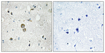
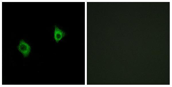
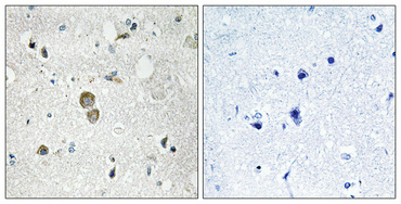
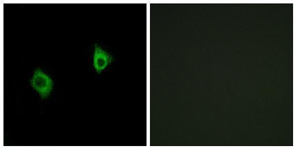


Comprehensive Human Protein Atlas IHC scoring per tissue (reliability: Approved — Medium consistency between antibody staining and RNA expression data.). Rows are taken directly from the HPA tissue chart — click any row's HPA link to view the source.
| Tissue | Cell type | Level | Evidence | Source |
|---|---|---|---|---|
| Bone marrow | Hematopoietic cells | High | Protein (IHC) | HPA → |
| Cervix | Glandular cells | High | Protein (IHC) | HPA → |
| Duodenum | Glandular cells | High | Protein (IHC) | HPA → |
| Gallbladder | Glandular cells | High | Protein (IHC) | HPA → |
| Pancreas | Exocrine glandular cells | High | Protein (IHC) | HPA → |
| Tissue | Cell type | Level | Evidence | Source |
|---|---|---|---|---|
| Adipose tissue | Adipocytes | Not detected | Protein (IHC) | HPA → |
| Cerebellum | Cells in granular layer | Not detected | Protein (IHC) | HPA → |
| Heart muscle | Cardiomyocytes | Not detected | Protein (IHC) | HPA → |
| Nasopharynx | Respiratory epithelial cells | Not detected | Protein (IHC) | HPA → |
| Ovary | Follicle cells | Not detected | Protein (IHC) | HPA → |
Troubleshoot RPS9 staining in paraffin section IHC using the catalog antibody’s tissue image, HPA tissue patterns, and known cellular localisation.
Three anti-RPS9 antibodies have IHC images from paraffin-embedded human brain or colorectal carcinoma (catalog image captions); two also have IF images from A549 cells (catalog IF captions). All list Human, Mouse and Rat reactivity (catalog).
A05633-1 and A05633S9 each show IHC in paraffin-embedded human brain and IF in A549 cells (respective catalog image captions); A05633-1 also lists ICC (catalog applications). A05633 shows IHC in paraffin-embedded human colorectal carcinoma at 1:50 (A05633 image caption); all three list Human, Mouse and Rat reactivity (catalog).
Which to pick: For tissue IHC, choose by the sample shown: A05633-1 or A05633S9 for paraffin-embedded human brain, each shown at 1:100 with Tris-EDTA pH 8.0 retrieval (respective IHC captions), or A05633 for paraffin-embedded human colorectal carcinoma (A05633 IHC caption). For IF, A05633-1 and A05633S9 have A549 images; choose A05633-1 if ICC is required because ICC appears in its application list (catalog IF captions; catalog applications). For other species, all three list Mouse and Rat reactivity and are rabbit polyclonals (catalog), but the supplied IHC images show human specimens only (respective IHC captions); the fixative is unreported in those captions.