RPTOR / Regulatory-associated protein of mTOR · IHC design guide

Design Immunohistochemistry for RPTOR

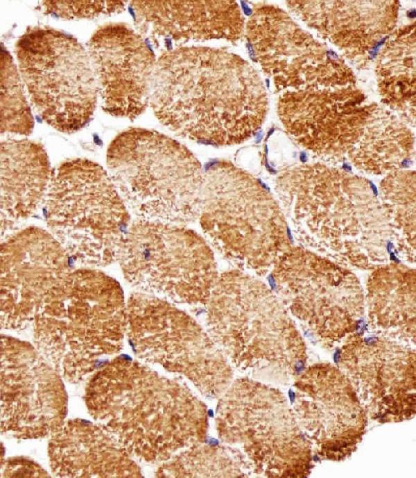
Plan paraffin-section RPTOR IHC around the general cytoplasmic tissue pattern (HPA tissue IHC). The catalog antibody M01463-1 has a 1:25 IHC-P dilution and a human skeletal-muscle DAB example (datasheet: M01463-1).

Evidence assembled Oct 2026 · For research use; verify linked source records and product datasheet before use
Immunohistochemistry protocol sheet for RPTOR (IHC for RPTOR): expected localisation General cytoplasmic staining (HPA tissue IHC), antibody M01463-1, validated IHC image, and IHC protocol steps
Printable RPTOR IHC protocol sheet — expected localisation General cytoplasmic staining (HPA tissue IHC), antibody M01463-1, controls and protocol steps. Open the full RPTOR IHC guide →

RPTOR Immunohistochemistry Experimental Design Guide

Expected localisation, validated protocols, controls and antibodies — the at-a-glance facts below, then the full design guide.

Must know before staining
Expected localisation General cytoplasmic staining (HPA tissue IHC)
Staining pattern Cytoplasmic staining across multiple cell types (HPA tissue IHC)
Antigen retrieval Citrate pH 6.0 HIER, 95–98 °C, 20 min (rule: cytoplasmic / membrane antigen)
Positive control ⓘ Adrenal gland+4 more · see all
Negative control ⓘ Fallopian tube
Important caveats
Reasons your staining may differ from the expected pattern.
Fixation Keep fixation consistent across sections. (standard IHC practice; not target-specific)
Caveat Amino acid availability changes lysosomal recruitment (UniProt)
Regulation Nutrient-responsive localization (UniProt)
Isoform / epitope 3 isoforms; epitope coverage is unspecified (UniProt)
Section 1

Recommended RPTOR IHC & IF Protocols

The catalog antibody’s IHC-P protocol is accompanied by one published RPTOR IHC protocol for mouse testes (PMC11405012).

Recommended immunohistochemistry (IHC-P) protocol parameters
SampleParaffin-embedded H. skeletal muscle tissue; fixative not specified (datasheet M01463-1)
FixationImage fixative and duration unreported (datasheet M01463-1); verify before use.
Sectioning4–5 µm sections on charged slides (standard)
DeparaffinisationXylene, graded ethanol series to water (standard)
Antigen retrievalHeat-induced epitope retrieval in citrate buffer, pH 6.0, 20 min at 95–98 °C (standard rule: cytoplasmic / membrane antigen)
Peroxidase block3% H2O2, 10 min, room temperature (standard)
Blocking10% normal serum of the secondary host, 30 min, room temperature (standard)
Primary antibodyMouse monoclonal (clone 1411CT316.2.151.34) anti-RPTOR, 1:25 (datasheet M01463-1)
Primary incubationOvernight at 4 °C (standard)
DetectionHRP-polymer secondary, DAB chromogen 5–10 min (standard)
CounterstainHematoxylin, blue, dehydrate and mount (standard)
Expected resultRPTOR-positive staining in glandular cells of adrenal gland (HPA tissue IHC: High). HPA tissue profile: General cytoplasmic expression. No signal in the no-primary control.
💡Decision noteStart with citrate pH 6.0 at 95–98 °C for 20 min (page retrieval rule). For mouse testes, Tris–EDTA pH 9 was published (PMC11405012).
Section 2

What Is the Expected RPTOR Staining Pattern?

RPTOR should give a predominantly cytoplasmic IHC pattern, including in HPA high staining glandular cells, cortical neurons and bone marrow hematopoietic cells (HPA tissue IHC). It can associate with lysosome membranes but has no transmembrane segment (UniProt Q8N122). HPA rates the tissue IHC pattern Approved, with medium consistency between antibody staining and RNA expression; interpret individual slides in that context (HPA tissue IHC).

What am I looking at on my slide?
Cytoplasmic staining in adrenal, breast or colon glandular cells, or cortical neurons.This fits the reported High staining in those cell types and the general cytoplasmic tissue pattern (HPA tissue IHC). Compare cells within the section before scoring: HPA's level describes its observed pattern, not an intensity threshold for every specimen (HPA tissue IHC).
Staining is predominantly nuclear, with little cytoplasmic signal.That compartment does not match HPA's general cytoplasmic tissue pattern or UniProt's cytoplasmic and lysosome membrane locations (HPA tissue IHC; UniProt Q8N122). Treat it as suspect and review controls and detection background before calling it RPTOR.
A cell population outside the expected stained cells is strongly positive.Check the cell identity and the relevant HPA tissue entry before assigning specificity: RPTOR has broad tissue RNA expression, while listed IHC levels vary by cell type (HPA tissue IHC). Unexpected staining may reflect cross-reactivity or endogenous chromogenic activity; morphology alone cannot distinguish them (general IHC practice).
Color spreads across stroma, lumens or most cells without clear cytoplasmic boundaries.A diffuse field is difficult to score as cell-associated RPTOR against HPA's general cytoplasmic pattern (HPA tissue IHC). Review the no-primary control and detection conditions for background, and assess only signal that can be assigned to cells (general IHC practice).
No signal appears in a section containing a listed high staining cell population.First confirm that the relevant cells are present and identifiable. HPA reports High staining in, for example, colon glandular cells and bone marrow hematopoietic cells (HPA tissue IHC). A blank result may reflect the assay or specimen; HPA does not establish a target-specific fixation sensitivity.
💡Expected RPTOR appearanceCall a positive when discernible cytoplasmic chromogen is present in the relevant cells, with stronger staining plausible in HPA High populations; predominantly nuclear color or diffuse staining without cell boundaries is suspect (HPA tissue IHC; UniProt Q8N122).
How each factor affects the staining
Cell type and tissueHPA reports High staining in several specific populations, Low staining in hepatocytes, and Not detected in fallopian tube glandular cells (HPA tissue IHC). Choose comparisons by cell type; a low staining population is a weak benchmark for a negative slide.
Subcellular distributionRPTOR is cytoplasmic and can be recruited to lysosome membranes according to amino acid availability (UniProt Q8N122). HPA describes tissue IHC broadly as cytoplasmic (HPA tissue IHC); do not require individually resolved lysosomes in a chromogenic section.
IHC evidence for the antibodyCAB013514 has IHC status Approved; HPA064306 has no IHC status listed in this payload (HPA antibodies). HPA describes medium agreement between tissue antibody staining and RNA expression, so an unusual result needs independent assessment rather than automatic acceptance (HPA tissue IHC).
IF/ICC Q: What pattern should I expect?A: Mainly vesicles and lysosomes, with additional cytosol; HPA marks these locations Supported in ICC-IF (HPA subcellular). This finer IF pattern can inform localisation, while the reported tissue IHC pattern remains generally cytoplasmic (HPA tissue IHC).
Why is my staining missing, weak or wrong?
SituationLikely causeNext action
Positive control cells show no chromogen.The relevant cells may be absent from the section, or an assay step may have failed (general IHC practice).Check morphology and a listed High population, then review the run's detection controls and documented antibody conditions (HPA tissue IHC; general IHC practice).
Signal is mainly nuclear.This conflicts with the reported cytoplasmic and lysosome-associated locations (HPA tissue IHC; UniProt Q8N122).Review localisation against a known positive section and inspect the no-primary control before scoring it as RPTOR (general IHC practice).
Strong color appears in an unexpected cell population.Possible cross-reactivity or endogenous chromogenic activity requires investigation (general IHC practice).Identify the cells morphologically, compare their HPA entry, and check detection controls; report any unresolved staining as uncertain (HPA tissue IHC; general IHC practice).
Background obscures cell boundaries.Nonspecific reagent staining or endogenous detection activity can obscure interpretation (general IHC practice).Compare no-primary and detection controls, then adjust blocking, washing or detection conditions as indicated by those controls (general IHC practice).
A low staining tissue looks negative.HPA reports Low staining in hepatocytes, seminal vesicle glandular cells and ovarian stroma cells (HPA tissue IHC).Use a listed High population to assess whether the run detects RPTOR; avoid declaring assay failure from a low staining population alone (HPA tissue IHC).
A weak section lacks distinct lysosomal puncta.HPA reports general cytoplasmic tissue IHC, whereas vesicle and lysosome localisation comes from ICC-IF (HPA tissue IHC; HPA subcellular).Score identifiable cytoplasmic staining in the expected cells; assess the controls if overall signal is weak, without making puncta a requirement for chromogenic IHC (HPA tissue IHC; general IHC practice).

Sample controls for RPTOR IHC & IF

🧪Run bone marrow first: hematopoietic cells should stain (HPA: High in hematopoietic cells). Use fallopian tube glandular cells as the negative tissue (HPA: Not detected); on the bone marrow slide, morphologically distinct nonhematopoietic cells, if present, should show only background staining and must be assessed rather than assumed negative (standard IHC practice).
Positive control tissue: Adrenal gland (Glandular cells, HPA High)
Negative control tissue: Fallopian tube (HPA Not detected)
ICC-IF cell lines (HPA subcellular resource): HPA ICC-IF images show RPTOR in U-251MG, U2OS, A-549, PC-3, with annotated localisation: Vesicles (supported), Lysosomes (supported) (HPA subcellular).
Technical controls: Include a no-primary, secondary-only control; an isotype control matched to the primary antibody’s host species and clonality; and RPTOR knockout tissue or a validated immunizing-peptide block as a biological specificity control (standard IHC practice). Block endogenous peroxidase in bone marrow before chromogenic detection, since hematopoietic cells can produce peroxidase-associated background (standard IHC practice).
⚠️Feasibility: A target-specific fixation window or fixation effect is unreported in the supplied evidence; the selected M01463-1 paraffin-section caption also leaves the fixative unreported (selected tissue-IHC caption). Antigen retrieval dependence is unreported, so optimize retrieval empirically for the catalog antibody (standard IHC practice); the supplied evidence does not establish whether frozen-section IHC or IF is easier. Interpret bone marrow chromogenic signal alongside the peroxidase control (standard IHC practice).

HPA tissue IHC evidence for RPTOR

Comprehensive Human Protein Atlas IHC scoring per tissue (reliability: Approved — Medium consistency between antibody staining and RNA expression data.). Rows are taken directly from the HPA tissue chart — click any row's HPA link to view the source.

Positive expression · recommended positive controls

TissueCell typeLevelEvidenceSource
Adrenal gland Glandular cells High Protein (IHC) HPA →
Appendix Glandular cells High Protein (IHC) HPA →
Bone marrow Hematopoietic cells High Protein (IHC) HPA →
Breast Glandular cells High Protein (IHC) HPA →
Cerebral cortex Neuronal cells High Protein (IHC) HPA →

Undetected expression · recommended negative controls

TissueCell typeLevelEvidenceSource
Fallopian tube Glandular cells Not detected Protein (IHC) HPA →
Section 3

Advanced RPTOR IHC Tips

Troubleshoot RPTOR staining in paraffin sections by checking retrieval, cell compartment, tissue controls, and scoring before interpreting changes in mTORC1 biology.

How should I retrieve RPTOR in paraffin sections with weak staining?
Use citrate pH 6.0 heat induced epitope retrieval at 95–98 °C for 20 min (page retrieval rule: cytoplasmic / membrane antigen). Let sections cool in retrieval buffer, then compare the same tissue and antibody dilution across runs so that heating is the variable under test (standard IHC practice). If staining remains weak, test a more alkaline buffer on adjacent sections as a fallback, while watching for tissue damage and increased background (standard IHC practice). The skeletal muscle image establishes staining at 1:25 with DAB, but its caption does not report a retrieval method (M01463-1 tissue-IHC caption).
Can fixation explain inconsistent RPTOR staining between paraffin blocks?
Target-specific fixation sensitivity is unknown: the selected paraffin-section caption does not state a fixative or fixation duration (M01463-1 tissue-IHC caption). Record the fixative and time for each block, and compare sections with matched processing before changing the RPTOR antibody dilution (standard IHC practice). For a controlled comparison, process matched tissue pieces with documented fixation intervals, such as 6 and 24 h, then stain them in the same run (standard IHC practice). Interpret any difference as a result of that local comparison, rather than inferring fixation tolerance from RPTOR localisation or tissue staining profiles (UniProt Q8N122; HPA tissue IHC).
What staining compartment should I expect for RPTOR by chromogenic IHC?
Expect predominantly cytoplasmic staining at tissue-IHC resolution, with possible punctate enrichment, because the reported tissue profile is generally cytoplasmic (HPA tissue IHC). RPTOR is recruited to lysosome membranes in an amino acid dependent manner and has no annotated transmembrane segment (UniProt Q8N122 subcellular location and topology). Supported cellular imaging also places it in vesicles, lysosomes, and cytosol, although fine puncta may be difficult to resolve with DAB (HPA subcellular; standard IHC practice). Compare suspicious nuclear or surface-only signal with an adjacent section lacking primary antibody and with the expected cytoplasmic pattern in an appropriate positive tissue (standard IHC practice; HPA tissue IHC).
Could isoforms or epitope accessibility change my RPTOR IHC result?
RPTOR has 3 listed isoforms, but the supplied antibody caption does not identify its epitope or establish isoform coverage (UniProt Q8N122 isoforms; M01463-1 tissue-IHC caption). Ask whether the immunogen maps to a sequence shared by the isoforms before interpreting a negative section as absence of all RPTOR protein (standard IHC practice; UniProt Q8N122 isoforms). Annotated modifications include phosphorylation near residues 696, 706, and 719, so epitope location matters when assessing a site specific reagent (UniProt Q8N122 modified residues). Compare retrieval conditions and an independently mapped antibody on adjacent sections if epitope masking remains plausible (standard IHC practice).
How can I check an IHC pattern with multiplex IF?
Use IF as a separate validation experiment: supported cellular images place RPTOR in vesicles, lysosomes, and cytosol (HPA subcellular). Pair RPTOR with a marker for the expected cell type in the sampled tissue, and include single stain controls to assess channel bleed through (HPA tissue IHC; standard IF practice). Choose a far red fluorophore when the tissue shows strong shorter wavelength autofluorescence, and inspect an unstained section before interpreting puncta (standard IF practice). Because RPTOR lacks a transmembrane segment and can associate with lysosome membranes, titrate mild detergent permeabilisation to expose its cytosolic epitope while preserving puncta (UniProt Q8N122 topology and subcellular location; standard IF practice).
How do I reduce diffuse or punctate DAB background?
Compare a no-primary control with the complete stain to identify signal from detection reagents or endogenous peroxidase (standard IHC practice). Apply a peroxidase block before the DAB step, and optimise protein blocking and washes if diffuse staining persists (standard IHC practice). The selected image used a 1:25 primary dilution, an undiluted biotinylated goat polyvalent secondary, and DAB; treat that combination as image-specific evidence rather than a universal setting (M01463-1 tissue-IHC caption). Titrate the primary on adjacent sections and watch for staining that rises equally in expected cells and tissue edges, which can indicate background (standard IHC practice; HPA tissue IHC).
How should I score RPTOR DAB staining across samples? ⚠ ANSWER MARKED FOR VERIFICATION
Define the cell compartment and sampled region before scoring, because the tissue-IHC profile describes general cytoplasmic expression (HPA tissue IHC). Score cytoplasmic intensity on a 0–3 scale and record the percentage of cells at each intensity to calculate an H-score, or report percentage positive cells under a fixed threshold (standard IHC practice). Normalise to the number of evaluable cells in the same annotated cell population, and keep section thickness, DAB development, and image acquisition consistent (standard IHC practice). Report punctate signal separately if it is a study endpoint, since RPTOR recruitment to lysosome membranes depends on amino acid availability (UniProt Q8N122 subcellular location).
How can I distinguish true RPTOR staining from tissue artefact?
A plausible positive is cytoplasmic staining in an appropriate cell population, potentially with puncta, consistent with reported tissue and cellular localisation (HPA tissue IHC; HPA subcellular). For a positive comparison, HPA reports high staining in cerebral cortex neuronal cells and kidney glomerular cells, while fallopian tube glandular cells were not detected (HPA tissue IHC). Treat isolated nuclear-only signal, section-edge enrichment, and staining confined to necrotic material as suspect until controls support them (UniProt Q8N122 subcellular location; standard IHC practice). Check a no-primary control and peroxidase block for endogenous enzyme signal before attributing brown deposits to RPTOR or mTORC1 activity (standard IHC practice; UniProt Q8N122 function).
Boster reagents

Best RPTOR / Regulatory-associated protein of mTOR IHC Antibodies

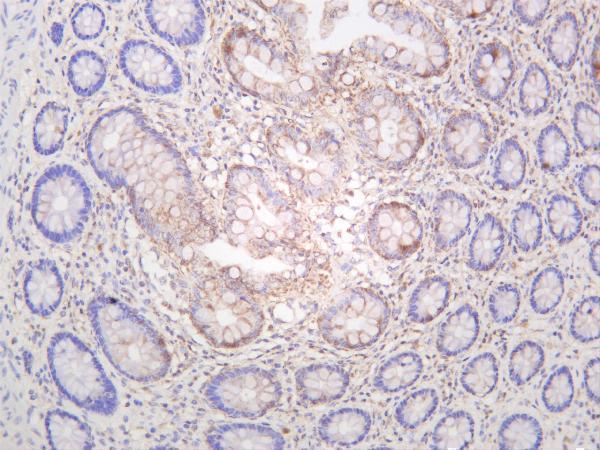
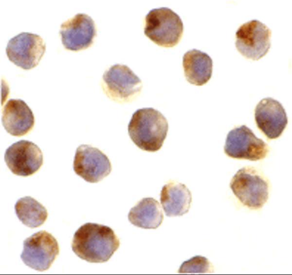
Anti-RPTOR antibodies have IHC images from paraffin-embedded human skeletal muscle and human colon (catalog images), plus IF/ICC images from L1210 cells (catalog images). Listed reactivity spans human, mouse and rat (catalog reactivity).

Real IHC data Immunohistochemical analysis of paraffin-embedded H. skeletal muscle section using RPTOR Antibody. M01463-1 was diluted at 1:25 dilution. A undiluted biotinylated goat polyvalent antibody was used as the secondary, followed by DAB staining.
Anti-RPTOR Antibody
Cat # M01463-1
Real IHC data Human colon was stained with anti-Raptor rabbit antibody
Anti-Raptor Rabbit Monoclonal Antibody
Cat # M01463-2
Real IF data Immunocytochemistry of RAPTOR in L1210 cells with RAPTOR antibody at 10 μg/mL.
Anti-Raptor Rptor Antibody
Cat # A01463-1

M01463-1 is listed for IHC-P in human, mouse and rat samples (catalog applications/reactivity), with DAB staining shown in paraffin-embedded human skeletal muscle at 1:25 (image caption). M01463-2 is listed for IHC and IF in human and mouse samples, with an IHC image from human colon (catalog applications/reactivity; image caption); A01463-1 is listed for IF/ICC in human and mouse samples, with L1210-cell IF/ICC images at 10 μg/mL (catalog applications/reactivity; image captions).

Which to pick: For paraffin-section IHC, choose the mouse monoclonal M01463-1: its own caption shows paraffin-embedded skeletal muscle with DAB detection, and its listed reactivity includes human, mouse and rat for broader species coverage (catalog host/clone/reactivity; M01463-1 image caption). The rabbit monoclonal M01463-2 offers an IHC image in human colon and a listed IHC dilution of 1:200–1:1000; the colon caption does not report tissue processing or fixative (catalog antibody details; M01463-2 image caption). For IF/ICC, choose A01463-1, which has L1210-cell images at 10 μg/mL; the fixative is unreported in those captions (catalog applications; A01463-1 image captions).

Each figure is that product's own IHC / IF validation image from its datasheet.

References

  1. UniProt Consortium. UniProt entry Q8N122 (RPTOR_HUMAN, Regulatory-associated protein of mTOR).
  2. Human Protein Atlas. RPTOR tissue IHC expression (reliability: Approved).
  3. Human Protein Atlas. RPTOR subcellular location (ICC-IF): Mainly localized to vesicles and lysosomes. In addition localized to the cytosol..
  4. Human Protein Atlas. RPTOR antibody validation summary (2 antibodies).
  5. RPTOR blockade suppresses brain metastases of NSCLC by interfering the ceramide metabolism via hijacking YY1 binding. Journal of experimental & clinical cancer research : CR 2024 — PMC10759737.
  6. mTORC1-dependent increase in oxidative metabolism in POMC neurons regulates food intake and action of leptin. Molecular metabolism 2018 — PMC6001919.
  7. mTORC1 as a metabolic rheostat coordinates hormonal signals and cellular proliferation to sustain endometrial competence. Cellular & molecular biology letters 2026 — PMC13101190.
  8. Mechanistic target of rapamycin (mTOR) pathway in Sertoli cells regulates age-dependent changes in sperm DNA methylation. eLife 2024 — PMC11405012.
  9. PubMed PMID:12150925 — UniProt-cited evidence.
  10. PubMed PMID:12150926 — UniProt-cited evidence.
  11. PubMed PMID:16625196 — UniProt-cited evidence.