This website uses cookies to ensure you get the best experience on our website.


- Table of Contents
Plan chromogenic RSPO1 IHC on paraffin sections with the catalog antibody at 2.5–5 μg/mL (datasheet A02857-1). Assess cytoplasmic staining in glandular cells while allowing for uncertainty from secreted variants (HPA tissue IHC).

Expected localisation, validated protocols, controls and antibodies — the at-a-glance facts below, then the full design guide.
| Expected localisation | Cytoplasmic staining in tissue sections (HPA tissue IHC) | |
| Staining pattern | Glandular cells show cytoplasmic staining in several tissues (HPA tissue IHC) | |
| Antigen retrieval | Citrate pH 6 HIER, heat-mediated (datasheet A02857-1) | |
| Positive control | Adrenal gland+4 more · see all | |
| Negative control | Bone marrow+3 more · see all |
| Fixation | Formaldehyde-fixed paraffin sections were used (selected-SKU IHC image A02857-1) | |
| Caveat | Secreted variants may make RNA and protein locations differ (HPA tissue IHC) | |
| Regulation | No specific expression inducer is reported (UniProt) | |
| Isoform / epitope | 3 isoforms; the signal peptide is cleaved, and there is no cytoplasmic domain (UniProt) |
Compare the catalog antibody’s IHC-P protocol with two published RSPO1 chromogenic IHC protocols (datasheet A02857-1; PMC4496395; PMC7065809).
| Sample | Formaldehyde-fixed, paraffin-embedded mouse spleen tissue (datasheet A02857-1) |
| Fixation | Image formalin-fixed; duration unreported (datasheet A02857-1); verify before use. |
| Sectioning | 4–5 µm sections on charged slides (standard) |
| Deparaffinisation | Xylene, graded ethanol series to water (standard) |
| Antigen retrieval | Heat retrieval: Citrate pH 6 (datasheet A02857-1); 20 min, 95–100 °C (standard) |
| Peroxidase block | 3% H2O2, 10 min, room temperature (standard) |
| Blocking | 10% serum (datasheet A02857-1) |
| Primary antibody | Rabbit anti-RSPO1, 2.5-5 μg/mL (datasheet A02857-1) |
| Primary incubation | Overnight at 4 °C (standard) |
| Detection | HRP-polymer secondary, DAB chromogen 5–10 min (standard) |
| Counterstain | Hematoxylin, blue, dehydrate and mount (standard) |
| Expected result | RSPO1-positive staining in glandular cells of adrenal gland (HPA tissue IHC: High). HPA tissue profile: Cytoplasmic expression in several different tissues. No signal in the no-primary control. |
RSPO1 is secreted, has no transmembrane segment, and may also localize to nucleoli (UniProt Q2MKA7). In paraffin sections, expect chiefly cytoplasmic staining in glandular cells and selected epithelial or tubular cells (HPA: tissue IHC). Treat this as a comparison pattern, not proof of specificity: HPA rates the tissue profile Uncertain because secreted protein can appear apart from its RNA source (HPA: reliability).
| Cytoplasmic staining in adrenal glandular cells, esophageal squamous epithelium, or kidney tubules (HPA: High). | This matches reported high tissue IHC staining in the named cell types (HPA: tissue IHC). Compare signal with an appropriate negative control; the HPA tissue profile remains Uncertain (HPA: reliability). |
| Dominant, uniform nuclear staining with little cytoplasmic signal in an otherwise positive section. | This diverges from the reported tissue IHC pattern (HPA: cytoplasmic expression). Check for artefact before scoring it as RSPO1; UniProt also notes possible nucleolar localization, so a nuclear focus alone cannot be dismissed (UniProt Q2MKA7). |
| Strong staining of marrow hematopoietic cells or smooth muscle cells (HPA: Not detected). | These are poor fits for the reported tissue pattern (HPA: tissue IHC). Cross-reactivity or endogenous detection activity is possible; compare antibody omission and detection controls before attributing the signal to RSPO1 (general IHC practice). |
| Diffuse chromogen over tissue and blank areas, with little cell-type contrast. | Interpretation is unreliable because the expected pattern has defined cellular contrast (HPA: tissue IHC). Review blocking, washing, detection background, and counterstain against controls (general IHC practice); diffuse staining does not establish secreted RSPO1 deposition (UniProt Q2MKA7). |
| No staining in adrenal glandular cells or kidney tubules (HPA: High). | The run has missed an expected positive pattern (HPA: tissue IHC). Check tissue preservation, retrieval, antibody dilution, and detection with run controls (general IHC practice); the HPA rating of Uncertain limits any single specimen as a definitive control (HPA: reliability). |
| Secreted protein and location (UniProt Q2MKA7). | The signal peptide spans residues 1–20, and the mature chain begins at 21 (UniProt Q2MKA7). Extracellular distribution is biologically plausible, but HPA's observed tissue pattern is cytoplasmic (HPA: tissue IHC); do not require membrane staining. |
| Tissue and cell type (HPA: tissue IHC). | High staining is reported in adrenal, appendix, breast, colon, duodenal, and gallbladder glandular cells; esophageal squamous cells; and kidney tubules (HPA: High). Ovary follicle cells and fallopian tube glandular cells are reported low (HPA: Low). |
| Protein versus RNA location (HPA: reliability). | HPA calls tissue RNA enhanced in cervix, endometrium, and fallopian tube, yet warns that secreted protein and RNA locations can differ (HPA: RNA specificity; reliability). Select positive IHC controls from observed protein staining, not RNA enrichment alone. |
| Isoforms and molecular processing (UniProt Q2MKA7). | UniProt lists 3 isoforms and glycosylation sites at 137, 153, and 156 (UniProt Q2MKA7). Epitope dependence cannot be predicted without antibody epitope information; these annotations do not establish an antigen-retrieval or fixation effect. |
| Subcellular evidence differs by assay (HPA: tissue IHC; HPA: ICC-IF). | HPA reports cytoplasmic tissue staining but approved vesicle localization in THP-1 ICC-IF images (HPA: tissue IHC; ICC-IF). Compare each result with its own assay and specimen context; neither observation proves that all tissue staining must be vesicular. |
| Antibody evidence (HPA: antibody validation). | HPA046154 has Uncertain IHC status, whereas HPA062923 has Approved ICC status (HPA: antibodies). ICC approval does not validate an IHC result; interpret a new paraffin-section pattern alongside controls and the tissue profile. |
| Situation | Likely cause | Next action |
|---|---|---|
| Expected high-staining cells are blank (HPA: High). | The run may lack usable antigen signal or detection; HPA's tissue profile also carries uncertainty (general IHC practice; HPA: reliability). | Check a same-run positive section, detection reagents, retrieval conditions, and the antibody's documented IHC dilution (general IHC practice). Do not assign a target-specific fixation effect without evidence. |
| Marrow hematopoietic cells or splenic red-pulp cells stain strongly (HPA: Not detected). | The pattern conflicts with HPA tissue observations; cross-reactivity or endogenous detection activity is possible (HPA: tissue IHC; general IHC practice). | Run antibody-omission and detection controls, then compare staining in reported positive cells on the same run (general IHC practice; HPA: High). |
| Chromogen spreads across the section or appears outside tissue. | Background can obscure cell boundaries and the cytoplasmic pattern reported for RSPO1 (general IHC practice; HPA: tissue IHC). | Inspect wash, blocking, chromogen development, and counterstain controls before scoring; document the tissue compartment that remains interpretable (general IHC practice). |
| Colon glandular cells stain despite UniProt listing colon among tissues without abundant expression. | The sources disagree: HPA reports High colon glandular-cell IHC, while UniProt does not list colon as abundantly expressed (HPA: tissue IHC; UniProt Q2MKA7). | Record the cell type and controls, and describe the result as consistent with HPA IHC while acknowledging the source discrepancy; avoid treating either source as proof of antibody specificity. |
| Only nuclear or nucleolar signal is visible. | HPA describes cytoplasmic tissue staining, while UniProt notes possible nucleolar localization (HPA: tissue IHC; UniProt Q2MKA7). | Review positive and omission controls and score nuclear signal separately from cytoplasmic signal; do not call a nuclear-only tissue pattern confirmed from these sources. |
| Q: Should an IF/ICC vesicle image dictate paraffin IHC scoring? | HPA's approved vesicle localization comes from THP-1 ICC-IF, whereas tissue IHC is rated Uncertain and described as cytoplasmic (HPA: ICC-IF; tissue IHC). | A: No. Use the tissue IHC pattern and controls for paraffin-section interpretation (HPA: tissue IHC; general IHC practice). Consult the separate IF/ICC guide for that application. |


Comprehensive Human Protein Atlas IHC scoring per tissue (reliability: Uncertain — At least one protein variant secreted, tissue location of RNA and protein might differ and correlation is complex. Pending external verification.). Rows are taken directly from the HPA tissue chart — click any row's HPA link to view the source.
| Tissue | Cell type | Level | Evidence | Source |
|---|---|---|---|---|
| Adrenal gland | Glandular cells | High | Protein (IHC) | HPA → |
| Appendix | Glandular cells | High | Protein (IHC) | HPA → |
| Breast | Glandular cells | High | Protein (IHC) | HPA → |
| Colon | Glandular cells | High | Protein (IHC) | HPA → |
| Duodenum | Glandular cells | High | Protein (IHC) | HPA → |
| Tissue | Cell type | Level | Evidence | Source |
|---|---|---|---|---|
| Bone marrow | Hematopoietic cells | Not detected | Protein (IHC) | HPA → |
| Lymph node | Germinal center cells | Not detected | Protein (IHC) | HPA → |
| Smooth muscle | Smooth muscle cells | Not detected | Protein (IHC) | HPA → |
| Spleen | Cells in red pulp | Not detected | Protein (IHC) | HPA → |
Troubleshoot RSPO1 staining in paraffin sections by checking tissue controls, compartment patterns, processing conditions and the reliability of each evidence source.
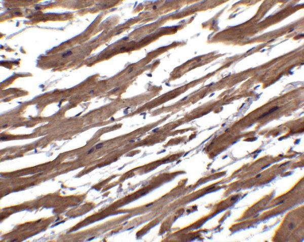
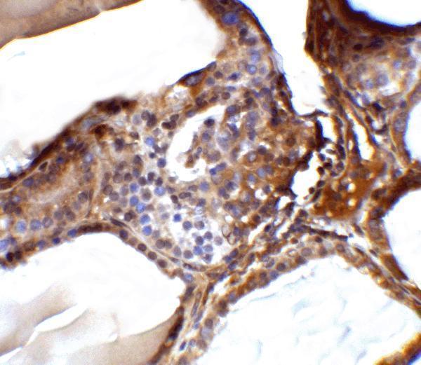
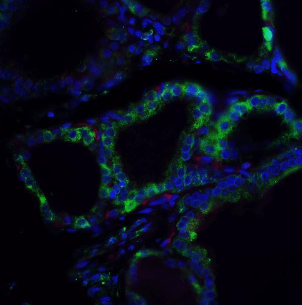
The catalog antibody A02857-1 has IHC images of paraffin-embedded human and mouse spleen and an IF image of human spleen (catalog image captions).
A02857-1 is listed for IHC-P and IF in human and mouse samples (catalog applications and reactivity). Its images show IHC in paraffin-embedded human and mouse spleen and IF in human spleen (catalog image captions).
Which to pick: For tissue IHC, choose A02857-1: its spleen images use paraffin sections fixed with formaldehyde, citrate retrieval at pH 6, and primary antibody at 2.5–5 μg/mL (A02857-1 IHC captions). For IF, the same SKU has a human spleen image at 5 μg/mL; ICC would need separate validation (A02857-1 IF caption; catalog applications). For cross-species work, A02857-1 lists human and mouse reactivity and shows IHC images from both species; the catalog describes IHC validation in human samples (catalog reactivity; A02857-1 IHC captions; catalog validation statement).