This website uses cookies to ensure you get the best experience on our website.


- Table of Contents
Plan chromogenic RUNX1 IHC in paraffin sections around the expected nuclear signal (HPA tissue IHC). Use HPA-positive basal or eccrine gland cells as reference material, and check antibody specificity before scoring (HPA tissue IHC; datasheet M00086).

Expected localisation, validated protocols, controls and antibodies — the at-a-glance facts below, then the full design guide.
| Expected localisation | Nuclear staining in several tissues (HPA tissue IHC) | |
| Staining pattern | Nuclear signal in basal and eccrine gland cells (HPA tissue IHC) | |
| Antigen retrieval | Citrate pH 6 HIER, heat-mediated (datasheet PB9157) | |
| Positive control | Nasopharynx+4 more · see all | |
| Negative control | Adipose tissue+4 more · see all |
| Fixation | Keep fixation consistent across paraffin sections (standard IHC practice; not target-specific) | |
| Caveat | M00086 recognizes RUNX1/2/3; verify specificity (datasheet M00086) | |
| Regulation | Highest in thymus, marrow and blood (UniProt) | |
| Isoform / epitope | 11 isoforms; verify epitope coverage (UniProt) |
The catalog antibody protocol uses citrate pH 6 heat retrieval (datasheet: PB9157). The published IHC protocols below provide sample-specific staining conditions (PMC4481134; PMC4420729; PMC13040063; PMC5725115).
| Sample | Paraffin-embedded rat thymus tissue; fixative not specified (datasheet PB9157) |
| Fixation | Image fixative and duration unreported (datasheet PB9157); verify before use. |
| Sectioning | 4–5 µm sections on charged slides (standard) |
| Deparaffinisation | Xylene, graded ethanol series to water (standard) |
| Antigen retrieval | Heat retrieval: Citrate pH 6, 20 min (datasheet PB9157) |
| Peroxidase block | 3% H2O2, 10 min, room temperature (standard) |
| Blocking | 10% goat serum (datasheet PB9157) |
| Primary antibody | Rabbit anti-RUNX1, 0.5-1μg/ml (datasheet PB9157) |
| Primary incubation | Overnight at 4 °C (datasheet PB9157) |
| Detection | Streptavidin-biotin complex (SABC), DAB chromogen (datasheet PB9157) |
| Counterstain | Hematoxylin, blue, dehydrate and mount (standard) |
| Expected result | RUNX1-positive staining in basal cells of nasopharynx (HPA tissue IHC: High). HPA tissue profile: Nuclear expression in several tissues. No signal in the no-primary control. |
RUNX1 should appear mainly in nuclei in paraffin-section IHC (UniProt Q01196: nucleus; HPA: nuclear expression in several tissues). Look for staining in nasopharyngeal basal cells, skin eccrine glands and bone-marrow hematopoietic cells (HPA: High, High and Medium, respectively). RUNX1 has no transmembrane segment (UniProt Q01196: topology). HPA rates the tissue IHC profile Enhanced, with medium consistency between antibody staining and RNA expression (HPA: tissue IHC).
| Distinct nuclear staining in nasopharyngeal basal cells or skin eccrine glands. | This matches two reported High cell populations and the expected compartment (HPA: tissue IHC; UniProt Q01196: nucleus). Judge the named cells, since staining across an entire tissue is not the reported pattern (HPA: cell-specific tissue IHC). |
| Strong cytoplasmic staining with little or no nuclear staining in an expected-positive cell population. | Treat this as an IHC localisation problem and review specificity and detection controls (UniProt Q01196: nucleus; general IHC practice). HPA also reports vesicles in ICC-IF, but that additional IF location does not establish a predominantly cytoplasmic chromogenic IHC pattern (HPA: subcellular ICC-IF). |
| Signal in adipocytes or skeletal-muscle myocytes while the expected-positive cells are unstained. | HPA reports both cell types as Not detected (HPA: tissue IHC). Suspect cross-reactivity or endogenous detection activity and compare control sections (general IHC practice); the unexpected signal alone does not identify its cause. |
| Broad, diffuse colour obscures nuclei and tissue boundaries. | The slide cannot be scored reliably for the reported nuclear pattern (HPA: tissue IHC; general IHC practice). Nonspecific binding, excess detection signal or endogenous enzyme activity are possible workflow causes; inspect controls before calling cells positive (general IHC practice). |
| No nuclear signal in nasopharyngeal basal cells, skin eccrine glands or bone-marrow hematopoietic cells. | These are reported High, High and Medium populations, respectively (HPA: tissue IHC). Check that the chosen population is present and that the IHC detection run worked (general IHC practice). An absent signal on one slide does not by itself establish absent RUNX1. |
| Cell population selected for scoring | HPA reports High nasopharyngeal basal cells and skin eccrine glands, but Medium bone-marrow hematopoietic cells and Not detected adipocytes (HPA: tissue IHC). Select and score the named cells; these labels do not promise uniform staining within a whole section. |
| Tissue expression versus slide intensity | UniProt reports highest expression in thymus, bone marrow and peripheral blood (UniProt Q01196: tissue specificity). HPA assigns bone-marrow hematopoietic cells Medium by IHC (HPA: tissue IHC). Do not convert the UniProt expression ranking into an expected chromogenic intensity. |
| Antibody validation | HPA004176 is IHC Enhanced, a status supported by independent antibody agreement or orthogonal data (HPA: antibody validation). HPA037912 has an ICC Enhanced entry but no IHC rating in the supplied record (HPA: antibody validation); its IF result is not IHC validation. |
| Isoform and epitope coverage | UniProt lists 11 RUNX1 isoforms and the Runt domain at residues 50–178 (UniProt Q01196: isoforms and domain). The supplied record gives no antibody epitope, so isoform recognition cannot be inferred; consult the IHC-validated antibody's documentation before interpreting a missing population. |
| Processing and staining compartment | UniProt reports no signal peptide, propeptide or transmembrane segment and lists one RUNX1 chain spanning residues 1–453 (UniProt Q01196: processing and topology). These facts support a nuclear expectation, but do not establish fixation sensitivity or predict how a particular epitope survives processing. |
| IF/ICC Q&A: what location should IF show? | Mainly nucleoplasmic staining, with an additional vesicular location reported by HPA ICC-IF (HPA: subcellular ICC-IF). Interpret that observation within the separate IF/ICC guide; this section offers no IF protocol option. |
| Situation | Likely cause | Next action |
|---|---|---|
| Expected-positive cells show no nuclear colour. | The cells may be absent from the section, or the IHC run may have failed (general IHC practice). HPA reports High nasopharyngeal basal and skin eccrine cells and Medium bone-marrow hematopoietic cells (HPA: tissue IHC). | Confirm the named cells on the section; review the IHC-validated antibody's documented conditions and a run control (general IHC practice). Do not assign a RUNX1-specific retrieval or fixation failure from these sources. |
| Signal is mostly cytoplasmic rather than nuclear. | Possible nonspecific staining or detection artefact (general IHC practice); a mostly cytoplasmic IHC result conflicts with the reported nuclear pattern (UniProt Q01196: nucleus; HPA: tissue IHC). | Compare the same cell population with a control section and inspect nuclear counterstain and signal distribution (general IHC practice). HPA's additional vesicular ICC-IF location does not validate diffuse cytoplasmic IHC (HPA: subcellular ICC-IF). |
| Negative-reference cells stain strongly. | Adipocytes and skeletal-muscle myocytes are Not detected in the supplied HPA tissue IHC record (HPA: tissue IHC). Cross-reactivity or endogenous detection activity is possible, but the appearance alone cannot distinguish them (general IHC practice). | Compare reagent controls and expected-positive cells on the run; investigate endogenous detection activity and nonspecific binding using standard IHC controls (general IHC practice). |
| Colour is diffuse across tissue or obscures cell boundaries. | Background or excessive detection signal can prevent compartment-level scoring (general IHC practice). HPA describes nuclear expression, so diffuse colour is not sufficient evidence of a RUNX1-positive cell (HPA: tissue IHC). | Inspect control sections and review blocking, antibody dilution and detection conditions against the IHC-validated antibody's documentation (general IHC practice). Score only cells with separable nuclear signal. |
| Bone marrow looks weaker than nasopharynx or skin. | HPA calls bone-marrow hematopoietic cells Medium, versus High nasopharyngeal basal cells and skin eccrine glands (HPA: tissue IHC). UniProt's high bone-marrow expression is a different type of evidence (UniProt Q01196: tissue specificity). | Score the identified hematopoietic cells against local background and the run control (general IHC practice). Avoid requiring identical intensity across the three HPA populations. |
| An IF image shows puncta in addition to nuclear signal. | HPA reports nucleoplasm as the main ICC-IF location and vesicles as an additional location (HPA: subcellular ICC-IF). The IF observation alone does not establish the expected chromogenic IHC appearance. | Use the separate IF/ICC guide to evaluate the image (HPA: subcellular ICC-IF). For this IHC section, base the positive call on discernible nuclear staining in the HPA-reported cell populations (HPA: tissue IHC). |


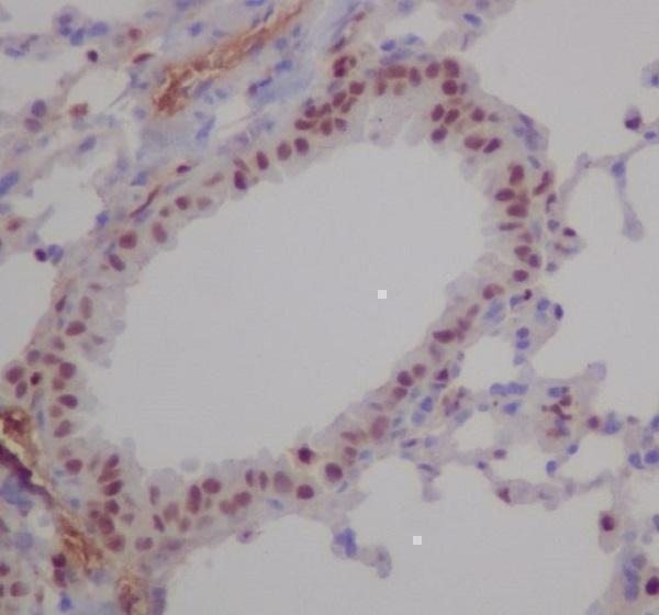


Comprehensive Human Protein Atlas IHC scoring per tissue (reliability: Enhanced — Medium consistency between antibody staining and RNA expression data.). Rows are taken directly from the HPA tissue chart — click any row's HPA link to view the source.
| Tissue | Cell type | Level | Evidence | Source |
|---|---|---|---|---|
| Nasopharynx | Basal cells | High | Protein (IHC) | HPA → |
| Skin | Eccrine glands | High | Protein (IHC) | HPA → |
| Appendix | Lymphoid tissue | Medium | Protein (IHC) | HPA → |
| Bone marrow | Hematopoietic cells | Medium | Protein (IHC) | HPA → |
| Breast | Glandular cells | Medium | Protein (IHC) | HPA → |
| Tissue | Cell type | Level | Evidence | Source |
|---|---|---|---|---|
| Adipose tissue | Adipocytes | Not detected | Protein (IHC) | HPA → |
| Esophagus | Squamous epithelial cells | Not detected | Protein (IHC) | HPA → |
| Liver | Cholangiocytes | Not detected | Protein (IHC) | HPA → |
| Oral mucosa | Squamous epithelial cells | Not detected | Protein (IHC) | HPA → |
| Parathyroid gland | Glandular cells | Not detected | Protein (IHC) | HPA → |
Troubleshoot RUNX1 staining in paraffin sections by checking nuclear localisation, cell identity and matched controls before comparing staining intensity.
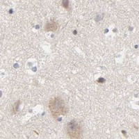
Real IHC figures show RUNX1 staining in rat thymus paraffin sections and RUNX-family staining in mouse lung paraffin sections (PB9157 and M00086 image captions); M00086 also lists IF/ICC (catalog applications).
PB9157 will render with a RUNX1 IHC figure from rat thymus paraffin sections; its listed IHC reactivity is human, mouse and rat (PB9157 image caption; catalog reactivity). M00086 will render with a RUNX1/RUNX2/RUNX3 IHC figure from mouse lung paraffin sections; it lists IHC and IF/ICC for human, mouse and rat, but supplies no IF figure (M00086 image caption; catalog applications and reactivity; IF image alts).
Which to pick: For RUNX1 tissue IHC, start with PB9157 at 0.5–1 μg/ml: its own figure shows rat thymus paraffin sections with citrate retrieval, and the fixative is unreported (PB9157 catalog dilution; PB9157 image caption). For IF/ICC, consider rabbit monoclonal M00086 at 1:50; IF/ICC is listed, while its own figure documents only paraffin-section IHC of mouse lung and reports no fixative (M00086 title; catalog applications and dilution; M00086 image caption). Both list human, mouse and rat reactivity for cross-species work; M00086 targets RUNX1/RUNX2/RUNX3, so choose PB9157 when RUNX1 specificity is the priority (catalog titles and reactivity).