This website uses cookies to ensure you get the best experience on our website.


- Table of Contents
Plan RUNX1T1 staining in paraffin sections using the catalog antibody at 0.5–1 μg/mL (datasheet A01792-1). Use the reported tissue patterns to choose controls, and interpret staining in light of the low consistency with RNA data (HPA tissue IHC).

Expected localisation, validated protocols, controls and antibodies — the at-a-glance facts below, then the full design guide.
| Expected localisation | Low nucleolar tissue staining (HPA tissue IHC); nuclear protein (UniProt) | |
| Staining pattern | Most tissues show low nucleolar staining (HPA tissue IHC) | |
| Antigen retrieval | Citrate pH 6 HIER, heat-mediated (datasheet A01792-1) | |
| Positive control | Bronchus+3 more · see all | |
| Negative control | Adrenal gland+4 more · see all |
| Fixation | Keep fixation consistent across sections (standard IHC practice; not target-specific) | |
| Caveat | Antibody staining has low consistency with RNA data (HPA tissue IHC) | |
| Regulation | Expression is enriched in brain (UniProt) | |
| Isoform / epitope | Six isoforms; check antibody epitope coverage (UniProt) |
The catalog antibody’s IHC-P protocol is accompanied by three published RUNX1T1 IHC protocols covering pancreatic endocrine neoplasm, gastric, and neuroblastoma sections (PMC4732279; PMC5143359; PMC11239676).
| Sample | Paraffin-embedded human mammary cancer tissue; fixative not specified (datasheet A01792-1) |
| Fixation | Image fixative and duration unreported (datasheet A01792-1); verify before use. |
| Sectioning | 4–5 µm sections on charged slides (standard) |
| Deparaffinisation | Xylene, graded ethanol series to water (standard) |
| Antigen retrieval | Heat retrieval: Citrate pH 6, 20 min (datasheet A01792-1) |
| Peroxidase block | 3% H2O2, 10 min, room temperature (standard) |
| Blocking | 10% goat serum (datasheet A01792-1) |
| Primary antibody | Rabbit anti-RUNX1T1, 0.5-1μg/ml (datasheet A01792-1) |
| Primary incubation | Overnight at 4 °C (datasheet A01792-1) |
| Detection | Streptavidin-biotin complex (SABC), DAB chromogen (datasheet A01792-1) |
| Counterstain | Hematoxylin, blue, dehydrate and mount (standard) |
| Expected result | RUNX1T1-positive staining in ciliated cells (ciliary rootlets) of bronchus (HPA tissue IHC: Medium). HPA tissue profile: Low nucleolar expression in most tissues. No signal in the no-primary control. |
RUNX1T1 is a nuclear protein without a transmembrane segment (UniProt Q06455: subcellular location and topology). In paraffin-section IHC, expect low nucleolar staining in most tissues, with medium staining reported in ciliary rootlets of bronchus, fallopian tube and nasopharynx, and in testis Leydig cells (HPA: tissue IHC). Treat these as reference patterns: HPA rates the tissue IHC evidence Approved but reports low consistency between antibody staining and RNA expression (HPA: reliability).
| Low staining within nucleoli across a tissue section, with cell-to-cell variation. | This matches the broad tissue IHC profile (HPA: low nucleolar expression in most tissues). Score the stained compartment and cell type alongside intensity; a weak, restricted signal can fit this reference better than uniformly strong staining. The HPA Approved rating carries a low RNA–staining consistency caveat (HPA: reliability). |
| Medium staining concentrated at ciliary rootlets in respiratory or fallopian-tube epithelium. | This matches the reported ciliated-cell pattern in bronchus, nasopharynx and fallopian tube (HPA: Medium in ciliary rootlets). Record the rootlet location explicitly. It differs from the broad low nucleolar profile (HPA: tissue IHC), so evaluate each compartment against its corresponding reference rather than imposing one pattern on every cell. |
| Strong, widespread cytoplasmic or membrane-like chromogen that obscures nuclei and cell boundaries. | That distribution warrants review against the nuclear UniProt annotation and low nucleolar tissue profile (UniProt Q06455: nucleus; HPA: tissue IHC). HPA also lists cytosol as an additional ICC-IF location, so cytoplasmic signal alone is not proof of artefact (HPA: subcellular). Check tissue morphology and controls before assigning RUNX1T1 specificity (standard IHC practice). |
| Intense staining in a cell population reported as not detected, such as bone-marrow hematopoietic cells. | This conflicts with that cell-specific tissue IHC observation (HPA: bone marrow hematopoietic cells, Not detected). Consider cross-reactivity or endogenous detection activity as possibilities, then inspect the negative control and a known-positive reference on the same run (standard IHC practice). One discrepant section does not establish which cause is responsible. |
| No staining in an otherwise well-preserved testis section's Leydig cells. | Leydig cells are a reported medium-staining reference (HPA: testis Leydig cells, Medium). A blank result can reflect a failed staining run, unsuitable assay conditions or sample variation (standard IHC practice). Check a positive control and the detection workflow before interpreting this section as biological absence; the HPA pattern is a reference, not a guarantee for every specimen. |
| Which reference cell population is being scored? | HPA reports Medium signal in ciliated-cell rootlets in bronchus, fallopian tube and nasopharynx and in testis Leydig cells; it reports Not detected in bone-marrow hematopoietic cells (HPA: tissue IHC). State the cell population when scoring, because a whole-section positive or negative label loses this distinction. |
| How much weight should RNA expression carry? | UniProt describes strongest tissue expression in brain, and HPA calls brain RNA tissue enhanced (UniProt Q06455: tissue specificity; HPA: RNA specificity). HPA nevertheless reports low staining in cerebral-cortex endothelial cells and cerebellar Purkinje cells, and none in hippocampal glial cells (HPA: tissue IHC). Do not infer uniform brain-cell IHC positivity from tissue-level RNA. |
| What does antibody validation establish? | The IHC antibody HPA070951 is Approved, while HPA049852 has Supported ICC validation and no listed IHC status (HPA: antibodies). HPA describes low consistency between antibody staining and RNA expression (HPA: reliability). Use the tissue images as pattern references, with controls for the specific assay; these labels do not establish equivalent IHC performance for the two antibodies. |
| Does IF/ICC show the same pattern? | IF/ICC has its own guide page. Its supported main location is nucleoplasm, with cytosol listed as an additional location; images are listed for HEK293, U2OS and NIH 3T3 (HPA: subcellular ICC-IF). UniProt also notes discrete nuclear dots with ATN1 (UniProt Q06455: subcellular location). These annotations inform localisation, without defining an IF/ICC protocol or overriding the tissue IHC patterns. |
| Situation | Likely cause | Next action |
|---|---|---|
| The positive-reference Leydig cells appear blank. | The result differs from the reported Medium Leydig-cell signal (HPA: testis tissue IHC); the cause is unresolved. | Check section morphology, primary-antibody application, chromogenic detection and a run-level positive control (standard IHC practice). If the run works, document the specimen-level discrepancy rather than assuming RUNX1T1 is absent. |
| Chromogen covers nearly every cell or fills empty tissue spaces. | Diffuse background can arise from nonspecific binding, incomplete washing or detection chemistry (standard IHC practice). | Compare with a no-primary control, inspect blocking and washes, and reassess dilution under the assay's established conditions (standard IHC practice). Re-evaluate against the low nucleolar and cell-restricted reference patterns (HPA: tissue IHC). |
| Hematopoietic cells show strong staining in bone marrow. | That conflicts with the reported Not detected result (HPA: bone marrow hematopoietic cells); cross-reactivity or endogenous detection activity is possible (standard IHC practice). | Compare a no-primary control and a positive reference, then review the chromogenic detection controls before calling the signal RUNX1T1 (standard IHC practice). |
| Brain sections show less staining than expected from RNA data. | Brain is tissue enhanced by RNA, but the listed brain-cell IHC observations are low or not detected (HPA: RNA specificity and tissue IHC). | Score the specified cells and compartment: cerebral-cortex endothelial cells and cerebellar Purkinje cells are Low; hippocampal glial cells are Not detected (HPA: tissue IHC). Avoid using bulk brain RNA as a cell-level IHC threshold. |
| The signal seems cytoplasmic rather than nuclear or nucleolar. | Tissue IHC is broadly low nucleolar, while ICC-IF lists cytosol as an additional location (HPA: tissue IHC; HPA: subcellular). The discrepancy alone cannot identify the cause. | Inspect focus, counterstain and compartment boundaries, then compare an appropriate positive tissue and no-primary control (standard IHC practice). Record the actual distribution rather than relabeling it nuclear. |
| Different antibodies give different tissue patterns. | HPA lists Approved IHC for HPA070951 and Supported ICC for HPA049852, with no IHC status listed for the latter (HPA: antibodies). Equivalent paraffin-section performance is unestablished. | Check each antibody's stated application and validation before comparing results (HPA: antibodies). For IHC interpretation, use an IHC-validated antibody with matched positive and negative controls (standard IHC practice). |

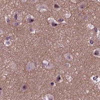


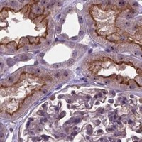


Comprehensive Human Protein Atlas IHC scoring per tissue (reliability: Approved — Low consistency between antibody staining and RNA expression data.). Rows are taken directly from the HPA tissue chart — click any row's HPA link to view the source.
| Tissue | Cell type | Level | Evidence | Source |
|---|---|---|---|---|
| Bronchus | Ciliated cells (ciliary rootlets) | Medium | Protein (IHC) | HPA → |
| Fallopian tube | Ciliated cells (ciliary rootlets) | Medium | Protein (IHC) | HPA → |
| Nasopharynx | Ciliated cells (ciliary rootlets) | Medium | Protein (IHC) | HPA → |
| Testis | Leydig cells | Medium | Protein (IHC) | HPA → |
| Tissue | Cell type | Level | Evidence | Source |
|---|---|---|---|---|
| Adrenal gland | Glandular cells | Not detected | Protein (IHC) | HPA → |
| Bone marrow | Hematopoietic cells | Not detected | Protein (IHC) | HPA → |
| Epididymis | Glandular cells | Not detected | Protein (IHC) | HPA → |
| Heart muscle | Cardiomyocytes | Not detected | Protein (IHC) | HPA → |
| Hippocampus | Glial cells | Not detected | Protein (IHC) | HPA → |
Troubleshoot RUNX1T1 staining by checking retrieval, nuclear localisation, antibody specificity and the cell types being scored (UniProt Q06455; HPA tissue IHC).
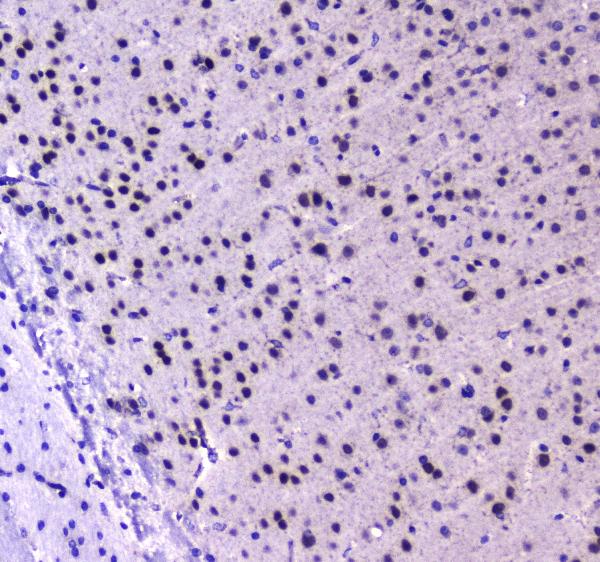
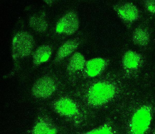
A01792-1 has IHC images from human mammary cancer and mouse brain paraffin sections (catalog IHC captions), plus IF data from U20S cells (catalog IF caption).
A01792-1 has documented paraffin-section IHC in human mammary cancer and mouse brain (catalog IHC captions). The same SKU has documented IF in U20S cells and lists human, mouse, and rat reactivity (catalog IF caption; catalog reactivity).
Which to pick: For tissue IHC, choose A01792-1: its paraffin-section captions document 1 μg/ml antibody after citrate retrieval at pH 6 (catalog IHC captions). For IF/ICC, A01792-1 lists both applications and shows IF in U20S cells at 2 μg/ml (catalog applications; catalog IF caption). For cross-species planning, A01792-1 lists human, mouse, and rat reactivity, while its IHC images document human and mouse tissue only; clone and tissue fixative are unreported (catalog reactivity; catalog IHC captions; catalog clone field).