Click image to see more details
-

-
-
-
-
+2
Product Info Summary
| SKU: | A01792-1 |
|---|---|
| Size: | 100 μg/vial |
| Reactive Species: | Human, Mouse, Rat |
| Host: | Rabbit |
| Application: | ELISA, Flow Cytometry, IP, IF, IHC, ICC, WB |
Customers Who Bought This Also Bought

Product info
Product Name
Anti-RUNX1T1/ETO Antibody Picoband®
SKU/Catalog Number
A01792-1
Size
100 μg/vial
Form
Lyophilized
Description
Boster Bio Anti-RUNX1T1/ETO Antibody Picoband® catalog # A01792-1. Tested in ELISA, Flow Cytometry, IP, IF, IHC, ICC, WB applications. This antibody reacts with Human, Mouse, Rat. The brand Picoband indicates this is a premium antibody that guarantees superior quality, high affinity, and strong signals with minimal background in Western blot applications. Only our best-performing antibodies are designated as Picoband, ensuring unmatched performance.
Storage & Handling
Store at -20˚C for one year from date of receipt. After reconstitution, at 4˚C for one month. It can also be aliquotted and stored frozen at -20˚C for six months. Avoid repeated freeze-thaw cycles.
Cite This Product
Anti-RUNX1T1/ETO Antibody Picoband® (Boster Biological Technology, Pleasanton CA, USA, Catalog # A01792-1)
Host
Rabbit
Contents
Each vial contains 4mg Trehalose, 0.9mg NaCl, 0.2mg Na2HPO4, 0.05mg NaN3.
Clonality
Polyclonal
Isotype
Rabbit IgG
Immunogen
E. coli-derived human RUNX1T1/ETO recombinant protein (Position: T335-D510).
Cross-reactivity
No cross-reactivity with other proteins.
Reactive Species
A01792-1 is reactive to RUNX1T1 in Human, Mouse, Rat
Observed Molecular Weight
67 kDa
Calculated molecular weight
67.6 kDa
Background of RUNX1T1
Protein CBFA2T1 is a protein that in humans is encoded by the RUNX1T1 gene. This gene encodes a member of the myeloid translocation gene family which interact with DNA-bound transcription factors and recruit a range of corepressors to facilitate transcriptional repression. The t (8;21) (q22;q22) translocation is one of the most frequent karyotypic abnormalities in acute myeloid leukemia. The translocation produces a chimeric gene made up of the 5'-region of the runt-related transcription factor 1 gene fused to the 3'-region of this gene. The chimeric protein is thought to associate with the nuclear corepressor/histone deacetylase complex to block hematopoietic differentiation. Alternative splicing results in multiple transcript variants.
Antibody Validation
Boster validates all antibodies on WB, IHC, ICC, Immunofluorescence, and ELISA with known positive control and negative samples to ensure specificity and high affinity, including thorough antibody incubations.
Application & Images
Applications
A01792-1 is guaranteed for ELISA, Flow Cytometry, IP, IF, IHC, ICC, WB Boster Guarantee
Recommend Dilution
| Application | Dilution | Species |
|---|---|---|
| Western blot | 0.1-0.5μg/ml | Human, Mouse |
| Immunohistochemistry (Paraffin-embedded Section) | 0.5-1μg/ml | Human, Mouse |
| Immunocytochemistry/Immunofluorescence | 2μg/ml | Human |
| Immunoprecipitation | 0.5-2 μg/ml | Human |
| Flow Cytometry (Fixed) | 1-3μg/1x106 cells | Human |
| ELISA | 0.1-0.5μg/ml | - |
Tested application
Suggested blocking solution with 5% non-fat milk or BSA; (*)Recommended protein loading: 20-40 µg per lane
Use TE buffer pH 9.0 for antigen retrieval; (*) citrate buffer pH 6.0 is an alternative.
Validation Images & Assay Conditions
Click image to see more details
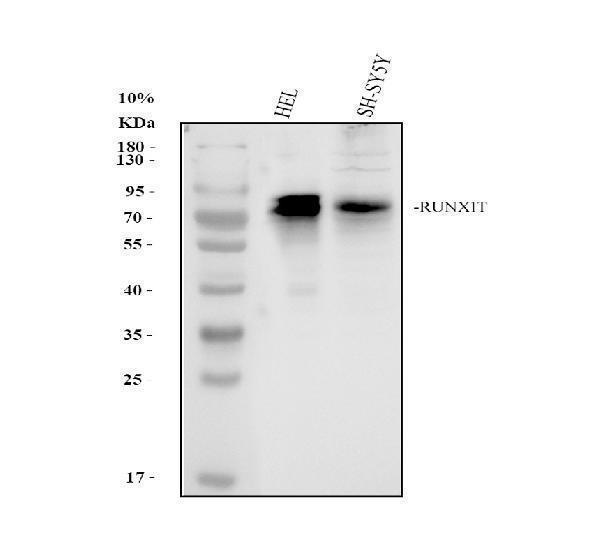
Western blot analysis of RUNX1T1 using anti-RUNX1T1 antibody (A01792-1).
Electrophoresis was performed on a 10% SDS-PAGE gel at 80V (Stacking gel) / 120V (Resolving gel) for 2 hours. The sample well of each lane was loaded with 30 ug of sample under reducing conditions.
Lane 1: human Hela whole cell lysates,
Lane 2: human SH-SY5Y whole cell lysates.
After electrophoresis, proteins were transferred to a nitrocellulose membrane at 150 mA for 50-90 minutes. Blocked the membrane with 5% non-fat milk/TBS for 1.5 hour at RT. The membrane was incubated with rabbit anti-RUNX1T1 antigen affinity purified polyclonal antibody (A01792-1) at 0.5 μg/mL overnight at 4°C, then washed with TBS-0.1%Tween 3 times with 5 minutes each and probed with a goat anti-rabbit IgG-HRP secondary antibody (Catalog # BA1054) at a dilution of 1:5000 for 1.5 hour at RT. The signal is developed using an ECL Plus Western Blotting Substrate (Catalog # AR1196-200) with Tanon 5200 system. A specific band was detected for RUNX1T1 at approximately 70-80 kDa. The expected band size for RUNX1T1 is at 67 kDa.

Click image to see more details
IHC analysis of RUNX1T1/ETO using anti-RUNX1T1/ETO antibody (A01792-1).
RUNX1T1/ETO was detected in paraffin-embedded section of human mammary cancer tissue. Heat mediated antigen retrieval was performed in citrate buffer (pH6, epitope retrieval solution) for 20 mins. The tissue section was blocked with 10% goat serum. The tissue section was then incubated with 1μg/ml rabbit anti-RUNX1T1/ETO Antibody (A01792-1) overnight at 4°C. Biotinylated goat anti-rabbit IgG was used as secondary antibody and incubated for 30 minutes at 37°C. The tissue section was developed using Strepavidin-Biotin-Complex (SABC)(Catalog # SA1022) with DAB as the chromogen.

Click image to see more details
IHC analysis of RUNX1T1/ETO using anti-RUNX1T1/ETO antibody (A01792-1).
RUNX1T1/ETO was detected in paraffin-embedded section of mouse brain tissue . Heat mediated antigen retrieval was performed in citrate buffer (pH6, epitope retrieval solution) for 20 mins. The tissue section was blocked with 10% goat serum. The tissue section was then incubated with 1μg/ml rabbit anti-RUNX1T1/ETO Antibody (A01792-1) overnight at 4°C. Biotinylated goat anti-rabbit IgG was used as secondary antibody and incubated for 30 minutes at 37°C. The tissue section was developed using Strepavidin-Biotin-Complex (SABC)(Catalog # SA1022) with DAB as the chromogen.

Click image to see more details
IF analysis of RUNX1T1 using anti-RUNX1T1 antibody (A01792-1).
RUNX1T1 was detected in immunocytochemical section of U20S cell. Enzyme antigen retrieval was performed using IHC enzyme antigen retrieval reagent (AR0022) for 15 mins. The cells were blocked with 10% goat serum. And then incubated with 2μg/mL rabbit anti-RUNX1T1 Antibody (A01792-1) overnight at 4°C. DyLight®488 Conjugated Goat Anti-Rabbit IgG (BA1127) was used as secondary antibody at 1:100 dilution and incubated for 30 minutes at 37°C. Visualize using a fluorescence microscope and filter sets appropriate for the label used.

Click image to see more details
Immunoprecipitating (IP) RUNX1T1 in SH-SY5Y whole cell lysate.
Western blot analysis of RUNX1T1 using anti-RUNX1T1 antibody (A01792-1);
Lane 1: SH-SY5Y whole cell lysates (30ug);
Lane 2: Rabbit control IgG instead of anti-RUNX1T1 antibody in SH-SY5Y whole cell lysate;
Lane 3: anti-RUNX1T1 antibody (2μg) + SH-SY5Y whole cell lysate (500μg).
After electrophoresis, proteins were transferred to a membrane. Then the membrane was incubated with rabbit anti-RUNX1T1 antigen affinity purified polyclonal antibody (A01792-1) at a dilution of 0.5 μg/mL and probed with a goat anti-rabbit IgG-HRP secondary antibody (Catalog # BA1054). The signal is developed using ECL Plus Western Blotting Substrate (Catalog # AR1196-200). A specific band was detected for RUNX1T1 at approximately 70-80 kDa. The expected band size for RUNX1T1 is at 67 kDa.

Click image to see more details
Flow Cytometry analysis of U20S cells using anti-RUNX1T1 antibody (A01792-1).
Overlay histogram showing U20S cells stained with A01792-1 (Blue line). To facilitate intracellular staining, cells were fixed with 4% paraformaldehyde and permeabilized with permeabilization buffer. The cells were blocked with 10% normal goat serum. And then incubated with rabbit anti-RUNX1T1 Antibody (A01792-1,1μg/1x106 cells) for 30 min at 20°C. DyLight®488 conjugated goat anti-rabbit IgG (BA1127, 5-10μg/1x106 cells) was used as secondary antibody for 30 minutes at 20°C. Isotype control antibody (Green line) was rabbit IgG (1μg/1x106) used under the same conditions. Unlabelled sample (Red line) was also used as a control.
Specific Publications For Anti-RUNX1T1/ETO Antibody Picoband® (A01792-1)
Loading publications
Recommended Resources
Here are featured tools and databases that you might find useful.
- Boster's Pathways Library
- Protein Databases
- Bioscience Research Protocol Resources
- Data Processing & Analysis Software
- Photo Editing Software
- Scientific Literature Resources
- Research Paper Management Tools
- Molecular Biology Software
- Primer Design Tools
- Bioinformatics Tools
- Phylogenetic Tree Analysis
Customer Reviews
Have you used Anti-RUNX1T1/ETO Antibody Picoband®?
Share your experimental results or join a short interview to earn up to $1,000 in product credits or other rewards.
0 Reviews For Anti-RUNX1T1/ETO Antibody Picoband®
Customer Q&As
Have a question?
Find answers in Q&As, reviews.
Can't find your answer?
Submit your question
4 Customer Q&As for Anti-RUNX1T1/ETO Antibody Picoband®
Question
I am interested in to test anti-RUNX1T1/ETO antibody A01792-1 on human brain for research purposes, then I may be interested in using anti-RUNX1T1/ETO antibody A01792-1 for diagnostic purposes as well. Is the antibody suitable for diagnostic purposes?
Verified Customer
Verified customer
Asked: 2020-03-26
Answer
The products we sell, including anti-RUNX1T1/ETO antibody A01792-1, are only intended for research use. They would not be suitable for use in diagnostic work. If you have the means to develop a product into diagnostic use, and are interested in collaborating with us and develop our product into an IVD product, please contact us for more discussions.
Boster Scientific Support
Answered: 2020-03-26
Question
We are currently using anti-RUNX1T1/ETO antibody A01792-1 for human tissue, and we are content with the IF results. The species of reactivity given in the datasheet says human, mouse, rat. Is it likely that the antibody can work on pig tissues as well?
Verified Customer
Verified customer
Asked: 2019-06-06
Answer
The anti-RUNX1T1/ETO antibody (A01792-1) has not been validated for cross reactivity specifically with pig tissues, though there is a good chance of cross reactivity. We have an innovator award program that if you test this antibody and show it works in pig you can get your next antibody for free. Please contact me if I can help you with anything.
Boster Scientific Support
Answered: 2019-06-06
Question
We appreciate helping with my inquiry over the phone. Here are the WB image, lot number and protocol we used for brain using anti-RUNX1T1/ETO antibody A01792-1. Let me know if you need anything else.
Verified Customer
Verified customer
Asked: 2018-05-03
Answer
We appreciate the data. You have provided everything we needed. Our lab team are working to resolve your inquiry as quickly as possible, and we appreciate your patience and understanding! Please let me know if there is anything you need in the meantime.
Boster Scientific Support
Answered: 2018-05-03
Question
Will A01792-1 anti-RUNX1T1/ETO antibody work on parafin embedded sections? If so, which fixation method do you recommend we use (PFA, paraformaldehyde, other)?
D. Jackson
Verified customer
Asked: 2016-12-01
Answer
It shows on the product datasheet, A01792-1 anti-RUNX1T1/ETO antibody as been tested on ELISA. It is best to use PFA for fixation because it has better tissue penetration ability. PFA needs to be prepared fresh before use. Long term stored PFA turns into formalin, as the PFA molecules congregate and become formalin.
Boster Scientific Support
Answered: 2016-12-01
