S100B / Protein S100-B · IHC design guide

Design Immunohistochemistry for S100B

Plan S100B paraffin-section IHC around nuclear and cytoplasmic staining in CNS glia and peripheral nerves (HPA tissue IHC). Keep fixation consistent, use appropriate controls, and interpret differences from RNA expression in light of reported secretion (HPA tissue IHC).

Evidence assembled Oct 2026 · For research use; verify linked source records and product datasheet before use
Immunohistochemistry protocol sheet for S100B (IHC for S100B): expected localisation Nuclear and cytoplasmic tissue staining (HPA tissue IHC), antibody M00979-1, validated IHC image, and IHC protocol steps
Printable S100B IHC protocol sheet — expected localisation Nuclear and cytoplasmic tissue staining (HPA tissue IHC), antibody M00979-1, controls and protocol steps. Open the full S100B IHC guide →

S100B Immunohistochemistry Experimental Design Guide

Expected localisation, validated protocols, controls and antibodies — the at-a-glance facts below, then the full design guide.

Must know before staining
Expected localisation Nuclear and cytoplasmic tissue staining (HPA tissue IHC)
Staining pattern Nuclear/cytoplasmic CNS glia, nerves, adipocytes and lymphoid cells (HPA tissue IHC)
Antigen retrieval Tris-EDTA pH 9.0 HIER, 95–98 °C, 20 min (rule: nuclear antigen)
Positive control ⓘ Breast+4 more · see all
Negative control ⓘ Adrenal gland+4 more · see all
Important caveats
Reasons your staining may differ from the expected pattern.
Fixation Keep paraffin-section fixation consistent across samples. (standard IHC practice; not target-specific)
Caveat Secreted variants may shift protein staining relative to RNA (HPA tissue IHC)
Regulation CLSTN3beta promotes secretion (UniProt)
Isoform / epitope 0 isoforms; extracellular versus cytoplasmic epitope effects unknown (UniProt)
Section 1

Recommended S100B IHC & IF Protocols

The catalog antibody’s IHC-P protocol is paired with published S100B staining methods for rat gastric tissue (PMC4003764), human brain (PMC1769505), and human liver (PMC5339465).

Recommended immunohistochemistry (IHC-P) protocol parameters
SampleParaffin-embedded Rat cerebral cortex tissue; fixative not specified (datasheet M00979-1)
FixationImage fixative and duration unreported (datasheet M00979-1); verify before use.
Sectioning4–5 µm sections on charged slides (standard)
DeparaffinisationXylene, graded ethanol series to water (standard)
Antigen retrievalHeat-induced epitope retrieval in Tris-EDTA buffer, pH 9.0, 20 min at 95–98 °C (standard rule: nuclear antigen)
Peroxidase block3% H2O2, 10 min, room temperature (standard)
Blocking10% normal serum of the secondary host, 30 min, room temperature (standard)
Primary antibodyRabbit monoclonal (clone BCB-19) anti-S100B, 1:50 recommended; image 1:500 (datasheet M00979-1)
Primary incubationOvernight at 4 °C (standard)
DetectionHRP-polymer secondary, DAB chromogen 5–10 min (standard)
CounterstainHematoxylin, blue, dehydrate and mount (standard)
Expected resultS100B-positive staining in myoepithelial cells of breast (HPA tissue IHC: High). HPA tissue profile: Nuclear and cytoplasmic expression mainly in CNS, peripheral nerves, adipocytes and subset of lymphoid cells. No signal in the no-primary control.
💡Decision noteStart with Tris-EDTA pH 9.0 at 95–98 °C for 20 min (page retrieval); citrate pH 6.0 is a published alternative (PMC1769505).
Section 2

What Is the Expected S100B Staining Pattern?

Glial S100B is nuclear and cytoplasmic (HPA: Enhanced); it has no TM segment (UniProt P04271 topology).

What am I looking at on my slide?
Strong glial nuclear and cytoplasmic stain (HPA: High in glia).Expected pattern in CNS sections (HPA: nuclear/cytoplasmic profile).
Cell-surface rim alone, without nuclear or cytoplasmic stain.Suspect artefact; S100B has no TM segment (UniProt P04271 topology).
Adrenal glandular cells stain.Investigate cross-reactivity or endogenous detection activity (HPA: not detected in these cells).
Diffuse stain obscures cell boundaries.Background may reflect excess antibody or incomplete blocking (standard IHC practice).
No stain in caudate glia.Check the assay: caudate glia stain High (HPA tissue IHC).
💡Expected S100B appearancePositive: strong glial nuclear/cytoplasmic stain (HPA: High); adrenal glandular staining is suspect (HPA: not detected).
How each factor affects the staining
Tissue controlsCaudate glia: High; adrenal glandular cells: not detected (HPA tissue IHC).
Secreted fractionS100B can be secreted (UniProt P04271); tissue RNA and protein may differ (HPA tissue IHC).
Antibody validationThree listed antibodies have Enhanced IHC validation (HPA antibodies).
IF/ICC: where is signal seen?Mainly vesicles, cytosol and primary cilium; some sites are uncertain (HPA subcellular).
Why is my staining missing, weak or wrong?
SituationLikely causeNext action
No signal in caudate glia (HPA: High).Assay sensitivity or retrieval may be inadequate (standard IHC practice).Check positive control and antibody IHC-P instructions (standard IHC practice).
Only a cell-surface rim appears.Possible edge artefact; S100B lacks a TM segment (UniProt P04271 topology).Review morphology and detection controls (standard IHC practice).
Adrenal glandular cells stain (HPA: not detected).Possible cross-reactivity or endogenous detection activity (standard IHC practice).Run a no-primary control and review blocking (standard IHC practice).
Diffuse background obscures cells.Excess antibody or incomplete blocking may contribute (standard IHC practice).Adjust antibody dilution and blocking with controls (standard IHC practice).
Only nuclei stain in glia.Expected cytoplasmic signal may be missed (HPA: nuclear/cytoplasmic profile).Compare with a known-positive glial section (HPA: High in glia).

Sample controls for S100B IHC & IF

🧪Run cerebral cortex first: glial cells should stain (HPA: High in cerebral cortex glial cells). Use adrenal gland glandular cells as the negative tissue (HPA: Not detected in adrenal gland glandular cells); on the cortex slide, assess morphologically identified non-glial cells that lack staining as an internal background check.
Positive control tissue: Breast (Myoepithelial cells, HPA High)
Negative control tissue: Adrenal gland (HPA Not detected)
ICC-IF cell lines (HPA subcellular resource): HPA ICC-IF images show S100B in SK-MEL-30, U-251MG, U2OS, ASC52telo, RPTEC/TERT1, hTERT-RPE1 (serum starved), with annotated localisation: Vesicles (approved), Primary cilium (uncertain), Cytosol (supported) (HPA subcellular).
Technical controls: Include no-primary (secondary-only) and host-species- and clonality-matched isotype controls, plus S100B knockout tissue or a peptide-block control when suitable material is available (standard IHC practice). Quench endogenous peroxidase for chromogenic cortex IHC, and assess tissue autofluorescence if using IF (standard IHC/IF practice).
⚠️Feasibility: No target-specific fixation window or retrieval dependency is reported in the supplied evidence; optimize antigen retrieval on paraffin sections (standard IHC practice). The M00979-1 rat cerebral cortex paraffin-section caption reports 1:500 but no fixative, so the fixative is unreported and fixation is unconfirmed (selected product caption). The supplied evidence does not establish whether frozen sections or IF are easier; assess cortex autofluorescence if using IF (standard IF practice). The selected M00979-1 tissue-IHC caption documents paraffin sections, but does not specify the fixative (selected-SKU IHC image M00979-1).

HPA tissue IHC evidence for S100B

Comprehensive Human Protein Atlas IHC scoring per tissue (reliability: Enhanced — Medium consistency between antibody staining and RNA expression data. At least one protein variant secreted, tissue location of RNA and protein might differ and correlation is complex.). Rows are taken directly from the HPA tissue chart — click any row's HPA link to view the source.

Positive expression · recommended positive controls

TissueCell typeLevelEvidenceSource
Breast Myoepithelial cells High Protein (IHC) HPA →
Caudate Glial cells High Protein (IHC) HPA →
Cerebellum Bergmann glia - cytoplasm/membrane High Protein (IHC) HPA →
Cerebral cortex Glial cells High Protein (IHC) HPA →
Hippocampus Glial cells High Protein (IHC) HPA →

Undetected expression · recommended negative controls

TissueCell typeLevelEvidenceSource
Adrenal gland Glandular cells Not detected Protein (IHC) HPA →
Appendix Glandular cells Not detected Protein (IHC) HPA →
Bone marrow Hematopoietic cells Not detected Protein (IHC) HPA →
Bronchus Respiratory epithelial cells Not detected Protein (IHC) HPA →
Cervix Glandular cells Not detected Protein (IHC) HPA →
Section 3

Advanced S100B IHC Tips

Use cell type, compartment, and matched controls to troubleshoot S100B staining in paraffin sections; optimise chromogenic IHC before comparing specimens.

What should I change when S100B staining is weak after retrieval?
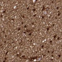
Start with Tris-EDTA at pH 9.0, heated to 95–98 °C for 20 min (page retrieval specification). If staining remains weak, compare a shorter heating time on adjacent sections while holding antibody concentration and detection conditions constant; excessive retrieval can damage morphology and complicate scoring (standard IHC practice). The catalog image shows S100B staining in paraffin-embedded rat cerebral cortex at 1:500, but its caption reports neither retrieval conditions nor fixative (catalog caption M00979-1). Judge any improvement in glial cells and nuclear or cytoplasmic signal, rather than overall slide darkness (HPA tissue IHC; UniProt P04271 subcellular location).
Could fixation explain weak or uneven S100B staining in paraffin sections?
Target-specific sensitivity of S100B to fixation is unknown from the supplied evidence, and the paraffin-section caption does not state a fixative (catalog caption M00979-1). Document the actual fixative and fixation duration for each specimen, then compare sections processed together under the same Tris-EDTA pH 9.0 retrieval and detection conditions (page retrieval specification; standard IHC practice). Check tissue preservation before increasing antibody exposure, since damaged or detached areas make staining comparisons unreliable (standard IHC practice). Use the caption's 1:500 dilution only as a documented starting point for that catalog antibody, and assess matched positive tissue alongside each run (catalog caption M00979-1; standard IHC practice).
Which S100B staining compartments should I accept in chromogenic IHC?
Evaluate nuclear and cytoplasmic staining in the relevant cells; both compartments are reported for S100B in tissue IHC (HPA tissue IHC; UniProt P04271 subcellular location). Glial cells in cerebral cortex and hippocampus, Bergmann glia in cerebellum, and peripheral nerves provide documented tissue patterns for comparison (HPA tissue IHC). S100B has no transmembrane segment, so a crisp membrane-only outline without cellular signal warrants scrutiny rather than automatic acceptance (UniProt P04271 topology; standard IHC practice). Compare localisation at the documented 1:500 catalog-antibody starting dilution on adjacent sections, checking that counterstain and DAB development leave nuclei and cell borders readable (catalog caption M00979-1; standard IHC practice).
How should epitope uncertainty affect interpretation of S100B staining?
The supplied S100B record lists 0 isoforms, two EF-hand regions at residues 13–48 and 49–84, and amino-terminal modification at residue 2 (UniProt P04271). Those features do not identify this catalog antibody's epitope or establish whether fixation and Tris-EDTA pH 9.0 retrieval preserve it (UniProt P04271; page retrieval specification). Treat a change in staining after retrieval as an assay effect until it reproduces in matched sections with intact morphology and an expected cellular distribution (standard IHC practice; HPA tissue IHC). If epitope discrimination matters, obtain antibody-specific epitope information or compare an independently validated antibody before assigning a molecular explanation (standard IHC practice).
How can I check S100B by multiplex IF without mistaking autofluorescence for signal?
For a separate IF experiment, pair S100B with a validated marker of the expected cell type and inspect whether their cellular patterns agree; glial cells are documented S100B-positive in cerebral cortex (HPA tissue IHC; standard IF practice). Select a fluorophore channel after imaging an unstained section, because tissue autofluorescence can overlap a reporter signal (standard IF practice). If the antibody recognises an intracellular epitope, optimise permeabilisation to reach cytoplasm or nucleus; the supplied record places S100B in both and lists no transmembrane segment (UniProt P04271 subcellular location and topology; standard IF practice). Check single-stain controls and image channels with identical acquisition settings; the 1:500 paraffin IHC caption supplies no IF dilution or IF fixation condition (standard IF practice; catalog caption M00979-1).
How do I troubleshoot diffuse brown background around S100B-positive cells?
First compare the stained section with a no-primary control and inspect whether brown material follows tissue edges, damaged regions, or cellular structures (standard IHC practice). Quench endogenous peroxidase before DAB detection, block nonspecific binding, and check wash stringency; these are general chromogenic IHC steps, not S100B-specific validation (standard IHC practice). Optimise primary-antibody exposure around the documented 1:500 catalog-image dilution while keeping Tris-EDTA pH 9.0 retrieval consistent between comparisons (catalog caption M00979-1; page retrieval specification; standard IHC practice). Score signal in expected glial cells separately from diffuse extracellular colour, since S100B can be secreted and tissue protein location may differ from RNA location (HPA tissue IHC; UniProt P04271 subcellular location).
How should I quantify S100B-positive cells across paraffin sections? ⚠ ANSWER MARKED FOR VERIFICATION
Define the tissue compartment and cell type before scoring, then record the percentage of positive cells and staining intensity on a 0–3 scale for an H-score (standard IHC practice). Alternatively, report positive-cell density per mm² of evaluable tissue, excluding tears, necrosis, and section edges by a prespecified rule (standard IHC practice). Normalise counts to the number of eligible cells or measured tissue area, and apply the same positivity threshold, counterstain, and DAB development criteria across specimens (standard IHC practice). Report nuclear and cytoplasmic results separately where both are visible, because both locations are documented for S100B in tissue IHC (HPA tissue IHC; UniProt P04271 subcellular location).
When is apparent S100B positivity likely to be an artefact?
Give greatest weight to cellular nuclear or cytoplasmic staining in a documented positive population, such as cortical glial cells, with preserved morphology (HPA tissue IHC; UniProt P04271 subcellular location; standard IHC practice). Question a membrane-only outline, staining confined to section edges or necrosis, or colour in a no-primary control; S100B has no transmembrane segment, and those patterns require artefact checks (UniProt P04271 topology; standard IHC practice). Check endogenous peroxidase when DAB colour persists without primary antibody, then review blocking, washing, and detection on matched sections (standard IHC practice). The catalog's 1:500 rat cerebral cortex image supports that specific paraffin-section example, but its caption does not establish a fixative or prove that every brown cell is S100B-positive (catalog caption M00979-1).
Boster reagents

Best S100B / Protein S100-B IHC Antibodies

Catalog antibodies cover human, mouse and rat samples, with IHC figures from tissue and an IF figure from cells (catalog applications/reactivity; IHC and IF image captions).

Real IHC data Immunohistochemical analysis of paraffin-embedded Rat cerebral cortex, using the Antibody at 1:500 dilution.
Anti-S100B/S100 Beta Rabbit Monoclonal Antibody
Cat # M00979-1
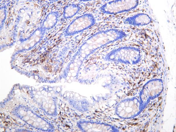
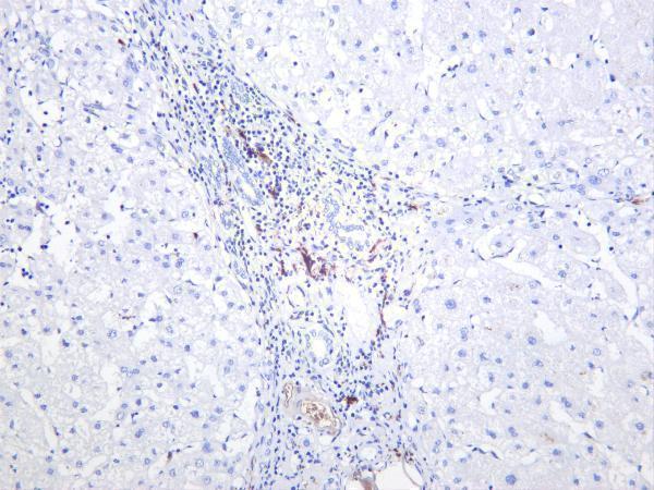
Real IHC data Human colon was stained with anti-S100B rabbit antibody
Anti-S100B Rabbit Monoclonal Antibody
Cat # M00979-4

M00979-1 has IHC images from paraffin-embedded rat cerebral cortex, mouse pancreas and hippocampus, and human testis cancer; its IF images include A375 cells (image captions). M00979-4 has IHC images from human colon and liver, mouse colon, and rat colon; IF is listed as an application, but no IF image is supplied (catalog applications; image captions).

Which to pick: For tissue IHC, choose M00979-1 when a paraffin-section example matters (M00979-1 IHC image captions); M00979-4 offers colon examples across three species (M00979-4 IHC image captions). For IF/ICC, choose M00979-1 because ICC and IF are listed and an A375 IF image is supplied (catalog applications; M00979-1 IF image caption). Both are rabbit monoclonals listed as reactive with human, mouse and rat, so either fits a cross-species study (catalog host, description and reactivity); the fixative is unreported in both products’ IHC captions (IHC image captions).

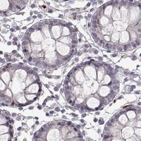
Each figure is that product's own IHC / IF validation image from its datasheet.

References

  1. UniProt Consortium. UniProt entry P04271 (S100B_HUMAN, Protein S100-B).
  2. Human Protein Atlas. S100B tissue IHC expression (reliability: Enhanced).
  3. Human Protein Atlas. S100B subcellular location (ICC-IF): Mainly localized to vesicles, cytosol and primary cilium. In addition localized to the nucleoplasm, microtubules, cytokinetic bridge, mitotic spindle and basal body..
  4. Human Protein Atlas. S100B antibody validation summary (3 antibodies).
  5. Synchronized dual pulse gastric electrical stimulation induces activation of enteric glial cells in rats with diabetic gastroparesis. Gastroenterology research and practice 2014 — PMC4003764.
  6. Evidence for a wide extra-astrocytic distribution of S100B in human brain. BMC neuroscience 2007 — PMC1769505.
  7. Bone marrow stem cells with or without superparamagnetic iron oxide nanoparticles as a magnetic targeting tool: Which is better in regeneration of neurolysed facial nerve? An experimental study. Heliyon 2024 — PMC10906296.
  8. Decreased S100B expression in chronic liver diseases. The Korean journal of internal medicine 2017 — PMC5339465.
  9. PubMed PMID:2394738 — UniProt-cited evidence.
  10. PubMed PMID:10830953 — UniProt-cited evidence.
  11. PubMed PMID:15489334 — UniProt-cited evidence.