This website uses cookies to ensure you get the best experience on our website.


- Table of Contents
Plan S100P IHC in paraffin sections using the cytoplasmic and nuclear tissue pattern reported in marrow, placenta and glandular epithelia (HPA tissue IHC). Compare staining across sections with consistent fixation, and interpret intensity in light of reported up-regulation in pancreatic ductal neoplasia (UniProt).

Expected localisation, validated protocols, controls and antibodies — the at-a-glance facts below, then the full design guide.
| Expected localisation | Cytoplasmic and nuclear staining (HPA tissue IHC) | |
| Staining pattern | Cytoplasmic/nuclear in marrow, placenta and glandular epithelia (HPA tissue IHC) | |
| Antigen retrieval | Tris-EDTA pH 9.0 HIER, 95–98 °C, 20 min (rule: nuclear antigen) | |
| Positive control | Bone marrow+4 more · see all | |
| Negative control | Adipose tissue+4 more · see all |
| Fixation | Keep fixation consistent across sections. (standard IHC practice; not target-specific) | |
| Caveat | Staining consistency with RNA is medium; restaining is pending (HPA tissue IHC) | |
| Regulation | Up-regulated in pancreatic ductal neoplasia (UniProt) | |
| Isoform / epitope | No annotated isoforms or processing; one 1–95 chain (UniProt) |
Compare the catalog antibody's IHC-P protocol with four published S100P IHC protocols covering pancreatic, multiple human tissue, hepatolithiasis, and nasopharyngeal specimens (PMC12743368; PMC2254630; PMC8046297; PMC5494647).
| Sample | Paraffin-embedded human bladder carcinoma tissue; fixative not specified (datasheet M01963) |
| Fixation | Image fixative and duration unreported (datasheet M01963); verify before use. |
| Sectioning | 4–5 µm sections on charged slides (standard) |
| Deparaffinisation | Xylene, graded ethanol series to water (standard) |
| Antigen retrieval | Heat-induced epitope retrieval in Tris-EDTA buffer, pH 9.0, 20 min at 95–98 °C (standard rule: nuclear antigen) |
| Peroxidase block | 3% H2O2, 10 min, room temperature (standard) |
| Blocking | 10% normal serum of the secondary host, 30 min, room temperature (standard) |
| Primary antibody | Rabbit monoclonal (clone AOCH-19) anti-S100P, 1:50 (datasheet M01963) |
| Primary incubation | Overnight at 4 °C (standard) |
| Detection | HRP-polymer secondary, DAB chromogen 5–10 min (standard) |
| Counterstain | Hematoxylin, blue, dehydrate and mount (standard) |
| Expected result | S100P-positive staining in hematopoietic cells of bone marrow (HPA tissue IHC: High). HPA tissue profile: Cytoplasmic and nuclear expression mainly in bone marrow, placenta and glandular epithelia in several tissues, including stomach and urinary bladder. No signal in the no-primary control. |
S100P should appear in the cytoplasm and nuclei of selected cells, especially bone marrow hematopoietic cells, placental syncytiotrophoblasts, and stomach glandular cells (HPA: tissue IHC profile; High in each). HPA rates the tissue pattern Enhanced, while noting medium consistency with RNA data and pending restaining (HPA: reliability). S100P has no transmembrane segment, so a membrane-only pattern is unexpected (UniProt P25815: topology).
| Cytoplasmic and nuclear staining in the expected cells. | This matches the reported tissue pattern (HPA: tissue IHC profile). Strong staining is especially plausible in bone marrow hematopoietic cells, placental syncytiotrophoblasts, stomach glandular cells, and bladder urothelial cells (HPA: High in each). Compare cells within the section rather than assuming every cell should stain equally (HPA: cell-specific levels). |
| Predominantly membrane-only staining or signal confined to an unexpected compartment. | Recheck the result before scoring it as S100P: tissue IHC reports cytoplasmic and nuclear expression (HPA: tissue IHC profile), and the protein has no transmembrane segment (UniProt P25815: topology). UniProt also lists microvillus membrane localization, so focal projection-associated signal cannot be dismissed solely because it approaches a membrane (UniProt P25815: subcellular location). |
| Strong staining in a cell type reported as undetected. | For example, adipocytes or adrenal glandular cells are listed as Not detected (HPA: tissue IHC). Check the cell identification and controls; cross-reactivity or endogenous detection activity is possible (standard IHC practice). An HPA negative applies to the listed cell type and sampled context, not automatically to every cell in that organ. |
| Diffuse color over tissue, stroma, or an empty area, with little cell definition. | The distribution is difficult to reconcile with the reported cell-associated cytoplasmic and nuclear pattern (HPA: tissue IHC profile). Assess background in a matched negative control and review blocking, washes, and detection reagents (standard IHC practice). Do not use diffuse color alone to assign a positive S100P score. |
| No staining in a section containing a listed high-expression cell type. | First verify that the expected cells are present: HPA lists High staining in bone marrow hematopoietic cells, placental syncytiotrophoblasts, stomach glandular cells, and bladder urothelial cells (HPA: tissue IHC). If a suitable positive control also fails, review the assay setup and reagent performance (standard IHC practice). |
| Cell type and tissue | HPA reports High staining in four listed cell populations, Medium staining in several glandular or squamous epithelial populations, and Low or Not detected staining in others (HPA: tissue IHC). Choose a control by its named cell population, not by organ name alone. |
| Compartment | Tissue IHC describes cytoplasmic and nuclear expression (HPA: tissue IHC profile). UniProt also lists microvillus membrane localization and no transmembrane segment (UniProt P25815: subcellular location; topology). Interpret a putative membrane signal alongside the cellular pattern. |
| Evidence strength | The tissue IHC reliability is Enhanced, but its description notes medium consistency with RNA data and pending restaining (HPA: reliability). HPA019502 has Enhanced IHC validation (HPA: antibodies). These ratings support interpretation while leaving individual slides subject to control review. |
| Regional absence versus broad expression | UniProt reports detection in most tissues except brain, testis, and small intestine (UniProt P25815: tissue specificity). HPA records Low glial staining in cerebral cortex and hippocampus and Medium duodenal glandular staining (HPA: tissue IHC). Treat these as source-specific observations rather than forcing one universal negative-tissue rule. |
| Protein forms | UniProt lists one 1–95 chain, no signal peptide, no propeptide, no listed isoforms, and no glycosylation sites (UniProt P25815: processing; isoforms; glycosylation). The supplied record therefore gives no basis to predict a distinct IHC pattern from a processed or glycosylated S100P form. |
| Situation | Likely cause | Next action |
|---|---|---|
| A listed High cell population stains weakly or not at all. | The expected cells may be absent from the section, or the assay may have failed (HPA: High cell populations; standard IHC practice). | Confirm the cell type on the counterstained section, then check a suitable positive control, detection reagents, and the antibody's validated IHC-P conditions (standard IHC practice). No target-specific fixation sensitivity is supplied. |
| The whole section has broad brown haze. | Nonspecific background or endogenous detection activity can obscure cell boundaries (standard IHC practice). | Compare a matched negative control; review blocking, washes, detection chemistry, and antibody concentration under the validated IHC-P workflow (standard IHC practice). Score only interpretable cell-associated staining. |
| Signal appears only at cell borders. | This differs from the main cytoplasmic and nuclear tissue pattern (HPA: tissue IHC profile), although UniProt lists microvillus membrane localization (UniProt P25815: subcellular location). | Check whether the signal follows identifiable microvilli or broadly outlines unrelated cells; compare expected positive cells and controls before calling it S100P (UniProt P25815: subcellular location; standard IHC practice). |
| Unexpected cells stain more strongly than the expected population. | Misidentified cells, cross-reactivity, or endogenous detection activity may explain a mismatch (standard IHC practice); HPA levels are cell-specific (HPA: tissue IHC). | Reidentify stained cells using tissue morphology and counterstain, then compare an appropriate negative control and an HPA High cell population (HPA: tissue IHC; standard IHC practice). |
| A brain or duodenal section seems to contradict a presumed negative control. | UniProt's broad tissue statement and HPA's cell-level observations differ in scope (UniProt P25815: tissue specificity; HPA: Low glia and Medium duodenal glandular cells). | Use the specific cell population and observed intensity when judging the slide; avoid treating the entire organ as uniformly negative (HPA: tissue IHC). |
| IF/ICC question: where should S100P appear? | HPA reports supported nucleoplasmic localization in ICC-IF, which addresses a different assay from the tissue IHC pattern (HPA: subcellular ICC-IF; tissue IHC). | For IF/ICC interpretation, look for nucleoplasmic signal and use its own controls; use the separate IF/ICC guide for assay setup (HPA: supported nucleoplasm; standard IF practice). |
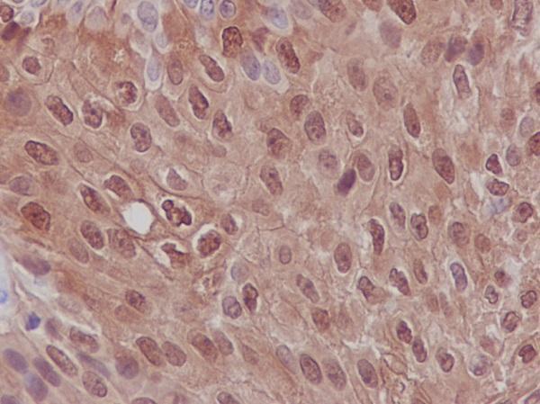
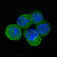
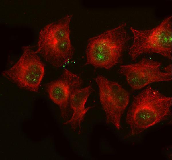


Comprehensive Human Protein Atlas IHC scoring per tissue (reliability: Enhanced — Medium consistency between antibody staining and RNA expression data. Pending restaining.). Rows are taken directly from the HPA tissue chart — click any row's HPA link to view the source.
| Tissue | Cell type | Level | Evidence | Source |
|---|---|---|---|---|
| Bone marrow | Hematopoietic cells | High | Protein (IHC) | HPA → |
| Placenta | Syncytiotrophoblasts - cell body | High | Protein (IHC) | HPA → |
| Stomach | Glandular cells | High | Protein (IHC) | HPA → |
| Urinary bladder | Urothelial cells | High | Protein (IHC) | HPA → |
| Cervix | Glandular cells | Medium | Protein (IHC) | HPA → |
| Tissue | Cell type | Level | Evidence | Source |
|---|---|---|---|---|
| Adipose tissue | Adipocytes | Not detected | Protein (IHC) | HPA → |
| Adrenal gland | Glandular cells | Not detected | Protein (IHC) | HPA → |
| Breast | Adipocytes | Not detected | Protein (IHC) | HPA → |
| Caudate | Glial cells | Not detected | Protein (IHC) | HPA → |
| Cerebellum | Cells in granular layer | Not detected | Protein (IHC) | HPA → |
Troubleshoot S100P staining in paraffin sections by checking retrieval, compartment, cell type, controls and scoring before interpreting chromogenic signal.
The catalog shows S100P staining in paraffin-embedded human bladder carcinoma and A549 cells (M01963 IHC caption; A01963-2 IF caption). Both antibodies list human reactivity (catalog reactivity).
M01963 will render with its IHC image of paraffin-embedded human bladder carcinoma; its applications include IHC (M01963 IHC caption; catalog applications). A01963-2 will render with its IF image of A549 cells; its applications include ICC/IF (A01963-2 IF caption; catalog applications).
Which to pick: Choose M01963 for paraffin-section IHC: it is monoclonal and lists IHC, with an image from paraffin-embedded human bladder carcinoma (catalog clone and applications; M01963 IHC caption). Choose A01963-2 for IF/ICC: it is polyclonal and lists ICC/IF, with an IF image from A549 cells (catalog dilution record and applications; A01963-2 IF caption). Neither has documented cross-species reactivity, since both list human only; the M01963 IHC caption does not report a fixative (catalog reactivity; M01963 IHC caption).