This website uses cookies to ensure you get the best experience on our website.


- Table of Contents
Plan chromogenic SERPINA3 IHC in paraffin sections using cervical glandular cells as a high-staining reference (HPA tissue IHC). Interpret extracellular deposits and limited cytoplasmic staining in light of secretion and inflammation-linked abundance (HPA tissue IHC; UniProt).

Expected localisation, validated protocols, controls and antibodies — the at-a-glance facts below, then the full design guide.
| Expected localisation | Extracellular deposits and cytoplasm in a few cell types (HPA tissue IHC) | |
| Staining pattern | Extracellular deposits with cytoplasmic staining in a few cells (HPA tissue IHC) | |
| Antigen retrieval | EDTA pH 8.0 HIER, heat-mediated (datasheet PA2217) | |
| Positive control | Cerebellum+4 more · see all | |
| Negative control | Adipose tissue+4 more · see all |
| Fixation | Keep fixation consistent across paraffin sections. (standard IHC practice; not target-specific) | |
| Caveat | Secretion can separate tissue RNA from protein staining (HPA tissue IHC) | |
| Regulation | Rises during inflammation or infection (UniProt) | |
| Isoform / epitope | 3 isoforms; mature N terminus varies (UniProt) |
The catalog antibody’s IHC-P protocol is accompanied by four published SERPINA3 IHC protocols (PMC9592916; PMC13094457; PMC5403010; PMC12808515).
| Sample | Tissue sections; selected-image fixative not specified (standard IHC workflow) |
| Fixation | Image fixative and duration unreported (datasheet PA2217); verify before use. |
| Sectioning | 4–5 µm sections on charged slides (standard) |
| Deparaffinisation | Xylene, graded ethanol series to water (standard) |
| Antigen retrieval | Heat retrieval: EDTA pH 8.0 (datasheet PA2217); 20 min, 95–100 °C (standard) |
| Peroxidase block | 3% H2O2, 10 min, room temperature (standard) |
| Blocking | 10% goat serum (datasheet PA2217) |
| Primary antibody | Rabbit anti-SERPINA3, 0.5-1μg/ml (datasheet PA2217) |
| Primary incubation | Overnight at 4 °C (datasheet PA2217) |
| Detection | HRP-conjugated secondary, DAB chromogen (datasheet PA2217) |
| Counterstain | Hematoxylin, blue, dehydrate and mount (standard) |
| Expected result | SERPINA3-positive staining in granular cells - nucleus of cerebellum (HPA tissue IHC: High). HPA tissue profile: Positivity in extracellular deposits and cytoplasmic expression in a few cell types. No signal in the no-primary control. |
SERPINA3 is secreted and has no transmembrane segment (UniProt P01011). In paraffin-section IHC, expect extracellular deposits and cytoplasmic staining in selected cells (HPA tissue IHC: Enhanced). Cervical glandular cells stain strongly, while hepatocyte staining is low in the HPA survey (HPA: High in cervical glandular cells; Low in hepatocytes).
| Strong staining in cervical glandular cells, with some extracellular deposits. | This fits the reported pattern (HPA: High in cervical glandular cells; extracellular deposits). Score cellular staining and deposits separately because secreted SERPINA3 can be present outside its producing cells (UniProt P01011: Secreted). |
| Staining is confined to an unexpected compartment in a cell type otherwise reported as positive. | Check for artefact and compare the cell type with HPA before interpreting it (HPA tissue IHC: cytoplasmic expression in a few cell types). A blanket rule that nuclear staining is false would be misleading: HPA reports High nuclear staining in cerebellar granular cells. |
| Strong staining appears in cells reported as undetected, such as adipocytes. | Treat the result as unconfirmed; cross-reactivity or endogenous detection activity is possible (HPA: adipocytes Not detected; standard IHC practice). Review the cell identity, detection controls, and whether the signal follows tissue structure before assigning it to SERPINA3. |
| Broad, fairly uniform color covers cells and extracellular spaces. | This differs from the selected-cell and deposit pattern (HPA tissue IHC: positivity in extracellular deposits and cytoplasmic expression in a few cell types). Background from the detection workflow is possible; compare a control section processed without primary antibody (standard IHC practice). |
| No staining appears in cervical glandular cells on a test section. | The result needs a control and a check of section quality, retrieval, antibody dilution, and detection (HPA: High in cervical glandular cells; standard IHC practice). An absent signal alone cannot establish that the specimen lacks SERPINA3. |
| Secretion and tissue location (UniProt P01011: Secreted; HPA tissue IHC: extracellular deposits). | Interpret deposits alongside stained cells; extracellular signal need not identify the cell that made the protein (UniProt P01011: Secreted). |
| Cell-specific reference pattern (HPA tissue IHC: Enhanced). | Use the named cell population, not the organ alone, as the comparison: cervical glandular cells are High, whereas hepatocytes are Low (HPA tissue IHC). |
| Processing and glycosylation (UniProt P01011: signal peptide 1–23; six glycosylation sites). | The mature annotated chains begin at residue 24 or 26 (UniProt P01011). Epitope accessibility in paraffin sections cannot be inferred without the antibody's epitope information. |
| Isoforms and antibody validation (UniProt P01011: three isoforms; HPA antibodies: IHC Enhanced or Supported). | The supplied record does not show which isoforms each antibody recognizes (UniProt P01011; HPA antibodies). Compare staining with the chosen antibody's IHC validation. |
| IF/ICC Q: Is a subcellular IF pattern established? (HPA subcellular: Secreted). | A: HPA gives no main intracellular location or cell-line ICC-IF images, and the listed antibodies have no ICC status (HPA subcellular; HPA antibodies). Do not assign a validated organelle pattern. |
| Situation | Likely cause | Next action |
|---|---|---|
| Cervical glandular cells lack signal (HPA: High). | The staining run may have failed, or the tested section may differ from the HPA reference (standard IHC practice; HPA tissue IHC). | Run an appropriate known-positive section alongside it; review tissue preservation, retrieval, antibody dilution, and detection before interpreting absence (standard IHC practice). |
| The whole section is diffusely colored (HPA tissue IHC: selected cells and deposits). | Nonspecific binding or detection background may obscure the reported pattern (standard IHC practice; HPA tissue IHC). | Compare a section processed without primary antibody; review blocking, washes, detection conditions, and exposure time (standard IHC practice). |
| Strong color appears in adipocytes (HPA: Not detected). | Cross-reactivity or endogenous detection activity is possible; the HPA finding alone cannot identify the cause (HPA: adipocytes Not detected; standard IHC practice). | Check the no-primary control and, where available, an independently validated antibody; assess whether color follows cell structures (standard IHC practice). |
| Only extracellular staining is visible (HPA tissue IHC: extracellular deposits). | Secreted protein can accumulate away from stained cells, but deposits alone do not establish their source (UniProt P01011: Secreted; HPA tissue IHC). | Record deposits separately and examine expected cellular staining in a suitable control section (HPA: High in cervical glandular cells; standard IHC practice). |
| Nuclear staining seems inconsistent with secretion (UniProt P01011: Secreted). | Compartment expectations vary by reported cell type; HPA specifically records nuclear signal in cerebellar granular cells (HPA: High in granular-cell nuclei). | Confirm the cell type, inspect the no-primary control, and compare the finding with HPA's tissue-specific pattern before calling it artefact (HPA tissue IHC; standard IHC practice). |
| A low-staining liver section is used to judge a failed run (HPA: Low in hepatocytes). | Low hepatocyte staining is the HPA protein-IHC observation, despite liver synthesis and liver-enriched RNA (HPA tissue IHC; UniProt P01011: synthesized in liver). | Use a clearly stained reference such as cervical glandular cells to assess run performance; do not require strong hepatocyte staining (HPA: High in cervical glandular cells; Low in hepatocytes). |
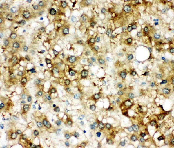
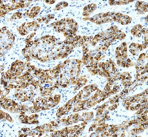



Comprehensive Human Protein Atlas IHC scoring per tissue (reliability: Enhanced — At least one protein variant secreted, tissue location of RNA and protein might differ and correlation is complex.). Rows are taken directly from the HPA tissue chart — click any row's HPA link to view the source.
| Tissue | Cell type | Level | Evidence | Source |
|---|---|---|---|---|
| Cerebellum | Granular cells - nucleus | High | Protein (IHC) | HPA → |
| Cervix | Glandular cells | High | Protein (IHC) | HPA → |
| Breast | Glandular cells | Medium | Protein (IHC) | HPA → |
| Kidney | Proximal tubules (cell body) | Medium | Protein (IHC) | HPA → |
| Placenta | Trophoblastic cells | Medium | Protein (IHC) | HPA → |
| Tissue | Cell type | Level | Evidence | Source |
|---|---|---|---|---|
| Adipose tissue | Adipocytes | Not detected | Protein (IHC) | HPA → |
| Appendix | Endocrine cells | Not detected | Protein (IHC) | HPA → |
| Bronchus | Respiratory epithelial cells | Not detected | Protein (IHC) | HPA → |
| Caudate | Glial cells | Not detected | Protein (IHC) | HPA → |
| Cerebral cortex | Endothelial cells | Not detected | Protein (IHC) | HPA → |
Troubleshoot SERPINA3 staining in paraffin sections by checking retrieval, tissue context, secreted protein localisation and the controls used for scoring.
The catalog covers human IHC and IF/ICC (catalog applications and reactivity). Real IHC images show paraffin sections of human liver and liver cancer (PA2217 image captions).
The PA2217 card renders with IHC data from a paraffin section of human liver cancer (PA2217 image caption). A second caption documents IHC in a paraffin section of human liver (PA2217 image caption).
Which to pick: Choose PA2217 for human tissue IHC: its images document paraffin sections, and the fixative is unreported (PA2217 image captions). For human IF/ICC, M02312 is a rabbit monoclonal listed for those applications, with no IF image supplied (M02312 catalog). Neither SKU lists cross-species reactivity; both list Human only (catalog reactivity).