SERPINB5 / Serpin B5 · IHC design guide

Design Immunohistochemistry for SERPINB5

Plan SERPINB5 chromogenic IHC around the cytoplasmic staining seen in tissue, especially squamous epithelia (HPA tissue IHC). Use esophageal squamous epithelium as a positive reference and adipocytes as a negative reference (HPA tissue IHC).

Evidence assembled Oct 2026 · For research use; verify linked source records and product datasheet before use
Immunohistochemistry protocol sheet for SERPINB5 (IHC for SERPINB5): expected localisation Cytoplasmic tissue staining (HPA tissue IHC); secreted (UniProt), antibody M03409, validated IHC image, and IHC protocol steps
Printable SERPINB5 IHC protocol sheet — expected localisation Cytoplasmic tissue staining (HPA tissue IHC); secreted (UniProt), antibody M03409, controls and protocol steps. Open the full SERPINB5 IHC guide →

SERPINB5 Immunohistochemistry Experimental Design Guide

Expected localisation, validated protocols, controls and antibodies — the at-a-glance facts below, then the full design guide.

Must know before staining
Expected localisation Cytoplasmic tissue staining (HPA tissue IHC); secreted (UniProt)
Staining pattern Variable cytoplasmic staining, strongest in squamous epithelia (HPA tissue IHC)
Antigen retrieval EDTA pH 8.0 HIER, heat-mediated (datasheet M03409)
Positive control ⓘ Esophagus+4 more · see all
Negative control ⓘ Adipose tissue+4 more · see all
Important caveats
Reasons your staining may differ from the expected pattern.
Fixation Keep paraffin-section fixation consistent across samples. (standard IHC practice; not target-specific)
Caveat A secreted pool may yield staining beyond the observed cytoplasm (UniProt; HPA tissue IHC)
Regulation Tissue-enhanced RNA in esophagus and skin (HPA tissue RNA)
Isoform / epitope 2 isoforms; epitope coverage is unspecified (UniProt)
Section 1

Recommended SERPINB5 IHC & IF Protocols

The catalog antibody protocol (datasheet M03409) is followed by published IHC examples using tumor or normal sections (PMC12921336), human stomach tissue (PMC10425712), and patient samples (PMC6995195).

Recommended immunohistochemistry (IHC-P) protocol parameters
SampleParaffin-embedded human tonsil tissue; fixative not specified (datasheet M03409)
FixationImage fixative and duration unreported (datasheet M03409); verify before use.
Sectioning4–5 µm sections on charged slides (standard)
DeparaffinisationXylene, graded ethanol series to water (standard)
Antigen retrievalHeat retrieval: EDTA pH 8.0 (datasheet M03409); 20 min, 95–100 °C (standard)
Peroxidase block3% H2O2, 10 min, room temperature (standard)
Blocking10% goat serum (datasheet M03409)
Primary antibodyMouse monoclonal (clone 7G4E1) anti-SERPINB5, 2 μg/ml (datasheet M03409)
Primary incubationOvernight at 4 °C (datasheet M03409)
DetectionHRP-conjugated secondary, DAB chromogen (datasheet M03409)
CounterstainHematoxylin, blue, dehydrate and mount (standard)
Expected resultSERPINB5-positive staining in squamous epithelial cells of esophagus (HPA tissue IHC: High). HPA tissue profile: Cytoplasmic expression with varying intensity in most tissues, most abundant in squamous epithelia and the gastrointestinal tract. No signal in the no-primary control.
💡Decision noteStart with heat-mediated EDTA retrieval at pH 8.0 (datasheet M03409); boiled 0.1 M sodium citrate is a published alternative (PMC10425712).
Section 2

What Is the Expected SERPINB5 Staining Pattern?

In paraffin-section IHC, expect SERPINB5 staining mainly in epithelial-cell cytoplasm, especially esophageal squamous cells, skin basal-layer cells, and urothelial cells (HPA: cytoplasmic expression; High in these cells). UniProt annotates SERPINB5 as secreted into extracellular space, with no transmembrane segment (UniProt P36952: subcellular location and topology). HPA rates its tissue IHC profile Enhanced, while reporting medium staining–RNA consistency and pending external verification (HPA: tissue reliability).

What am I looking at on my slide?
Clear cytoplasmic staining in esophageal squamous epithelium, skin basal-layer cells, or bladder urothelium.These are strong reference patterns (HPA: High in these cells). Compare cell-specific staining with adjacent structures; a dark field alone does not establish the expected pattern (general IHC practice).
Predominantly nuclear or sharply membrane-restricted staining, with little epithelial cytoplasmic signal.That distribution lacks support from the supplied tissue IHC profile (HPA: cytoplasmic expression). Treat it as suspect and review controls before assigning a new localisation; UniProt also reports no transmembrane segment (UniProt P36952: topology).
Strong staining mainly in adipocytes, hematopoietic cells, or lymph-node germinal-center cells.Those cell populations are reported as not detected (HPA: adipose tissue, bone marrow, lymph node). Consider nonspecific antibody binding or endogenous chromogen-generating activity, then inspect the appropriate controls (general IHC practice).
Diffuse color across epithelium, stroma, and blank areas, without recognizable cell boundaries.An indiscriminate field cannot be scored as the reported epithelial cytoplasmic pattern (HPA: tissue IHC profile). Uneven background, incomplete blocking, or overdeveloped chromogen are workflow possibilities to check (general IHC practice).
No epithelial signal in an esophagus, skin, or bladder section chosen as a positive control.This conflicts with the supplied High staining references (HPA: esophagus, skin, urinary bladder). Check the run and section quality before interpreting a study sample as negative (general IHC practice).
💡Expected SERPINB5 appearanceA convincing positive shows stronger epithelial cytoplasmic color in esophageal squamous cells, skin basal-layer cells, or urothelium (HPA: High); uniform color outside those cell patterns or dominant nuclear staining is suspect (HPA: cytoplasmic profile; general IHC practice).
How each factor affects the staining
Which tissue provides a useful reference?Esophagus, skin, and urinary bladder have High staining in specified epithelial cells; cervix, tonsil, and vagina have Medium squamous-cell staining (HPA: tissue IHC). Use the specified cell population, not the whole section, as the reference.
How should compartment calls be interpreted?HPA describes cytoplasmic tissue staining, while UniProt annotates extracellular secretion and no transmembrane segment (HPA: tissue profile; UniProt P36952: location and topology). Neither annotation alone proves that extracellular color in a section is specific.
What does Enhanced validation establish?The listed antibodies have Enhanced IHC status, indicating a pattern supported by independent antibodies or orthogonal data (HPA: antibody validation). The overall tissue profile still has medium staining–RNA consistency and awaits external verification (HPA: reliability description).
IF/ICC: where has localisation been reported?HPA reports mainly vesicular signal in ICC-IF, with vesicles as the supported main location (HPA: subcellular). This is a separate application observation; it does not replace the cytoplasmic pattern used to read paraffin-section IHC (HPA: tissue IHC).
Why is my staining missing, weak or wrong?
SituationLikely causeNext action
The positive-control epithelium is blank.A failed stain run or unsuitable section is possible; the chosen epithelial cells are reported High (HPA: esophagus, skin, urinary bladder; general IHC practice).Check control-slide integrity, reagent delivery, antigen-retrieval run, primary-antibody application, and detection reagents; repeat alongside a previously successful control (general IHC practice).
Signal is present but mainly nuclear or confined to a cell border.The observed compartment differs from the reported cytoplasmic tissue profile (HPA: tissue IHC). Specificity cannot be inferred from color intensity alone (general IHC practice).Inspect the negative control and compare with a High reference tissue; review whether the apparent boundary follows cells or chromogen deposits before scoring (HPA: High reference cells; general IHC practice).
Adipocytes or hematopoietic cells stain as strongly as the target epithelium.These populations are reported as not detected, so nonspecific binding or endogenous detection activity is plausible (HPA: adipose tissue and bone marrow; general IHC practice).Compare a no-primary control, check the detection-system blocking step, and reassess antibody concentration using positive and negative reference cells (general IHC practice).
The section has widespread haze or granular precipitate.Excess detection background or chromogen precipitate may obscure the cell-specific cytoplasmic pattern (HPA: tissue profile; general IHC practice).Inspect a no-primary control, review wash and blocking steps, and check chromogen preparation and development time before interpreting weak cellular color (general IHC practice).
Weak staining appears in a low-expression tissue, but the control is clear.Low reported levels make that tissue a poor sole test of run performance; lung alveolar cells and bronchial respiratory cells are listed Low (HPA: tissue IHC).Use an esophagus, skin, or bladder epithelial control reported High, then assess the study tissue within its own cell types (HPA: tissue IHC; general IHC practice).
Extracellular color seems stronger than epithelial cytoplasm.Secretion is annotated, but the supplied tissue IHC profile describes cytoplasmic expression; extracellular color alone is not validated here as a diagnostic pattern (UniProt P36952: location; HPA: tissue profile).Check whether extracellular color persists in negative controls and whether the expected epithelial cytoplasm stains in a High reference section before assigning it to SERPINB5 (HPA: tissue IHC; general IHC practice).

Sample controls for SERPINB5 IHC & IF

🧪Run esophagus first and require staining in squamous epithelial cells (HPA: High in esophageal squamous epithelial cells). Use adipose tissue as the negative tissue, checking adipocytes for background staining (HPA: Not detected in adipocytes); on the esophagus slide, assess non-squamous cells as internal background comparators without assuming they are target-negative from the HPA row.
Positive control tissue: Esophagus (Squamous epithelial cells, HPA High)
Negative control tissue: Adipose tissue (HPA Not detected)
ICC-IF cell lines (HPA subcellular resource): HPA ICC-IF images show SERPINB5 in A-431, PC-3, U2OS, with annotated localisation: Vesicles (supported) (HPA subcellular).
Technical controls: Include a no-primary (secondary-only) slide, a concentration-matched mouse IgG isotype control (selected-SKU caption: mouse primary antibody), and a validated SERPINB5 knockout sample as a biological negative. For HRP/DAB detection, quench endogenous peroxidase and check any signal in blood or inflammatory cells against the controls (selected-SKU caption: HRP/DAB detection; standard IHC practice).
⚠️Feasibility: A target-specific fixation window and fixation effect are unreported, and the selected-SKU paraffin-section caption does not report a fixative (selected-SKU caption: fixative not stated). The caption uses heat retrieval in EDTA at pH 8.0, so use that as a starting condition and compare with an unretrieved section; a requirement for retrieval has not been established (selected-SKU caption: EDTA retrieval). Paraffin IHC has a supplied tissue example, while the supplied ICC-IF evidence shows vesicular localisation in A-431, PC-3 and U2OS cells; neither source establishes that frozen sections or IF are easier (selected-SKU caption: tonsil paraffin section; HPA: supported vesicular ICC-IF localisation).

HPA tissue IHC evidence for SERPINB5

Comprehensive Human Protein Atlas IHC scoring per tissue (reliability: Enhanced — Medium consistency between antibody staining and RNA expression data. Pending external verification.). Rows are taken directly from the HPA tissue chart — click any row's HPA link to view the source.

Positive expression · recommended positive controls

TissueCell typeLevelEvidenceSource
Esophagus Squamous epithelial cells High Protein (IHC) HPA →
Skin Cells in basal layer High Protein (IHC) HPA →
Urinary bladder Urothelial cells High Protein (IHC) HPA →
Cervix Squamous epithelial cells Medium Protein (IHC) HPA →
Tonsil Squamous epithelial cells Medium Protein (IHC) HPA →

Undetected expression · recommended negative controls

TissueCell typeLevelEvidenceSource
Adipose tissue Adipocytes Not detected Protein (IHC) HPA →
Bone marrow Hematopoietic cells Not detected Protein (IHC) HPA →
Caudate Glial cells Not detected Protein (IHC) HPA →
Cerebellum Cells in granular layer Not detected Protein (IHC) HPA →
Hippocampus Glial cells Not detected Protein (IHC) HPA →
Section 3

Advanced SERPINB5 IHC Tips

Troubleshoot SERPINB5 staining in paraffin sections by checking retrieval, cellular context and controls before interpreting chromogenic signal.

What should I change if SERPINB5 staining is weak after retrieval?
Start with heat-mediated retrieval in EDTA, pH 8.0, for paraffin sections (datasheet M03409). The selected image used this retrieval before 2 μg/ml primary antibody overnight at 4°C, so compare those recorded conditions before changing several variables (datasheet M03409). If staining remains weak, check that heating and cooling were consistent across slides, then test a different retrieval buffer as a documented fallback on matched sections (standard IHC practice). Compare epithelial signal with adjacent negative cell populations; strong staining in esophageal squamous cells and absent staining in adipocytes are reported reference patterns (HPA: Esophagus high; adipocytes not detected).
Could fixation explain inconsistent SERPINB5 staining between paraffin blocks?
Target-specific fixation sensitivity is unknown because the selected paraffin-section caption does not state its fixative (datasheet M03409). Record each block’s fixative and fixation duration, and compare blocks only after matching section thickness, EDTA pH 8.0 retrieval, and antibody incubation conditions (datasheet M03409; standard IHC practice). If a block stains poorly, inspect morphology and include a process-matched positive control before attributing the loss to fixation (standard IHC practice). The catalog image used 2 μg/ml antibody overnight at 4°C; its successful staining cannot establish how SERPINB5 responds to any particular fixative (datasheet M03409).
How should I assess cytoplasmic, vesicular and extracellular SERPINB5 signal?
Assess staining by cell type and compartment: tissue IHC reports predominantly cytoplasmic expression, while cell imaging supports vesicular localisation (HPA: tissue profile; HPA: vesicles supported). SERPINB5 is annotated as secreted and has no transmembrane segment, so extracellular signal is biologically plausible but needs comparison with controls (UniProt P36952: subcellular location and topology). In paraffin sections, compare epithelial staining with nearby cells reported as negative, and distinguish discrete cellular signal from diffuse deposition (HPA: tissue profile; standard IHC practice). A crisp membrane-only rim should prompt review of detection background and antibody specificity before it is scored as SERPINB5 (UniProt P36952: topology; standard IHC practice).
Can a negative stain reflect epitope masking or isoform recognition?
SERPINB5 has 2 annotated isoforms, but the supplied product evidence does not map the antibody epitope or establish isoform recognition (UniProt P36952: isoforms; datasheet M03409). Four glycosylation sites are annotated at residues 99, 133, 188 and 361; their effects on this antibody’s staining are unknown (UniProt P36952: glycosylation). If staining is absent, first verify EDTA pH 8.0 retrieval and a positive tissue control before proposing epitope masking (datasheet M03409; standard IHC practice). To investigate isoform selectivity, obtain epitope mapping or compare independently validated antibodies; a single negative chromogenic section cannot resolve that question (standard IHC practice).
How should I investigate SERPINB5 localisation with multiplex IF?
For the separate IF/ICC workflow, pair SERPINB5 with an epithelial marker to identify the expected squamous cells, then assess whether the signal overlaps those cells (HPA: esophageal and tonsillar squamous epithelial staining; standard IF practice). Include single-colour controls and choose a fluorophore whose channel separates signal from the specimen’s measured autofluorescence, especially before interpreting weak puncta (standard IF practice). Permeabilise appropriately when testing intracellular vesicular staining; compare with non-permeabilised samples if asking whether an epitope is extracellularly accessible (HPA: vesicles supported; UniProt P36952: secreted; standard IF practice). The paraffin IHC caption supplies no IF/ICC fixation or antibody-validation conditions, so optimise and validate this application independently (datasheet M03409).
What explains diffuse DAB staining despite a reasonable epithelial pattern?
Run a no-primary control and inspect whether colour follows tissue edges, folds or broadly stained extracellular areas rather than epithelial cells (standard IHC practice). The selected paraffin-section workflow used 10% goat-serum block, a peroxidase-conjugated secondary and DAB; these details document that image, not a background-free condition for every specimen (datasheet M03409). Check peroxidase blocking and washing, then compare a lower primary concentration against the documented 2 μg/ml condition on adjacent sections (datasheet M03409; standard IHC practice). Retain a positive epithelial control while adjusting detection, since expected cytoplasmic staining can vary among tissues (HPA: tissue profile; standard IHC practice).
How should I score heterogeneous SERPINB5 staining across sections? ⚠ ANSWER MARKED FOR VERIFICATION
Define the scored cell population before counting: HPA reports high staining in esophageal squamous cells and skin basal cells, with variable cytoplasmic intensity across tissues (HPA: positive tissues; tissue profile). For chromogenic sections, record percentage of positive target cells and intensity, or calculate an H-score using fixed intensity categories across the cohort (standard IHC practice). Normalise each score to eligible epithelial cells or epithelial area, and report the denominator, since stromal or acellular area can distort whole-section averages (standard IHC practice). Use matched retrieval and detection conditions, including the documented EDTA pH 8.0 starting point, and exclude folds, necrosis and edge artefacts consistently (datasheet M03409; standard IHC practice).
When is an apparent SERPINB5-positive area likely to be artefactual?
Give greatest weight to reproducible cellular staining in expected epithelial populations: esophageal squamous cells and skin basal cells are reported high, whereas adipocytes are not detected (HPA: positive and negative tissues). Cytoplasmic staining fits the tissue profile, and punctate signal can be assessed against supported vesicular localisation (HPA: tissue profile; HPA: vesicles supported). Treat isolated membrane rims, section-edge gradients and necrotic deposits cautiously, and check a no-primary slide for endogenous peroxidase or detection artefact (UniProt P36952: no transmembrane segment; standard IHC practice). Interpret extracellular colour with control sections because SERPINB5 is annotated as secreted, but DAB deposition alone does not identify its source (UniProt P36952: subcellular location; standard IHC practice).
Boster reagents

Best SERPINB5 / Serpin B5 IHC Antibodies

IHC data cover human paraffin sections for both antibodies and mouse paraffin sections for PB9356; PB9356 also has IF data in A431 cells (M03409 and PB9356 image captions).

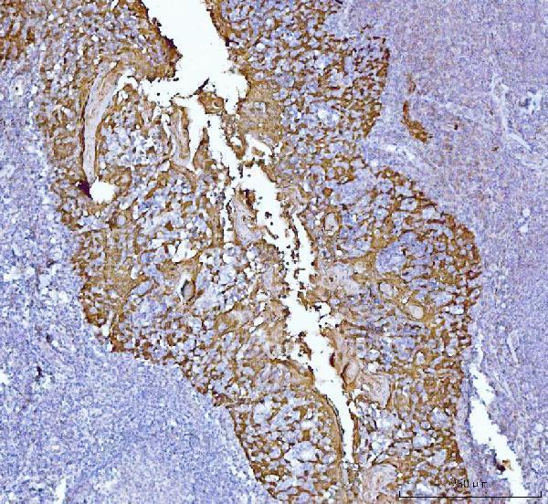
Real IHC data IHC analysis of MASPIN using anti-MASPIN antibody (M03409). MASPIN was detected in a paraffin-embedded section of human tonsil tissue. Heat mediated antigen retrieval was performed in EDTA buffer (pH 8.0, epitope retrieval solution). The tissue section was blocked with 10% goat serum. The tissue section was then incubated with 2 μg/ml mouse anti-MASPIN Antibody (M03409) overnight at 4°C. Peroxidase Conjugated Goat Anti-mouse IgG was used as secondary antibody and incubated for 30 minutes at 37°C. The tissue section was developed using HRP Conjugated Mouse IgG Super Vision Assay Kit (Catalog # SV0001) with DAB as the chromogen.
Anti-MASPIN Antibody ® (monoclonal, 7G4E1)
Cat # M03409
Real IHC data IHC analysis of MASPIN/SERPINB5 using anti-MASPIN/SERPINB5 antibody (PB9356). MASPIN/SERPINB5 was detected in a paraffin-embedded section of human colon cancer tissue. Heat mediated antigen retrieval was performed in EDTA buffer (pH 8.0, epitope retrieval solution). The tissue section was blocked with 10% goat serum. The tissue section was then incubated with 2 μg/ml rabbit anti-MASPIN/SERPINB5 Antibody (PB9356) overnight at 4°C. Peroxidase Conjugated Goat Anti-rabbit IgG was used as secondary antibody and incubated for 30 minutes at 37°C. The tissue section was developed using HRP Conjugated Rabbit IgG Super Vision Assay Kit (Catalog # SV0002) with DAB as the chromogen.
Anti-MASPIN/SERPINB5 Antibody ®
Cat # PB9356

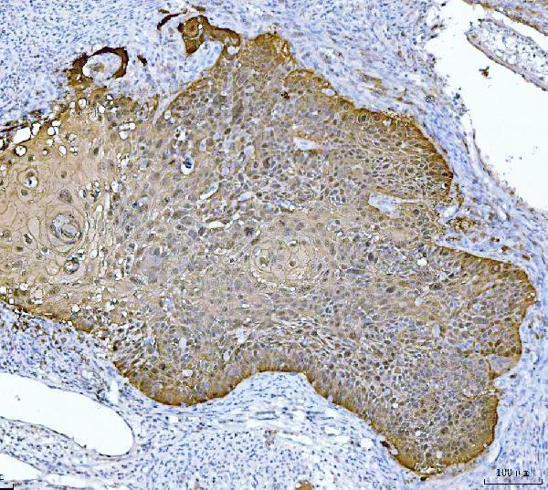
M03409 has IHC images of human tonsil and laryngeal squamous cell carcinoma paraffin sections (M03409 IHC image captions). PB9356 has IHC images of human colon cancer and mouse colon paraffin sections, plus an IF image of A431 cells (PB9356 IHC and IF image captions).

Which to pick: For human tissue IHC, consider monoclonal M03409, clone 7G4E1; its paraffin examples use EDTA pH 8 retrieval and 2 μg/ml primary antibody (M03409 catalog; M03409 IHC image captions). For IF/ICC, choose PB9356, which lists both applications and shows A431 cell IF at 5 μg/ml (PB9356 catalog; PB9356 IF image caption). For human and mouse tissue IHC, choose PB9356; its paraffin examples use EDTA pH 8 retrieval and 2 μg/ml primary antibody, and the fixative is unreported in both SKUs’ IHC captions (PB9356 catalog; PB9356 and M03409 IHC image captions).

Each figure is that product's own IHC / IF validation image from its datasheet.

References

  1. UniProt Consortium. UniProt entry P36952 (SPB5_HUMAN, Serpin B5).
  2. Human Protein Atlas. SERPINB5 tissue IHC expression (reliability: Enhanced).
  3. Human Protein Atlas. SERPINB5 subcellular location (ICC-IF): Mainly localized to vesicles..
  4. Human Protein Atlas. SERPINB5 antibody validation summary (4 antibodies).
  5. M(5)C-driven stabilization of SERPINB5 promotes cervical cancer progression and chemotherapy resistance. Cell death & disease 2026 — PMC12921336.
  6. SERPINB5 is a novel serum diagnostic biomarker for gastric high-grade intraepithelial neoplasia and plays a role in regulation of macrophage phenotypes. Translational oncology 2023 — PMC10425712.
  7. TRIM21-SERPINB5 aids GMPS repression to protect nasopharyngeal carcinoma cells from radiation-induced apoptosis. Journal of biomedical science 2020 — PMC6995195.
  8. BTG2 and SerpinB5, a novel gene pair to evaluate the prognosis of lung adenocarcinoma. Frontiers in immunology 2023 — PMC10064863.
  9. PubMed PMID:8290962 — UniProt-cited evidence.
  10. PubMed PMID:14702039 — UniProt-cited evidence.
  11. PubMed PMID:16177791 — UniProt-cited evidence.