This website uses cookies to ensure you get the best experience on our website.


- Table of Contents
Plan SFRP1 paraffin-section IHC around extracellular staining and a high-staining testis control with pachytene spermatocytes (HPA tissue IHC). The catalog antibody A01968-2 has an IHC working range of 0.5–1 μg/mL (datasheet).

Expected localisation, validated protocols, controls and antibodies — the at-a-glance facts below, then the full design guide.
| Expected localisation | Extracellular tissue staining (HPA tissue IHC) | |
| Staining pattern | Extracellular positivity in several tissues (HPA tissue IHC) | |
| Antigen retrieval | Citrate pH 6 HIER, heat-mediated (datasheet A01968-2) | |
| Positive control | Testis | |
| Negative control | Adipose tissue+4 more · see all |
| Fixation | Keep fixation conditions consistent across sections. (standard IHC practice; not target-specific) | |
| Caveat | Secreted SFRP1 may stain away from producing cells (UniProt) | |
| Regulation | Tissue-dependent expression (UniProt) | |
| Isoform / epitope | No isoforms annotated; mature chain spans aa 32–314 (UniProt) |
The catalog antibody’s IHC-P protocol is followed by published SFRP1 staining methods for large bowel, breast, prostate, and oral specimens (PMC2361362; PMC7565692; PMC4342152; PMC4610781).
| Sample | Paraffin-embedded human colon cancer tissue; fixative not specified (datasheet A01968-2) |
| Fixation | Image fixative and duration unreported (datasheet A01968-2); verify before use. |
| Sectioning | 4–5 µm sections on charged slides (standard) |
| Deparaffinisation | Xylene, graded ethanol series to water (standard) |
| Antigen retrieval | Heat retrieval: Citrate pH 6, 20 min (datasheet A01968-2) |
| Peroxidase block | 3% H2O2, 10 min, room temperature (standard) |
| Blocking | 10% goat serum (datasheet A01968-2) |
| Primary antibody | Rabbit anti-SFRP1, 0.5-1μg/ml (datasheet A01968-2) |
| Primary incubation | Overnight at 4 °C (datasheet A01968-2) |
| Detection | Streptavidin-biotin complex (SABC), DAB chromogen (datasheet A01968-2) |
| Counterstain | Hematoxylin, blue, dehydrate and mount (standard) |
| Expected result | SFRP1-positive staining in pachytene spermatocytes of testis (HPA tissue IHC: High). HPA tissue profile: extracellular positivity in several tissues. No signal in the no-primary control. |
SFRP1 is secreted and may remain associated with the cell surface or extracellular matrix; it has no transmembrane segment (UniProt Q8N474 topology). In paraffin sections, expect extracellular staining in several tissues and strong staining associated with pachytene spermatocytes in testis (HPA: Approved tissue IHC; High in pachytene spermatocytes). Because secreted protein can move from its producing cells, the site of staining need not match the site of RNA expression (HPA: tissue IHC reliability note).
| Extracellular staining near pachytene spermatocytes, with a strong signal in testis. | This fits the reported extracellular tissue profile and high testis staining (HPA: extracellular positivity; High in pachytene spermatocytes). Record the stained compartment as well as the associated cell type; secreted SFRP1 may be retained outside cells (UniProt Q8N474 subcellular location). |
| Predominantly crisp nuclear staining, especially without extracellular signal. | Treat this as unexpected: SFRP1 is secreted and has no transmembrane segment (UniProt Q8N474 topology). Nuclear color alone does not establish SFRP1 localisation. Compare with a known-positive testis section and a detection control before interpreting it as specific staining (HPA: High in pachytene spermatocytes; standard IHC practice). |
| Strong staining confined to a cell population reported as undetected, such as adipocytes in adipose tissue. | This conflicts with that specific HPA observation (HPA: Not detected in adipose-tissue adipocytes). Check morphology, antibody specificity and endogenous chromogen-generating activity. A negative call for one cell population does not make every structure in that tissue a validated negative control (HPA: cell-specific tissue IHC results). |
| Weak, widespread color across cells, matrix and empty areas of the section. | A uniform haze is difficult to reconcile with the reported extracellular pattern and distinct high testis signal (HPA: extracellular positivity; High in pachytene spermatocytes). Assess background with a no-primary control, then review blocking, washes, antibody concentration and chromogen development (standard IHC practice). |
| No convincing staining in a testis section containing pachytene spermatocytes. | The result fails to reproduce the supplied positive reference (HPA: High in pachytene spermatocytes). First confirm the relevant cells are present; then check that detection reagents work and review the antibody, retrieval and dilution conditions (standard IHC practice). HPA supplies no target-specific fixation-sensitivity finding. |
| Secretion and retention | SFRP1 is secreted but may associate with cell membranes or extracellular matrix and can be released by heparin binding (UniProt Q8N474 subcellular location). Interpret a pericellular or matrix deposit in anatomical context; staining need not sit entirely inside the producing cell (HPA: secreted-protein reliability note). |
| Processing and domain location | The annotated signal peptide spans residues 1–31; the mature chain spans 32–314, with FZ and NTR domains (UniProt Q8N474 processing and domains). Epitope position is not supplied for CAB008116, so these annotations cannot predict whether that antibody detects a particular processed form (HPA: CAB008116 listing). |
| Antibody evidence | CAB008116 is listed as rabbit polyclonal with IHC status Approved; ICC status is not supplied (HPA: antibody validation). Approved describes the reported IHC evidence; it should not be presented as Enhanced or as independent confirmation of every compartment or tissue result (HPA: validation status). |
| IF/ICC Q&A: what localisation is established? | No HPA ICC-IF main location or cell-line images are available (HPA: subcellular record). Extracellular or pericellular signal is biologically plausible from secretion and matrix association (UniProt Q8N474 subcellular location), but a specific IF/ICC staining pattern is unverified here. Use the separate IF/ICC guide for that application. |
| Situation | Likely cause | Next action |
|---|---|---|
| Testis positive control is blank. | The reference signal is not reproduced, or the section lacks identifiable pachytene spermatocytes (HPA: High in pachytene spermatocytes). | Verify the cell population and run a functioning detection control; review the antibody's IHC conditions, retrieval and dilution as general workflow variables (standard IHC practice). No SFRP1-specific retrieval or fixation effect is established here. |
| Adipose-tissue adipocytes stain strongly. | Those cells were reported as not detected; nonspecific antibody binding or endogenous detection activity is possible (HPA: Not detected in adipose-tissue adipocytes; standard IHC practice). | Confirm adipocyte identity, compare a no-primary control, and assess the detection-system block (standard IHC practice). Interpret other structures separately; the HPA negative designation applies to the listed cells. |
| Signal is nuclear with little extracellular color. | That distribution conflicts with the secreted annotation and reported extracellular tissue profile (UniProt Q8N474 topology; HPA: tissue IHC profile). | Check the testis positive control and no-primary control, then reassess morphology and antibody specificity before scoring the nuclear signal (HPA: High in pachytene spermatocytes; standard IHC practice). |
| Brown haze obscures tissue boundaries. | Excess antibody, incomplete washing or prolonged chromogen development can raise general IHC background (standard IHC practice). | Use a no-primary control to locate detection background; adjust blocking, washes, antibody concentration or development time one variable at a time (standard IHC practice). Preserve extracellular deposits that follow tissue structure (HPA: extracellular positivity). |
| Staining varies between cells and adjacent extracellular space. | Secreted SFRP1 may remain membrane or matrix-associated, so intracellular and extracellular distributions need careful separation (UniProt Q8N474 subcellular location). | Score cell-associated and extracellular signal separately, using tissue morphology and the testis reference (HPA: extracellular positivity; High in pachytene spermatocytes). Do not infer the producing cell solely from the location of a deposit (HPA: secreted-protein reliability note). |
| An IF/ICC image lacks a clear matching pattern. | The supplied subcellular record has no main location or ICC-IF images, and CAB008116 has no stated ICC validation (HPA: subcellular record; antibody listing). | Treat the IF/ICC result as unverified by these sources; assess it within the separate IF/ICC guide. Do not use the IHC Approved status as an IF/ICC validation claim (HPA: CAB008116 IHC Approved; ICC status not supplied). |


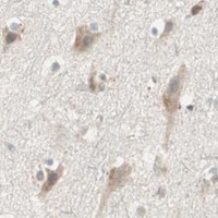


Comprehensive Human Protein Atlas IHC scoring per tissue (reliability: Approved — At least one protein variant secreted, tissue location of RNA and protein might differ and correlation is complex.). Rows are taken directly from the HPA tissue chart — click any row's HPA link to view the source.
| Tissue | Cell type | Level | Evidence | Source |
|---|---|---|---|---|
| Testis | Pachytene spermatocytes | High | Protein (IHC) | HPA → |
| Tissue | Cell type | Level | Evidence | Source |
|---|---|---|---|---|
| Adipose tissue | Adipocytes | Not detected | Protein (IHC) | HPA → |
| Adrenal gland | Glandular cells | Not detected | Protein (IHC) | HPA → |
| Appendix | Glandular cells | Not detected | Protein (IHC) | HPA → |
| Bone marrow | Hematopoietic cells | Not detected | Protein (IHC) | HPA → |
| Breast | Adipocytes | Not detected | Protein (IHC) | HPA → |
Troubleshoot SFRP1 staining in paraffin sections by checking retrieval, compartment, tissue context, and controls before comparing chromogenic signal across samples.
A01968-2 has IHC images from human colon and lung cancer and mouse intestine and kidney, plus an IF image from U20S cells (catalog image captions).
A01968-2 will render with a paraffin-section IHC figure; its captions also document IHC in human colon and lung cancer and mouse intestine and kidney, and IF in U20S cells (catalog image captions). M01968 will render with listed human reactivity and IHC, ICC and IF applications, but no IHC or IF image captions (catalog payload).
Which to pick: Choose A01968-2 for tissue IHC: its figure shows paraffin-section staining after citrate retrieval at pH 6, while the fixative is unreported (A01968-2 IHC caption). Choose A01968-2 for IF/ICC when an illustrated example matters; M01968 is a rabbit monoclonal with listed ICC/IF applications but no image caption (catalog payload). For work across species, A01968-2 lists human, mouse and rat reactivity, whereas M01968 lists human only; the IHC captions illustrate human and mouse tissue, with no rat example (catalog reactivity; A01968-2 IHC captions).