This website uses cookies to ensure you get the best experience on our website.


- Table of Contents
Plan paraffin SGK1 IHC using the general cytoplasmic tissue pattern (HPA tissue IHC) and a 0.5–1 μg/mL starting range for A00673-2 (datasheet). Score nuclear and cytoplasmic staining separately because SGK1 localization changes with cell cycle, growth factors, and stress (UniProt).

Expected localisation, validated protocols, controls and antibodies — the at-a-glance facts below, then the full design guide.
| Expected localisation | General cytoplasmic tissue staining (HPA tissue IHC) | |
| Staining pattern | Cytoplasmic staining in glandular and other cells (HPA tissue IHC) | |
| Antigen retrieval | EDTA pH 8.0 HIER, heat-mediated (datasheet A00673-2) | |
| Positive control | Adrenal gland+4 more · see all | |
| Negative control | Soft tissue |
| Fixation | Keep fixation consistent across sections (standard IHC practice; not target-specific). Selected-image fixative and duration unreported (datasheet A00673-2); verify before use. | |
| Caveat | Stress can shift SGK1 from nucleus to cytoplasm (UniProt) | |
| Regulation | Hormones and stress redistribute SGK1 (UniProt) | |
| Isoform / epitope | Five isoforms; antibody epitope coverage needs checking (UniProt) |
The catalog antibody’s IHC-P protocol uses EDTA pH 8.0 retrieval (datasheet A00673-2). The published SGK1 IHC protocols below cover human brain and mouse skin, lung, and other tissues (PMCs cited below).
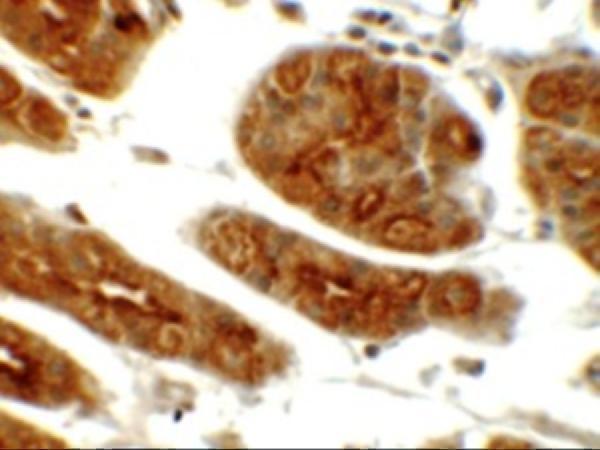
| Sample | Paraffin-embedded human mammary cancer tissue; fixative not specified (datasheet A00673-2) |
| Fixation | Image fixative and duration unreported (datasheet A00673-2); verify before use. |
| Sectioning | 4–5 µm sections on charged slides (standard) |
| Deparaffinisation | Xylene, graded ethanol series to water (standard) |
| Antigen retrieval | Heat retrieval: EDTA pH 8.0 (datasheet A00673-2); 20 min, 95–100 °C (standard) |
| Peroxidase block | 3% H2O2, 10 min, room temperature (standard) |
| Blocking | 10% goat serum (datasheet A00673-2) |
| Primary antibody | Rabbit anti-SGK1, 0.5-1μg/ml (datasheet A00673-2) |
| Primary incubation | Overnight at 4 °C (datasheet A00673-2) |
| Detection | Streptavidin-biotin complex (SABC), DAB chromogen (datasheet A00673-2) |
| Counterstain | Hematoxylin, blue, dehydrate and mount (standard) |
| Expected result | SGK1-positive staining in glandular cells of adrenal gland (HPA tissue IHC: High). HPA tissue profile: General cytoplasmic expression. No signal in the no-primary control. |
SGK1 staining in paraffin sections is expected to be mainly cytoplasmic, including glandular, hematopoietic and neuronal cells in tissues where those populations stain (HPA tissue IHC). Nuclear staining can also be plausible because SGK1 changes location with cell state (UniProt O00141 subcellular location). SGK1 has no transmembrane segment (UniProt O00141 topology). HPA rates its tissue IHC as Approved but reports low consistency between staining and RNA expression (HPA tissue IHC).
| Cytoplasmic staining is strongest in adrenal or colon glandular cells, bone marrow hematopoietic cells, or cerebral cortical neurons. | This matches cell populations scored High by HPA; assess the stated cell population within each tissue, rather than treating every cell on the section as an expected positive (HPA tissue IHC). |
| Nuclear staining accompanies cytoplasmic staining or varies between otherwise comparable cells. | A nuclear component can be biologically plausible: UniProt places SGK1 in both compartments and describes cell cycle, hormone and stress dependent shifts. A purely nuclear result still needs tissue and antibody context before acceptance (UniProt O00141 subcellular location; HPA tissue IHC). |
| Strong staining outlines extracellular spaces or glandular lumens, with little signal in the expected cells. | That distribution does not match HPA’s general cytoplasmic tissue profile. Check the section and detection controls for artefact; do not call the outlined spaces SGK1 positive solely because adjacent glandular cells are expected to stain (HPA tissue IHC; general IHC practice). |
| Strong signal appears in soft tissue fibroblasts, or color appears broadly across unrelated cell types. | HPA lists soft tissue fibroblasts as Not detected. Investigate antibody cross-reactivity or endogenous detection activity with controls before interpreting this as SGK1; the HPA result is a reference observation, not proof that every fibroblast must be negative (HPA tissue IHC; general IHC practice). |
| No signal appears in a section containing a HPA High cell population. | First confirm that the expected cells are present and the staining run worked. A negative result alone cannot establish SGK1 absence, particularly given HPA’s reported low agreement between tissue staining and RNA expression (HPA tissue IHC; general IHC practice). |
| Tissue and cell selection | Adrenal, appendix, breast and colon glandular cells; bone marrow hematopoietic cells; and cerebral cortical neurons are High, whereas soft tissue fibroblasts are Not detected. Select and score the actual cell population named by HPA (HPA tissue IHC). |
| State dependent location | UniProt describes nuclear residence after serum or growth factor stimulation, cytoplasmic detection after stress or glucocorticoids, and possible mitochondrial association under certain stress conditions. The expected compartment therefore depends on biological context (UniProt O00141 subcellular location). |
| Membrane interpretation | SGK1 has no transmembrane segment. UniProt nevertheless describes location at the cytoplasmic face of the cell membrane during epithelial sodium channel regulation; a membrane associated component is possible, but an obligatory surface outline is unsupported (UniProt O00141 topology and subcellular location). |
| Isoform coverage | UniProt lists five SGK1 isoforms and reports stronger isoform 2 expression in brain and pancreas. An antibody’s isoform recognition cannot be inferred from those facts; check its documented target region before explaining tissue differences as isoform effects (UniProt O00141 isoforms and tissue specificity; general IHC practice). |
| Evidence across assays | HPA describes general cytoplasmic tissue IHC but mainly nuclear speckles in ICC-IF. These observations come from different preparations; use the paraffin tissue profile for the IHC call and treat ICC-IF as context, without assuming identical patterns (HPA tissue IHC; HPA subcellular ICC-IF). |
| Strength of tissue evidence | HPA rates tissue IHC Approved and explicitly notes low consistency between antibody staining and RNA expression. Its High and Not detected entries guide comparison, but a single discrepant section needs controls and cell level review before a biological conclusion (HPA tissue IHC; general IHC practice). |
| Situation | Likely cause | Next action |
|---|---|---|
| A HPA High cell population shows no chromogenic signal. | The expected cells may be absent from the examined area, or the staining run may have failed; this slide alone cannot distinguish those possibilities (HPA tissue IHC; general IHC practice). | Verify morphology and run an appropriate positive tissue and detection control in the same workflow. Review documented primary antibody dilution and antigen retrieval settings; no SGK1 specific retrieval condition is supplied here (general IHC practice). |
| Diffuse color covers the section, including spaces without cells. | Background from detection reagents, endogenous activity or nonspecific binding can obscure a cellular pattern (general IHC practice). | Compare primary antibody omission and detection controls, assess blocking and washing, and score only cell associated staining against HPA’s general cytoplasmic profile (general IHC practice; HPA tissue IHC). |
| Soft tissue fibroblasts stain strongly. | This conflicts with the HPA Not detected reference and may reflect cross-reactivity or endogenous detection activity; the stain alone does not identify which (HPA tissue IHC; general IHC practice). | Inspect controls and morphology, then compare with a HPA High cell population before accepting the fibroblast signal as SGK1 (HPA tissue IHC; general IHC practice). |
| Only nuclei stain in an otherwise expected tissue population. | SGK1 can enter nuclei, but HPA describes tissue IHC as generally cytoplasmic. Nuclear staining alone does not establish whether the pattern is biological or assay related (UniProt O00141 subcellular location; HPA tissue IHC). | Check cell identity and staining controls; interpret nuclear signal with the sample context and the HPA tissue profile, rather than rejecting or accepting it by compartment alone (HPA tissue IHC; UniProt O00141 subcellular location; general IHC practice). |
| A sharp membrane outline dominates the image. | UniProt permits association with the cytoplasmic membrane surface in one functional context, while reporting no transmembrane segment; a strong outline by itself is inconclusive (UniProt O00141 topology and subcellular location). | Assess accompanying cytoplasmic staining and cell identity, then inspect detection controls for edge artefact before assigning the outline to SGK1 (HPA tissue IHC; general IHC practice). |
| IF/ICC shows nuclear speckles while paraffin IHC looks cytoplasmic. | HPA reports mainly nuclear speckles in ICC-IF and general cytoplasmic expression in tissue IHC; UniProt also describes variable SGK1 location (HPA subcellular ICC-IF; HPA tissue IHC; UniProt O00141 subcellular location). | For this paraffin IHC section, judge the result against HPA tissue IHC and the sampled cell population. Use the ICC-IF observation as context, without transferring it into an IHC protocol requirement (HPA tissue IHC; HPA subcellular ICC-IF). |
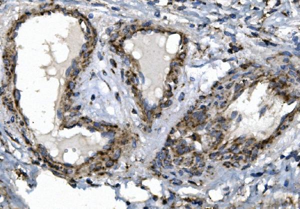
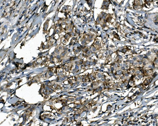
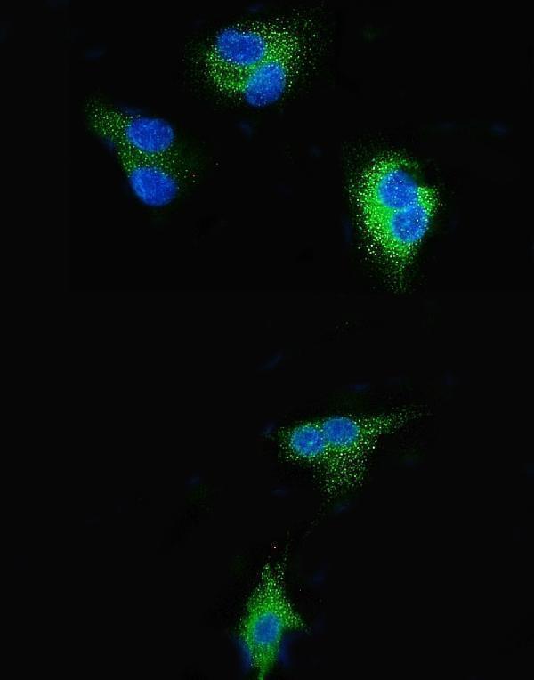
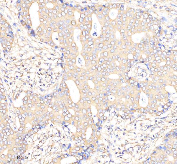
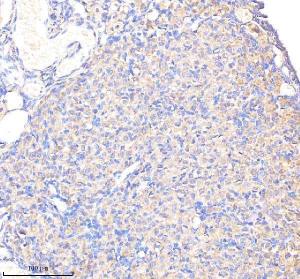
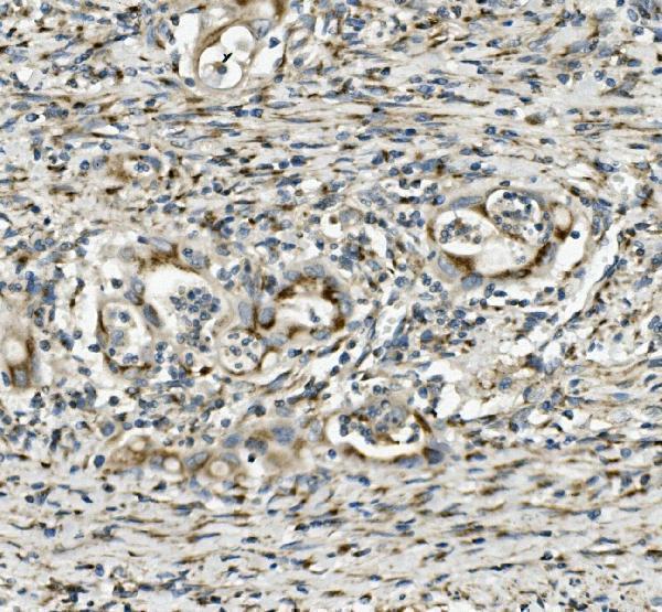
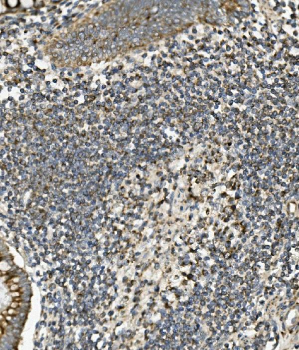
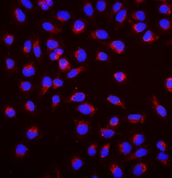


Comprehensive Human Protein Atlas IHC scoring per tissue (reliability: Approved — Low consistency between antibody staining and RNA expression data.). Rows are taken directly from the HPA tissue chart — click any row's HPA link to view the source.
| Tissue | Cell type | Level | Evidence | Source |
|---|---|---|---|---|
| Adrenal gland | Glandular cells | High | Protein (IHC) | HPA → |
| Appendix | Glandular cells | High | Protein (IHC) | HPA → |
| Bone marrow | Hematopoietic cells | High | Protein (IHC) | HPA → |
| Breast | Glandular cells | High | Protein (IHC) | HPA → |
| Cerebellum | Cells in molecular layer | High | Protein (IHC) | HPA → |
| Tissue | Cell type | Level | Evidence | Source |
|---|---|---|---|---|
| Soft tissue | Fibroblasts | Not detected | Protein (IHC) | HPA → |
Troubleshoot SGK1 staining in paraffin sections by tracking retrieval conditions, cellular compartment, controls and scoring across matched samples.
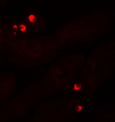
Anti-SGK1 antibodies have paraffin-section IHC images from human cancer and stomach tissues and mouse and rat ovary (catalog IHC captions); IF examples include human cells and stomach tissue (catalog IF captions).
A00673-2 shows human mammary cancer IHC, and A00673 shows human rectal cancer IHC, both in paraffin sections (respective catalog IHC captions). M00673 shows human breast cancer and mouse and rat ovary paraffin-section IHC (catalog IHC captions); A00673-1 shows human stomach IHC (catalog IHC caption).
Which to pick: For human paraffin-section IHC, choose A00673-2 for mammary cancer or A00673 for rectal cancer; each image caption documents EDTA pH 8.0 retrieval and leaves the fixative unreported (respective catalog IHC captions). For IF/ICC, A00673-2 has an A549 cell example at 4 μg/mL and A00673 has a U20S cell example at 2 μg/mL (respective catalog IF captions); A00673-1 also has a human stomach IF example (catalog IF caption). For cross-species IHC, choose rabbit monoclonal M00673, clone EHF-19 (catalog title and clone), whose paraffin-section captions show human breast cancer and mouse and rat ovary; the fixative is unreported (catalog IHC captions).