SHH / Sonic hedgehog protein · Western blot design guide

Design a Western Blot for SHH

Real validated SHH Western blot protocols, expected-band and isoform facts, troubleshooting for weak or shifted signal, and recommended anti-SHH WB antibodies. Everything you need to plan the experiment before you commit precious samples.

Evidence assembled September 2026 · For research use; verify linked source records and product datasheet before use
Western blot protocol sheet for SHH: expected band ~49.6 kDa, hero antibody PA1072-1, catalog values and labelled standard workflow; separate PMC comparisons on the guide
Printable SHH Western blot protocol sheet — expected band ~49.6 kDa, antibody PA1072-1, controls and PMC citations. Open the full SHH WB guide →

SHH Western Blot Experimental Design Guide

Expected bands, validated protocols, controls and antibodies — the at-a-glance facts below, then the full design guide.

Must know before running
Expected band ~49.6 kDa
Gel 12–15% (standard starting point)
Negative control ⓘ Suggested KO / knockdown lysate
Important caveats
Reasons your observed band may differ from the expected size.
PTM Glycosylated + Cleaved
Caveat Autocatalytic cleavage products
Gene-set association MSigDB Hallmark membership
Isoform 1 isoform(s)
Section 1

Real Curated SHH Western Blot Protocols

The PA1072-1 protocol combines labelled catalog values with standard starting conditions. Published comparisons retain their own sample, reagent and detection scope.

Recommended Western blot protocol parameters
Sample / lysateRat Liver, Rat Intestine, HELA (catalog PA1072-1)
Gel %12–15% (standard starting point)
Load20–30 µg total protein per lane; optimize for abundance (standard starting point)
TransferStandard semi-dry transfer; verify efficiency (standard starting point)
Membrane0.45 µm PVDF (standard starting point)
Blocking5% milk or 5% BSA in TBST (standard starting point)
Primary antibodyPA1072-1; use the WB datasheet starting dilution (standard starting point)
Primary incubationOvernight at 4 °C (standard starting point)
Secondary antibodySpecies-matched HRP conjugate at validated dilution (standard starting point)
Secondary incubation1 h at room temperature (standard starting point)
Wash3 × 5 min in TBST (standard starting point)
DetectionECL; bracket exposures to avoid saturation (standard starting point)
Section 2

What Is the Expected SHH Western Blot Band Size?

UniProt predicts a 49.6 kDa SHH precursor; cleavage and Asn278 glycosylation may affect migration, but no empirical band size or demonstrated shift is supplied.

What am I looking at on my blot?
Band near 49.6 kDaConsistent with the predicted full-length SHH precursor; confirm identity with antibody controls
Band below 49.6 kDaMay reflect signal peptide removal or autocatalytic processing; fragment size is unspecified
Band above 49.6 kDaAsn278 N-linked glycosylation could contribute, but a visible shift is not established
Little or no band in whole-cell lysateSHH is secreted, so cellular abundance may be low
💡Expected SHH appearanceUniProt predicts a 49.6 kDa full-length SHH precursor; no empirical band size is supplied, and processing or Asn278 glycosylation cannot establish a specific migration without band-identity controls.
How each factor affects band size
Full-length precursor massUniProt predicts 49.6 kDa before processing
Signal peptide at residues 1–23Its removal produces a smaller mature protein, with no measured band size supplied
N-linked glycosylation at Asn278Could increase apparent size, but a visible shift is not established
Autocatalytic cleavageCan yield products below precursor size; product masses are not supplied
Why is my band missing or off?
SituationLikely causeNext action
No band in lysateSHH is secretedCheck conditioned medium alongside lysate
Band higher than expectedAsn278 N-linked glycosylation is possible, but the band identity is unconfirmedCompare with a deglycosylated sample and verify antibody specificity
Band lower than expectedSignal peptide removal or autocatalytic processing may produce smaller speciesCheck the antibody epitope and compare with a full-length control
Multiple bandsPrecursor and processed SHH species may coexistCompare precursor and conditioned-medium samples with an independent SHH antibody
Weak or no signalSecreted SHH may be scarce in the tested sampleTest concentrated conditioned medium with a positive control
Fragments below expected sizeSHH has an autocatalytic cleavage annotationCheck which processed product the antibody recognizes and confirm with an independent antibody

Sample controls for SHH Western blot

🧪For positive controls for SHH in Western blot, you can use an HPA-verified positive sample once identified; none is supplied here.
Positive control: No high/medium HPA tissue identified
Negative control: Suggested KO / knockdown lysate
Loading controls: Run GAPDH, β-actin, and a total-protein stain such as Ponceau alongside SHH.
⚠️Feasibility: SHH is secreted, so conditioned medium may yield a stronger signal than whole-cell lysate; HPA supplies no tissue controls.

HPA tissue expression evidence for SHH

Comprehensive Human Protein Atlas IHC scoring per tissue. Rows are taken directly from the HPA tissue chart — click any row's HPA link to view the source.

Higher expression tissues · candidate positive controls from IHC

TissueCell typeLevelEvidenceSource
No high/medium HPA tissues identified in the supplied evidence.

Lower expression tissues · IHC evidence, not confirmed WB-negative controls

TissueCell typeLevelEvidenceSource
No lower-expression tissue rows available in the supplied evidence.
Section 3

Advanced SHH Western Blot Tips

Deeper troubleshooting and optimisation questions for SHH, answered from its protein features.

How should SHH band migration be interpreted?
Band shift · Use the separately labelled calculated mass and catalog-observed evidence above. A sequence annotation does not establish an observed migration shift. Verify target identity with orthogonal controls.
Should I expect multiple SHH isoforms?
Isoforms · The supplied record lists one isoform and no alternative sequence. Do not assign multiple bands to annotated isoforms on this evidence alone; consider the listed signal peptide and autocatalytic cleavage features when interpreting distinct forms.
Could glycosylation affect an SHH band?
PTM · UniProt lists one N-linked glycosylation site at Asn278, using canonical sequence numbering. Consider this site when assessing a band, but its presence alone does not establish a visible shift. Check whether the band you are measuring contains residue 278.
Does this guide establish induction of SHH?
Induction · No general induction response is established by this guide. A pathway or gene-set association is not evidence of induction in a particular specimen. Verify the relevant treatment and control in a target-specific experiment.
How should transfer be checked for SHH?
Transfer · Standard workflow guidance: verify transfer efficiency for the intended target size before interpreting a weak signal. Use total-protein assessment and optimize transfer for the membrane, gel and apparatus; the labelled catalog values take precedence.
How should blocking be optimized?
Blocking · Standard workflow guidance: follow the PA1072-1 datasheet where specified. Otherwise compare 5% milk or 5% BSA in TBST; for a phospho-specific assay start with BSA. Optimize background and specific signal with matched controls.
How should I quantify SHH when several bands appear?
Quantitation · Define which SHH form and sample compartment you intend to measure, then quantify the corresponding band consistently across samples. The record lists autocatalytic cleavage, an N-product, and both membrane-associated and secreted locations; it does not provide apparent band masses for assigning individual bands.
Why might SHH bands differ from the predicted 49.6 kDa?
Interpretation · The 49.6 kDa prediction is for the 462-residue sequence. SHH has a signal peptide at residues 1–23, and its features indicate autocatalytic cleavage and an N-product. These features suggest distinct protein forms, but they do not establish the apparent mass of any band or explain a particular difference.

The record places SHH at endoplasmic reticulum and Golgi membranes and also describes it as secreted. Choose cell-associated or secreted samples according to the form you intend to measure, and compare like sample types across conditions.

The SHH N-product is annotated as a multimer. If an additional band appears, keep multimerization in mind, but this annotation alone cannot identify that band or predict where it will migrate.

Compare its apparent mass with the 49.6 kDa full-sequence prediction cautiously. The record lists a signal peptide at 1–23, autocatalytic cleavage, an N-product, and N-linked glycosylation at canonical Asn278. None alone confirms the identity or mass of an unexpected band.
Boster reagents

SHH Western Blot Antibodies

Catalog antibodies with Western blot application and product-specific WB images. Evaluate suitability with the reported sample, controls and experimental conditions.

Real WB data Anti-Sonic Hedgehog antibody, PA1072-1, Western blotting Lane 1: Rat Liver Tissue Lysate Lane 2: Rat Intestine Tissue Lysate Lane 3: HELA Cell Lysate Lane 4: A549 Cell Lysate Lane 5: SMMC Cell Lysate Lane 6: MM231 Cell Lysate
Anti-Sonic Hedgehog/SHH Antibody Picoband®
Cat # PA1072-1
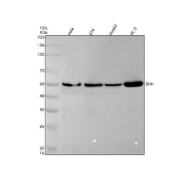
Real WB data Western blot analysis of SHH using anti-SHH antibody (M00058). Electrophoresis was performed on a 10% SDS-PAGE gel at 80V (Stacking gel) / 120V (Resolving gel) for 2 hours. The sample well of each lane was loaded with 30 ug of sample under reducing conditions. Lane 1: human Hela whole cell lysates, Lane 2: human RT4 whole cell lysates, Lane 3: human HepG2 whole cell lysates, Lane 4: human PC-3 whole cell lysates. After electrophoresis, proteins were transferred to a nitrocellulose membrane at 150 mA for 50-90 minutes. Blocked the membrane with 5% non-fat milk/TBS for 1.5 hour at RT. The membrane was incubated with rabbit anti-SHH antigen affinity purified monoclonal antibody (M00058) at 1: 500 overnight at 4°C, then washed with TBS-0.1%Tween 3 times with 5 minutes each and probed with a goat anti-rabbit IgG-HRP secondary antibody at a dilution of 1:5000 for 1.5 hour at RT. The signal is developed using an ECL Plus Western Blotting Substrate (Catalog # AR1196-200) with Tanon 5200 system. A specific band was detected for SHH at approximately 50 kDa. The expected band size for SHH is at 50 kDa.
Anti-Sonic Hedgehog SHH Rabbit Monoclonal Antibody
Cat # M00058

Both anti-SHH antibodies list human, mouse, and rat reactivity and include WB images. PA1072-1 shows rat tissue and cell lysates; M00058 shows human cell lysates with an approximately 50 kDa band. No publication evidence is supplied.

Which to pick: Choose M00058 for a human whole-cell lysate protocol with reported conditions and a 50 kDa band. Choose PA1072-1 for WB images of rat liver or intestine. Both list mouse reactivity, but neither supplied caption shows a mouse specimen.

Source: BosterBio SHH gene-info card — filtered to Western-blot-capable antibodies; each card shows that product's actual WB validation figure.