This website uses cookies to ensure you get the best experience on our website.


- Table of Contents
This guide helps plan paraffin SIGLEC7 IHC using immune-cell staining in bone marrow, lymph node, spleen and tonsil as reference patterns (HPA tissue IHC). It pairs those patterns with a 1:100 starting dilution for the IHC-validated antibody (datasheet: 1:100).

Expected localisation, validated protocols, controls and antibodies — the at-a-glance facts below, then the full design guide.
| Expected localisation | Immune-cell cytoplasm (HPA tissue IHC); membrane (UniProt) | |
| Staining pattern | Immune cells show cytoplasmic positivity (HPA tissue IHC) | |
| Antigen retrieval | Citrate pH 6.0 HIER, 95–98 °C, 20 min (rule: cytoplasmic / membrane antigen) | |
| Positive control | Bone marrow+3 more · see all | |
| Negative control | Adipose tissue+4 more · see all |
| Fixation | Keep fixation consistent across sections (standard IHC practice; not target-specific); Matched tissue-IHC evidence does not establish the fixation claim. Validate the specimen-specific method before use. (selected-SKU IHC image A06553) | |
| Caveat | Marrow/spleen peroxidase may mimic chromogenic signal (HPA tissue IHC; standard IHC practice) | |
| Regulation | Present in resting and activated NK cells (UniProt) | |
| Isoform / epitope | 4 isoforms; check whether the epitope is extracellular or cytoplasmic (UniProt) |
The catalog antibody’s IHC-P protocol (datasheet) is accompanied by a published SIGLEC7 protocol using glioma tissue sections (PMC11357930).
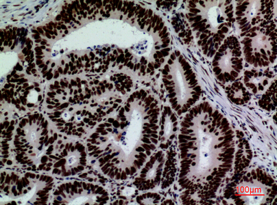
| Sample | Paraffin-embedded human colon-cancer tissue; fixative not specified (datasheet A06553) |
| Fixation | Image fixative and duration unreported (datasheet A06553); verify before use. |
| Sectioning | 4–5 µm sections on charged slides (standard) |
| Deparaffinisation | Xylene, graded ethanol series to water (standard) |
| Antigen retrieval | Heat-induced epitope retrieval in citrate buffer, pH 6.0, 20 min at 95–98 °C (standard rule: cytoplasmic / membrane antigen) |
| Peroxidase block | 3% H2O2, 10 min, room temperature (standard) |
| Blocking | 10% normal serum of the secondary host, 30 min, room temperature (standard) |
| Primary antibody | Rabbit anti-SIGLEC7, 1:100 (datasheet A06553) |
| Primary incubation | Overnight at 4 °C (standard) |
| Detection | HRP-polymer secondary, DAB chromogen 5–10 min (standard) |
| Counterstain | Hematoxylin, blue, dehydrate and mount (standard) |
| Expected result | SIGLEC7-positive staining in hematopoietic cells of bone marrow (HPA tissue IHC: Medium). HPA tissue profile: Cytoplasmic positivity in immune cells. No signal in the no-primary control. |
SIGLEC7 is a membrane protein with an extracellular region and a cytoplasmic tail (UniProt Q9Y286 topology). In paraffin-section IHC, expect staining in immune-cell populations, especially hematopoietic cells in bone marrow and cells in splenic red pulp; HPA describes cytoplasmic positivity in immune cells at these sites (HPA tissue IHC). HPA rates the tissue staining reliability Enhanced, while reporting medium consistency between antibody staining and RNA expression (HPA tissue IHC).
| Moderate staining in bone-marrow hematopoietic cells or splenic red-pulp cells, with other cells relatively pale (HPA tissue IHC). | This matches the reported IHC distribution and level: both populations are rated Medium, within an immune-cell profile described as cytoplasmic positivity (HPA tissue IHC). Evaluate the stained cells, rather than treating a whole positive tissue as uniformly positive (HPA tissue IHC). |
| Predominantly nuclear staining, without a convincing immune-cell pattern. | A nuclear-dominant result conflicts with the membrane topology of SIGLEC7 and HPA’s immune-cell IHC profile (UniProt Q9Y286 topology; HPA tissue IHC). Investigate staining artefact before calling it target-specific (general IHC practice). |
| Strong staining of adipocytes or appendix glandular cells. | HPA reports SIGLEC7 as Not detected in those cell populations (HPA tissue IHC). Such staining raises concern for cross-reactivity or endogenous detection activity; compare with a reagent-negative control and inspect nearby immune cells (general IHC practice). |
| Widespread, diffuse color obscures cell boundaries and tissue detail. | This cannot establish which cells express SIGLEC7 (general IHC practice). Check whether the color persists without primary antibody, and assess blocking, washes, and detection chemistry before scoring positive cells (general IHC practice). |
| No staining in bone-marrow hematopoietic cells or splenic red-pulp cells. | These are reported Medium positive populations, so a blank result warrants a technical check (HPA tissue IHC). It does not alone prove biological absence: inspect the control section and the same staining run before interpreting the specimen (general IHC practice). |
| Cell population and sampled site | Bone-marrow hematopoietic cells, lymph-node and tonsil non-germinal-center cells, and splenic red-pulp cells are rated Medium (HPA tissue IHC). Score the reported population within each section; adjacent cells need not share its staining (HPA tissue IHC). |
| Compartment and assay | SIGLEC7 has residues 19–353 extracellular, 354–376 transmembrane, and 377–467 cytoplasmic (UniProt Q9Y286 topology). HPA describes cytoplasmic positivity in tissue IHC, while its ICC-IF summary places signal mainly at the plasma membrane and additionally in vesicles (HPA tissue IHC; HPA subcellular). |
| Antibody evidence | The reported tissue profile has Enhanced reliability, with medium staining–RNA consistency (HPA tissue IHC). CAB080396 has Enhanced IHC validation; HPA075513 has Approved ICC validation but no reported IHC status (HPA antibodies). Interpret a new antibody’s pattern against its own controls (general IHC practice). |
| Sequence features and epitope uncertainty | UniProt lists a cleaved signal peptide at residues 1–18, eight glycosylation sites, and four isoforms (UniProt Q9Y286 processing, glycosylation, isoforms). Without an antibody epitope or isoform-specific validation in these sources, those features cannot predict this assay’s staining or retrieval needs (UniProt Q9Y286; HPA antibodies). |
| Antigen retrieval | Target-specific fixation effects are not established by the supplied assay evidence. Verify with a matched IHC source before attributing a result to fixation. |
| IF/ICC Q&A: where should signal appear? | HPA places ICC-IF signal mainly at the plasma membrane, with additional vesicular signal (HPA subcellular). That approved ICC-IF localization helps interpret an IF image; it is separate from HPA’s cytoplasmic tissue-IHC description and does not specify an IF protocol (HPA subcellular; HPA tissue IHC). |
| Situation | Likely cause | Next action |
|---|---|---|
| The positive-control section is blank. | A missed staining step, ineffective detection, or an unsuitable assay condition is possible (general IHC practice); bone-marrow hematopoietic and splenic red-pulp cells are reported Medium positive (HPA tissue IHC). | Target-specific fixation effects are not established by the supplied assay evidence. Verify with a matched IHC source before attributing a result to fixation. |
| Only nuclei are strongly colored. | This conflicts with the membrane topology and reported immune-cell IHC profile (UniProt Q9Y286 topology; HPA tissue IHC); background or detection artefact is possible (general IHC practice). | Compare the primary-omitted section, examine counterstain and tissue detail, and require a reproducible immune-cell pattern before scoring the signal (general IHC practice; HPA tissue IHC). |
| Adipocytes or appendix glandular cells stain strongly. | Those populations are listed as Not detected (HPA tissue IHC); cross-reactivity or endogenous detection activity is possible (general IHC practice). | Check a primary-omitted control, assess endogenous activity for the detection system, and compare staining with the expected immune-cell populations (general IHC practice; HPA tissue IHC). |
| Color spreads across most of the section. | Diffuse background may follow insufficient blocking, inadequate washing, or excess detection signal (general IHC practice). It prevents a reliable comparison with HPA’s cell-specific profile (HPA tissue IHC). | Inspect the primary-omitted control; optimize blocking, washes, and detection conditions, then score only distinguishable cells (general IHC practice). |
| A positive site stains, but the expected cells are hard to identify. | HPA reports positivity in specific populations within bone marrow, lymph node, spleen, and tonsil, rather than assigning one level to every cell in those tissues (HPA tissue IHC). | Use morphology and the counterstain to locate the reported population, then record its staining separately from surrounding cells (general IHC practice; HPA tissue IHC). |
| IHC appears cytoplasmic, while an IF image shows a membrane rim. | HPA reports cytoplasmic immune-cell positivity in tissue IHC and mainly plasma-membrane localization in ICC-IF; UniProt describes a transmembrane protein (HPA tissue IHC; HPA subcellular; UniProt Q9Y286 topology). | Judge each image against its assay-specific HPA description and controls; do not reject an otherwise matching tissue-IHC result solely because its chromogenic pattern lacks a sharp membrane rim (HPA tissue IHC; HPA subcellular). |


Comprehensive Human Protein Atlas IHC scoring per tissue (reliability: Enhanced — Medium consistency between antibody staining and RNA expression data.). Rows are taken directly from the HPA tissue chart — click any row's HPA link to view the source.
| Tissue | Cell type | Level | Evidence | Source |
|---|---|---|---|---|
| Bone marrow | Hematopoietic cells | Medium | Protein (IHC) | HPA → |
| Lymph node | Non-germinal center cells | Medium | Protein (IHC) | HPA → |
| Spleen | Cells in red pulp | Medium | Protein (IHC) | HPA → |
| Tonsil | Non-germinal center cells | Medium | Protein (IHC) | HPA → |
| Tissue | Cell type | Level | Evidence | Source |
|---|---|---|---|---|
| Adipose tissue | Adipocytes | Not detected | Protein (IHC) | HPA → |
| Adrenal gland | Glandular cells | Not detected | Protein (IHC) | HPA → |
| Appendix | Glandular cells | Not detected | Protein (IHC) | HPA → |
| Breast | Adipocytes | Not detected | Protein (IHC) | HPA → |
| Caudate | Glial cells | Not detected | Protein (IHC) | HPA → |
Troubleshoot SIGLEC7 staining in paraffin sections by checking retrieval, cell identity, subcellular pattern, and controls before interpreting signal intensity.
A06553 is a human-reactive anti-SIGLEC7 antibody with an IHC image from paraffin-embedded human colon cancer (catalog: reactivity; A06553 IHC image caption). IF is listed without an IF image (catalog: applications; IF image alts).
A06553 has IHC evidence from paraffin-embedded human colon cancer at 1:100 (A06553 IHC image caption). A06553 also lists IF for human samples, but supplies no IF image (catalog: applications, reactivity, IF image alts).
Which to pick: Choose A06553 for human tissue IHC because its own image shows staining of paraffin-embedded human colon cancer at 1:100; the fixative is unreported (A06553 IHC image caption). For IF, A06553 lists 1:50, but provides no IF figure or ICC validation (catalog: applications, IF dilution, IF image alts). No cross-species option is documented: A06553 lists Human reactivity only, and its clone is unreported (catalog: reactivity, clone).