This website uses cookies to ensure you get the best experience on our website.


- Table of Contents
Plan SIK2 paraffin IHC around cytoplasmic staining in most tissues (HPA tissue IHC). Compare medium staining in cerebral cortex neuronal cells with undetected lymph node germinal center cells (HPA tissue IHC), and interpret staining intensity cautiously because agreement with RNA expression is low (HPA tissue IHC).

Expected localisation, validated protocols, controls and antibodies — the at-a-glance facts below, then the full design guide.
| Expected localisation | Cytoplasmic tissue staining (HPA tissue IHC); ER membrane location (UniProt) | |
| Staining pattern | Cytoplasmic staining in most tissues (HPA tissue IHC) | |
| Antigen retrieval | EDTA pH 8.0 HIER, heat-mediated (datasheet A04515-1) | |
| Positive control | Adrenal gland+4 more · see all | |
| Negative control | Bone marrow+4 more · see all |
| Fixation | Keep fixation consistent across sections (standard IHC practice; not target-specific) | |
| Caveat | Staining has low consistency with RNA expression (HPA tissue IHC) | |
| Regulation | Low tissue specificity (HPA RNA specificity) | |
| Isoform / epitope | 0 isoforms; no processing or extracellular domain (UniProt) |
The catalog antibody’s IHC-P protocol is accompanied by published IHC methods for HCC specimens, paraffin pathology sections, spinal cord tissue, and tumor sections (PMC12528092; PMC5955149; PMC12676768; PMC9353504).
| Sample | Paraffin-embedded mouse brain tissue; fixative not specified (datasheet A04515-1) |
| Fixation | Image fixative and duration unreported (datasheet A04515-1); verify before use. |
| Sectioning | 4–5 µm sections on charged slides (standard) |
| Deparaffinisation | Xylene, graded ethanol series to water (standard) |
| Antigen retrieval | Heat retrieval: EDTA pH 8.0 (datasheet A04515-1); 20 min, 95–100 °C (standard) |
| Peroxidase block | 3% H2O2, 10 min, room temperature (standard) |
| Blocking | 10% goat serum (datasheet A04515-1) |
| Primary antibody | Rabbit anti-SIK2, 2-5 μg/ml (datasheet A04515-1) |
| Primary incubation | Overnight at 4 °C (datasheet A04515-1) |
| Detection | HRP-conjugated secondary, DAB chromogen (datasheet A04515-1) |
| Counterstain | Hematoxylin, blue, dehydrate and mount (standard) |
| Expected result | SIK2-positive staining in glandular cells of adrenal gland (HPA tissue IHC: Medium). HPA tissue profile: Cytoplasmic expression in most tissues. No signal in the no-primary control. |
In paraffin-section IHC, expect predominantly cytoplasmic SIK2 staining, including medium staining in adrenal glandular cells and cortical neurons (HPA tissue IHC). UniProt also places SIK2 at the endoplasmic reticulum membrane, with no transmembrane segment (UniProt Q9H0K1). HPA rates the tissue antibody Approved but reports low consistency between staining and RNA expression; use the listed cell patterns as guides, not absolute rules (HPA tissue IHC).
| Cytoplasmic signal in adrenal glandular cells or cerebral cortical neurons. | This matches HPA's medium-staining examples and its predominantly cytoplasmic tissue profile (HPA tissue IHC). Compare staining within the named cells before judging an entire section positive; adjacent cell populations need not share the same appearance (HPA tissue IHC). |
| Predominantly nuclear chromogen in a tissue section, with little cytoplasmic signal. | This departs from the reported tissue-IHC pattern and warrants a localisation check (HPA tissue IHC). Nuclear localisation alone does not settle specificity: nucleoplasm is an approved location in HPA ICC-IF images, a different application (HPA subcellular ICC-IF). |
| Strong signal in germinal-center cells or bone-marrow hematopoietic cells. | HPA lists these cells as not detected, so assess cross-reactivity and endogenous chromogenic activity with detection controls (HPA tissue IHC; general IHC practice). Because tissue-antibody staining has low consistency with RNA, an unexpected result is a prompt to verify, not proof of an artefact (HPA tissue IHC). |
| Uniform haze across cells, stroma, and nominally negative regions. | A pattern without cell or compartment definition is hard to interpret as SIK2; examine primary-omission and detection controls for nonspecific background (general IHC practice). HPA's reported pattern is cytoplasmic expression in most tissues, with differences among named cell populations (HPA tissue IHC). |
| No signal in adrenal glandular cells despite interpretable section morphology. | HPA reports medium staining there, making this a useful failed-signal check rather than a guaranteed positive control (HPA tissue IHC). Review the catalog antibody's IHC-P instructions, detection reagents, and a parallel control section (general IHC practice); note HPA's low RNA–staining consistency (HPA tissue IHC). |
| Tissue and cell choice | HPA reports medium staining in adrenal glandular cells, breast myoepithelial cells, and cortical neurons, but low staining in kidney tubule cells and adipocytes (HPA tissue IHC). Choose a named medium-staining population when checking assay performance. |
| Strength of tissue-IHC evidence | The tissue antibody is Approved, with low consistency between staining and RNA; its IHC status is Approved, while a separate antibody has Approved ICC status (HPA tissue IHC; HPA antibodies). Do not treat these statuses as interchangeable application validation. |
| Compartment and topology | HPA tissue IHC describes cytoplasmic expression in most tissues; UniProt lists cytoplasm and endoplasmic reticulum membrane and no transmembrane segment (HPA tissue IHC; UniProt Q9H0K1). These annotations guide interpretation but do not specify a chromogenic retrieval condition. |
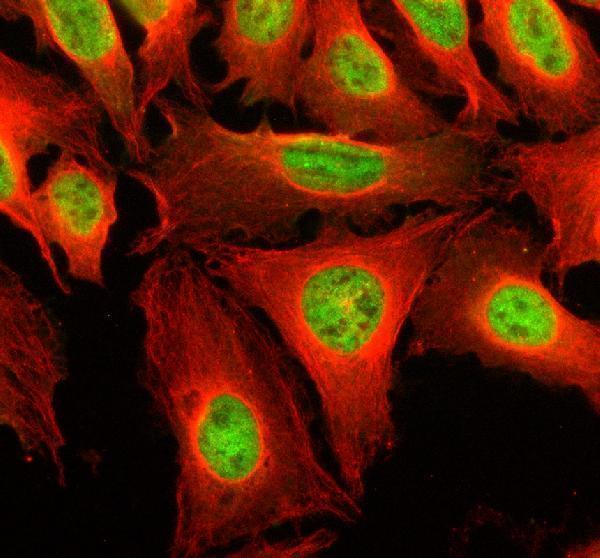
| IF/ICC Q&A: should its nuclear and vesicular pattern govern this IHC readout? | No. HPA approves nucleoplasm and vesicles as main ICC-IF locations, with cytosol among additional locations (HPA subcellular ICC-IF). Interpret paraffin-section chromogen against the tissue-IHC cytoplasmic profile; the applications show different reported patterns (HPA tissue IHC). |
| Processing and epitope uncertainty | UniProt lists one 1–926 chain, no signal peptide or propeptide, no annotated isoforms, and phosphorylation and acetylation sites (UniProt Q9H0K1). The payload does not locate the antibody epitope, so it cannot predict retrieval response or modification-dependent staining. |
| Chromogenic detection | Endogenous activity and nonspecific detection can create colour independent of primary-antibody binding (general IHC practice). Use appropriate detection controls when signal appears in HPA-listed undetected cells; those examples are reference observations, not universal biological negatives (HPA tissue IHC). |
| Situation | Likely cause | Next action |
|---|---|---|
| Adrenal glandular cells show no convincing cytoplasmic signal. | A missed assay step or weak detection is possible (general IHC practice); HPA's medium result is an observed reference, not a guarantee for every section (HPA tissue IHC). | Check the catalog antibody's IHC-P instructions and reagent performance, then compare a parallel named positive tissue and detection control (general IHC practice; HPA tissue IHC). |
| Chromogen appears mainly in nuclei. | The compartment differs from HPA's predominant tissue-IHC pattern, although nucleoplasm is approved in ICC-IF (HPA tissue IHC; HPA subcellular ICC-IF). | Check cell identity, counterstain, and primary-omission control before interpreting nuclear signal as SIK2 in paraffin sections (general IHC practice). |
| Germinal-center cells stain strongly. | HPA reports these cells as not detected; cross-reactivity or endogenous detection activity may explain discordant colour (HPA tissue IHC; general IHC practice). | Compare primary-omission and detection controls, then review whether cytoplasmic staining also occurs in a named medium-staining cell population (general IHC practice; HPA tissue IHC). |
| Colour coats most of the section without clear cell boundaries. | Diffuse background can arise from nonspecific antibody or detection binding (general IHC practice), obscuring the cell-level pattern reported by HPA (HPA tissue IHC). | Review blocking, washing, and antibody concentration under the catalog IHC-P instructions; judge the result against a primary-omission control (general IHC practice). |
| Kidney tubules look faint beside adrenal glandular cells. | This can reflect HPA's low kidney-tubule versus medium adrenal-glandular staining observations (HPA tissue IHC). | Score each named cell population against its own reference level; avoid using a low-staining population alone to declare assay failure (HPA tissue IHC). |
| Staining disagrees with an RNA-based expectation. | HPA explicitly reports low consistency between tissue-antibody staining and RNA expression despite an Approved reliability label (HPA tissue IHC). | Document cell type, compartment, and controls; interpret the observed IHC pattern cautiously and verify unexpected staining with an independent approach when available (HPA tissue IHC; general IHC practice). |


Comprehensive Human Protein Atlas IHC scoring per tissue (reliability: Approved — Low consistency between antibody staining and RNA expression data.). Rows are taken directly from the HPA tissue chart — click any row's HPA link to view the source.
| Tissue | Cell type | Level | Evidence | Source |
|---|---|---|---|---|
| Adrenal gland | Glandular cells | Medium | Protein (IHC) | HPA → |
| Appendix | Glandular cells | Medium | Protein (IHC) | HPA → |
| Breast | Myoepithelial cells | Medium | Protein (IHC) | HPA → |
| Caudate | Neuronal cells | Medium | Protein (IHC) | HPA → |
| Cerebellum | Cells in molecular layer | Medium | Protein (IHC) | HPA → |
| Tissue | Cell type | Level | Evidence | Source |
|---|---|---|---|---|
| Bone marrow | Hematopoietic cells | Not detected | Protein (IHC) | HPA → |
| Bronchus | Respiratory epithelial cells | Not detected | Protein (IHC) | HPA → |
| Lymph node | Germinal center cells | Not detected | Protein (IHC) | HPA → |
| Ovary | Follicle cells | Not detected | Protein (IHC) | HPA → |
| Smooth muscle | Smooth muscle cells | Not detected | Protein (IHC) | HPA → |
Troubleshoot SIK2 staining in paraffin sections by checking retrieval, tissue processing, cellular distribution, controls, and scoring before interpreting DAB signal.
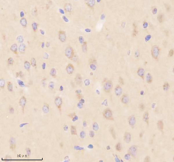
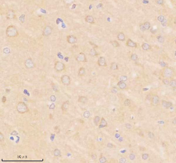
Anti-SIK2 antibodies have IHC images from paraffin-embedded mouse and rat brain sections (A04515-1 IHC captions) and IF/ICC images from human cells and rat brain (catalog IF/ICC captions).
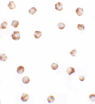
A04515-1 has IHC images from mouse and rat brain sections and IF images from human U2OS cells and rat brain (A04515-1 image captions). A04515 has ICC and IF images from human SW480 cells (A04515 image captions).
Which to pick: Choose A04515-1 for mouse or rat paraffin-section IHC: its listed dilution is 2–5 μg/ml, and its IHC captions show EDTA retrieval at pH 8.0; the fixative is unreported (A04515-1 dilution listing and IHC captions). For human-cell IF/ICC, A04515 has SW480 images, while A04515-1 has a U2OS IF image (A04515 and A04515-1 image captions). For cross-species planning, both list human, mouse and rat reactivity, but the tissue IHC images cover mouse and rat only; clonality is unreported for both (catalog reactivity and clone fields; A04515-1 IHC captions).