This website uses cookies to ensure you get the best experience on our website.


- Table of Contents
Plan SIK3 chromogenic IHC on paraffin sections using the catalog antibody A30762 at 1:100–1:300 (datasheet). Compare nuclear tissue staining (HPA tissue IHC) with the cytoplasmic location annotated for SIK3 (UniProt).

Expected localisation, validated protocols, controls and antibodies — the at-a-glance facts below, then the full design guide.
| Expected localisation | Nuclear in tissue (HPA tissue IHC); cytoplasmic (UniProt) | |
| Staining pattern | General nuclear staining across tissue cell types (HPA tissue IHC) | |
| Antigen retrieval | Citrate pH 6.0 HIER, 95–98 °C, 20 min (rule: cytoplasmic / membrane antigen) | |
| Positive control | Adrenal gland+4 more · see all | |
| Negative control | None in HPA (detected in all 45 tissues); use no-primary + isotype controls |
| Fixation | Keep fixation consistent across paraffin sections. (standard IHC practice; not target-specific) | |
| Caveat | Tissue nuclear staining differs from the cytoplasmic annotation (HPA tissue IHC; UniProt) | |
| Regulation | Expression regulation unspecified (UniProt) | |
| Isoform / epitope | Four isoforms; epitope coverage is unspecified (UniProt) |
Compare the catalog antibody’s IHC-P protocol with four published SIK3 IHC protocols (PMC6856590; PMC6825558; PMC4222365; PMC8411910).
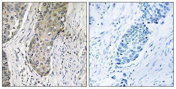
| Sample | Paraffin-embedded human lung carcinoma tissue; fixative not specified (datasheet A30762) |
| Fixation | Image fixative and duration unreported (datasheet A30762); verify before use. |
| Sectioning | 4–5 µm sections on charged slides (standard) |
| Deparaffinisation | Xylene, graded ethanol series to water (standard) |
| Antigen retrieval | Heat-induced epitope retrieval in citrate buffer, pH 6.0, 20 min at 95–98 °C (standard rule: cytoplasmic / membrane antigen) |
| Peroxidase block | 3% H2O2, 10 min, room temperature (standard) |
| Blocking | 10% normal serum of the secondary host, 30 min, room temperature (standard) |
| Primary antibody | Rabbit anti-SIK3, 1:100 - 1:300 (datasheet A30762) |
| Primary incubation | Overnight at 4 °C (standard) |
| Detection | HRP-polymer secondary, DAB chromogen 5–10 min (standard) |
| Counterstain | Hematoxylin, blue, dehydrate and mount (standard) |
| Expected result | SIK3-positive staining in glandular cells of adrenal gland (HPA tissue IHC: High). HPA tissue profile: General nuclear expression. No signal in the no-primary control. |
SIK3 is annotated in the cytoplasm, including puncta when bound to YWHAZ, and has no transmembrane segment (UniProt Q9Y2K2). In paraffin-section IHC, HPA instead reports general nuclear staining, with high staining in several glandular, hematopoietic, epithelial and neural cell populations (HPA tissue IHC). HPA rates the tissue profile Enhanced, while noting medium agreement with RNA data and pending external verification (HPA tissue IHC).
| Clear nuclear staining in adrenal or breast glandular cells, with restrained background. | This matches the reported general nuclear profile and high staining in those cells (HPA tissue IHC). Score the stained cell population as well as intensity; the HPA profile does not establish that every cell in a section must stain (HPA tissue IHC). |
| Predominantly cytoplasmic puncta, or staining confined to cell borders, with little nuclear signal. | Cytoplasmic puncta are biologically plausible through YWHAZ binding (UniProt Q9Y2K2), but they differ from the reported tissue IHC profile (HPA tissue IHC). Border-only staining is also unsupported by that profile; check morphology and controls before calling either pattern SIK3-positive. |
| Strong staining in cells outside the expected positive population, while adjacent expected cells are weak. | Compare cell identity with the tissue-specific HPA annotations before scoring (HPA tissue IHC). An unexpected pattern may reflect antibody cross-reactivity or endogenous detection activity (general IHC practice); morphology alone cannot distinguish those causes. |
| Uniform color across nuclei, stroma and spaces between cells. | A diffuse deposit obscures the cell-restricted nuclear pattern reported for SIK3 (HPA tissue IHC). In chromogenic IHC, incomplete blocking, excess detection reagent or inadequate washing can raise background (general IHC practice); interpret the slide only after controls clarify the signal. |
| No staining in a section containing a documented high-staining cell population. | Absence in, for example, adrenal glandular cells conflicts with the HPA high-staining reference (HPA tissue IHC). Review tissue identity, positive-control performance and the IHC workflow before concluding that the sample lacks detectable SIK3 (general IHC practice). |
| Choice of reference tissue and cell population | HPA reports high staining in adrenal, appendix and breast glandular cells; bone marrow hematopoietic cells; bronchial respiratory epithelial cells; caudate glial cells; cerebellar granular-layer cells; and cerebral-cortex endothelial cells (HPA tissue IHC). Adipocytes are listed as low (HPA tissue IHC). |
| Compartment evidence | General nuclear staining describes HPA tissue IHC, whereas UniProt places SIK3 in the cytoplasm and notes YWHAZ-associated puncta (HPA tissue IHC; UniProt Q9Y2K2). Record the observed compartment explicitly; neither source resolves this discrepancy for a particular section. |
| IHC antibody validation | HPA lists 2 rabbit polyclonal antibodies, HPA045245 and HPA048161, with Enhanced IHC status (HPA antibodies). The tissue profile still has medium RNA agreement and awaits external verification (HPA tissue IHC), so validation supports a comparison rather than certainty about each stained cell. |
| Protein form and epitope uncertainty | SIK3 has 4 isoforms, kinase and UBA domains, and several modified residues (UniProt Q9Y2K2). The supplied record gives no antibody epitope; these features cannot predict which isoforms stain or whether a particular modification changes IHC recognition. |
| Antigen retrieval and detection | Target-specific fixation effects are not established by the supplied assay evidence. Verify with a matched IHC source before attributing a result to fixation. |
| IF/ICC Q: Does its reported membrane location settle IHC scoring? | A: No. HPA's IF summary says membrane but provides no main location or cell-line images (HPA subcellular). Compare that limited summary with UniProt cytoplasmic localisation and the HPA nuclear tissue-IHC profile; interpret IF/ICC on its separate guide page (UniProt Q9Y2K2; HPA tissue IHC). |
| Situation | Likely cause | Next action |
|---|---|---|
| Nuclei stain in a reference tissue, but background prevents cell-level scoring. | Chromogen or detection background may obscure the HPA nuclear pattern (HPA tissue IHC; general IHC practice). | Compare reagent controls, then review blocking, washes and detection exposure; score only cells whose boundaries and nuclei remain interpretable (general IHC practice). |
| An HPA high-staining cell population is blank. | A failed staining run, unsuitable control section or missed target cell population can mimic a negative result (general IHC practice). | Confirm the named cells are present, inspect the run's positive control, and repeat with reviewed retrieval and detection settings before interpreting absence (HPA tissue IHC; general IHC practice). |
| Only cytoplasmic puncta are visible. | Puncta can fit the UniProt YWHAZ-associated localisation but differ from the HPA nuclear tissue profile (UniProt Q9Y2K2; HPA tissue IHC). | Document both compartment and cell type, compare a high-staining reference tissue, and avoid converting a plausible cellular location into an automatically validated IHC result (HPA tissue IHC). |
| Staining is strongest in an unexpected cell type. | The pattern may reflect cross-reactivity, endogenous activity or incorrect cell identification (general IHC practice). | Recheck morphology against the tissue's HPA cell annotation and inspect detection controls; retain uncertainty if the expected high-staining cells remain weak (HPA tissue IHC; general IHC practice). |
| Adipocytes stain as strongly as nearby high-staining reference cells. | HPA lists adipocytes as low, so this relative pattern deserves scrutiny rather than automatic acceptance (HPA tissue IHC). | Compare cells within the same section, examine background and controls, and report the departure from the HPA reference without treating low as an absolute negative control (HPA tissue IHC; general IHC practice). |
| A different antibody yields a different compartment or cell pattern. | The available records show Enhanced IHC status for 2 antibodies but do not guarantee identical staining in every specimen (HPA antibodies; HPA tissue IHC). | Compare matched sections, cell identities and controls; report each observed pattern and the nuclear-versus-cytoplasmic source disagreement instead of forcing agreement (HPA tissue IHC; UniProt Q9Y2K2; general IHC practice). |
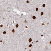

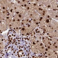

Comprehensive Human Protein Atlas IHC scoring per tissue (reliability: Enhanced — Medium consistency between antibody staining and RNA expression data. Pending external verification.). Rows are taken directly from the HPA tissue chart — click any row's HPA link to view the source.
| Tissue | Cell type | Level | Evidence | Source |
|---|---|---|---|---|
| Adrenal gland | Glandular cells | High | Protein (IHC) | HPA → |
| Appendix | Glandular cells | High | Protein (IHC) | HPA → |
| Bone marrow | Hematopoietic cells | High | Protein (IHC) | HPA → |
| Breast | Glandular cells | High | Protein (IHC) | HPA → |
| Bronchus | Respiratory epithelial cells | High | Protein (IHC) | HPA → |
| Tissue | Cell type | Level | Evidence | Source |
|---|---|---|---|---|
| None in HPA: SIK3 is detected in all 45 scored tissues. Use a no-primary (secondary-only) and an isotype control instead. | ||||
Troubleshoot SIK3 staining in paraffin sections by checking retrieval, compartment, cell identity and controls before comparing signal across samples.
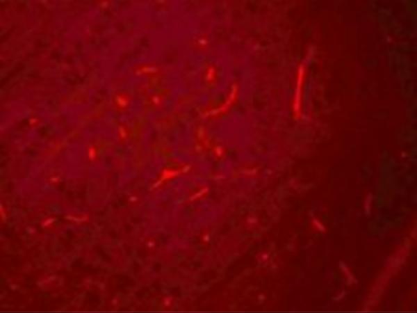
Explore SIK3 antibodies with IHC data from paraffin-embedded human lung carcinoma tissue (A30762 image caption) and IF data from rat brain cells (A05580-1 image caption).
A30762 is listed for IHC and IF in human and mouse samples, with an IHC image from paraffin-embedded human lung carcinoma tissue (A30762 applications, reactivity and image caption). A05580-1 is listed for IF in human, mouse and rat samples, with an IF image from rat brain cells (A05580-1 applications, reactivity and image caption).
Which to pick: Choose polyclonal A30762 for tissue IHC at 1:100–1:300; its image documents paraffin sections, but the fixative is unreported (A30762 dilution_raw and image caption). For ICC, choose A30762, which lists ICC and IF; for IF in rat brain cells, A05580-1 has image evidence at 20 μg/mL (A30762 applications; A05580-1 image caption). A05580-1 has the broader listed species reactivity—human, mouse and rat—while its supplied IF image documents rat cells only (A05580-1 reactivity and image caption).