This website uses cookies to ensure you get the best experience on our website.


- Table of Contents
Plan SIRT1 IHC-P around nuclear staining in several tissues, with high signal reported in adrenal glandular and lymph node germinal center cells (HPA tissue IHC). Score nuclear signal by cell type and use controls to assess unexpected staining, given the report of presumed off-target binding (HPA tissue IHC).

Expected localisation, validated protocols, controls and antibodies — the at-a-glance facts below, then the full design guide.
| Expected localisation | Nuclear staining observed in several tissues (HPA tissue IHC) | |
| Staining pattern | Nuclear staining across several tissues (HPA tissue IHC) | |
| Antigen retrieval | Tris-EDTA pH 9.0 HIER, 95–98 °C, 20 min (rule: nuclear antigen) | |
| Positive control | Adrenal gland+4 more · see all | |
| Negative control | Adipose tissue+4 more · see all |
| Fixation | Matched tissue-IHC evidence does not establish the fixation claim. Validate the specimen-specific method before use. (selected-SKU IHC image M00018-2) | |
| Caveat | Presumed off-target staining may confound scoring (HPA tissue IHC) | |
| Regulation | Expression regulation is not specified (UniProt) | |
| Isoform / epitope | 2 isoforms and a 2–533 fragment; epitope coverage is unknown (UniProt) |
The catalog antibody protocol is accompanied by one published SIRT1 IHC protocol for human articular cartilage (PMC5116180).
| Sample | Tissue sections; selected-image fixative not specified (standard IHC workflow) |
| Fixation | Image fixative and duration unreported (datasheet M00018-2); verify before use. |
| Sectioning | 4–5 µm sections on charged slides (standard) |
| Deparaffinisation | Xylene, graded ethanol series to water (standard) |
| Antigen retrieval | Heat-induced epitope retrieval in Tris-EDTA buffer, pH 9.0, 20 min at 95–98 °C (standard rule: nuclear antigen) |
| Peroxidase block | 3% H2O2, 10 min, room temperature (standard) |
| Blocking | 10% normal serum of the secondary host, 30 min, room temperature (standard) |
| Primary antibody | Rabbit anti-SIRT1, 1:100-1:200 (datasheet M00018-2) |
| Primary incubation | Overnight at 4 °C (standard) |
| Detection | HRP-polymer secondary, DAB chromogen 5–10 min (standard) |
| Counterstain | Hematoxylin, blue, dehydrate and mount (standard) |
| Expected result | SIRT1-positive staining in glandular cells of adrenal gland (HPA tissue IHC: High). HPA tissue profile: Nuclear expression in several tissues. No signal in the no-primary control. |
SIRT1 should stain predominantly in nuclei in paraffin sections, especially adrenal glandular cells, lymph node germinal center cells, and cells in testicular seminiferous ducts (HPA: High; nuclear expression in several tissues). Cytoplasmic staining can occur, but SIRT1 has no transmembrane segment (UniProt Q96EB6: localization and topology). Treat unexpected staining cautiously: HPA rates tissue IHC Approved with medium staining–RNA consistency and notes presumed off-target binding.
| Clear nuclear chromogen in adrenal glandular cells, lymph node germinal center cells, or cells in seminiferous ducts (HPA: High). | This matches the reported IHC distribution and predominant nuclear pattern (HPA: tissue IHC). Compare the signal with adjacent cells and the negative control before scoring it as SIRT1 (general IHC practice). |
| Strong, widespread cytoplasmic staining with little or no nuclear signal. | This conflicts with the predominant nuclear IHC pattern (HPA: tissue IHC). Cytoplasmic SIRT1 is possible (UniProt Q96EB6: localization), so judge this pattern against a known-positive section and background controls rather than rejecting every cytoplasmic signal. |
| Prominent staining in alveolar cells or adipocytes, with weak staining in an expected positive population. | HPA reports alveolar cells and adipocytes as Not detected in its sampled tissues (HPA: tissue IHC). Cross-reactivity or endogenous chromogenic activity is possible (general IHC practice); the HPA result does not prove these cells can never express SIRT1. |
| Diffuse chromogen over cells and extracellular areas, without identifiable nuclear boundaries. | A field-wide deposit cannot establish the reported nuclear distribution (HPA: tissue IHC). Review the negative control, blocking, wash stringency, and detection chemistry to locate nonspecific staining (general IHC practice). |
| No nuclear signal in a section containing expected positive cells. | First confirm that the relevant cells are present: adrenal glandular, lymph node germinal center, and seminiferous duct cells are reported High (HPA: tissue IHC). Then review assay controls and IHC-P handling (general IHC practice); one negative slide does not establish SIRT1 absence. |
| Compartment and topology | Nuclear staining is the main tissue IHC expectation (HPA: tissue IHC). UniProt also lists cytoplasm, PML bodies, nucleoplasm, and mitochondrion, with no transmembrane segment (UniProt Q96EB6: localization and topology); a membrane outline is therefore unexpected. |
| Cell population and tissue context | HPA calls adrenal glandular cells, lymph node germinal center cells, and seminiferous duct cells High; marrow hematopoietic and placental decidual cells are Medium (HPA: tissue IHC). Score the identified cell population, since neighboring populations need not share its staining category. |
| Antibody evidence | HPA006295 and CAB003855 have Approved IHC status, while HPA006295 and HPA052351 have Supported ICC status (HPA: antibody validation). HPA's tissue IHC summary still reports medium staining–RNA consistency and presumed off-target binding; validation does not make every positive deposit specific. |
| Isoforms and processing | UniProt lists two isoforms and chains spanning residues 2–747 and 2–533 (UniProt Q96EB6: isoforms and processing). The supplied evidence gives no antibody epitope, so it cannot establish which forms a particular IHC stain detects or explain a negative slide. |
| What should IF/ICC show? | Mainly nucleoplasmic signal, with additional nucleoli fibrillar center, mitochondrial, and cytosolic localization reported (HPA: subcellular ICC-IF). This is an IF/ICC interpretation cue; tissue IHC still has a predominantly nuclear expectation (HPA: tissue IHC). |
| Situation | Likely cause | Next action |
|---|---|---|
| Known-positive cells are unstained. | The positive population may be absent from the section, or the assay may have failed (HPA: High populations; general IHC practice). | Verify cell identity and section integrity, then check positive and negative controls and review the IHC-P retrieval and detection workflow (general IHC practice). |
| Signal appears only in cytoplasm. | Cytoplasmic localization is possible, but a cytoplasm-only tissue pattern differs from HPA's nuclear IHC profile (UniProt Q96EB6: localization; HPA: tissue IHC). | Compare nuclear counterstain and matched controls; assess whether cytoplasmic signal exceeds nonspecific background (general IHC practice). |
| Many HPA Not detected cells stain strongly. | Off-target staining is plausible because HPA reports presumed off-target binding in its tissue assessment (HPA: tissue IHC). Endogenous detection activity is another general IHC possibility. | Inspect a negative control and repeat with an independently validated antibody if available (general IHC practice; HPA: IHC Approved antibodies). |
| The whole section has hazy chromogen. | Poorly localized background can arise from blocking, washing, or detection conditions (general IHC practice). | Check reagent-only or primary-omission controls, wash steps, and chromogen development; score nuclei only after the background is resolved (general IHC practice). |
| A Medium or Low population stains less than a High population. | HPA reports differing cell-level staining categories across tissues (HPA: tissue IHC). | Use the appropriate cell population as the comparator and avoid calling a weak section negative solely because another tissue stains more strongly (HPA: tissue IHC; general IHC practice). |
| Different antibodies give different patterns. | HPA reports Approved IHC validation but also medium staining–RNA consistency and presumed off-target binding (HPA: antibody validation and tissue IHC). Epitope information is absent from the supplied evidence. | Compare matched sections, controls, and cell-level localization; report the antibody identifier and unresolved discrepancy (general IHC practice). |




Comprehensive Human Protein Atlas IHC scoring per tissue (reliability: Approved — Medium consistency between antibody staining and RNA expression data. Presumed off target binding observed and disregarded.). Rows are taken directly from the HPA tissue chart — click any row's HPA link to view the source.
| Tissue | Cell type | Level | Evidence | Source |
|---|---|---|---|---|
| Adrenal gland | Glandular cells | High | Protein (IHC) | HPA → |
| Lymph node | Germinal center cells | High | Protein (IHC) | HPA → |
| Testis | Cells in seminiferous ducts | High | Protein (IHC) | HPA → |
| Bone marrow | Hematopoietic cells | Medium | Protein (IHC) | HPA → |
| Placenta | Decidual cells | Medium | Protein (IHC) | HPA → |
| Tissue | Cell type | Level | Evidence | Source |
|---|---|---|---|---|
| Adipose tissue | Adipocytes | Not detected | Protein (IHC) | HPA → |
| Breast | Adipocytes | Not detected | Protein (IHC) | HPA → |
| Bronchus | Respiratory epithelial cells | Not detected | Protein (IHC) | HPA → |
| Cerebral cortex | Endothelial cells | Not detected | Protein (IHC) | HPA → |
| Epididymis | Glandular cells | Not detected | Protein (IHC) | HPA → |
Troubleshoot SIRT1 staining in paraffin sections by checking retrieval, cell identity, subcellular pattern and controls before interpreting chromogenic signal.
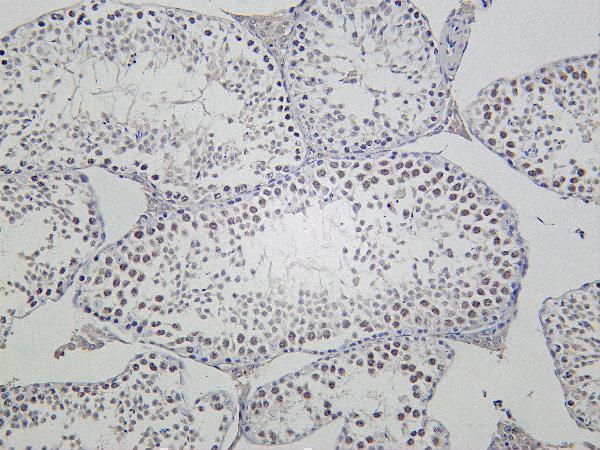
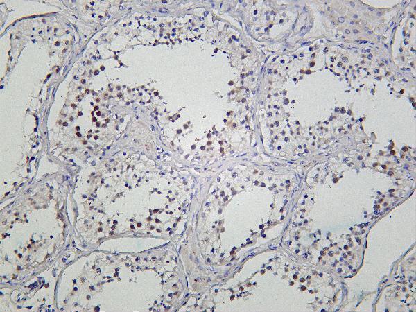
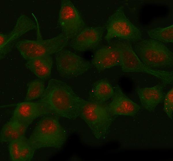
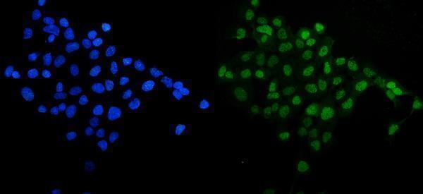
Validated anti-SIRT1 antibodies have IHC images from mouse testis and human kidney and IF images from HeLa and A431 cells (catalog image captions). Listed reactivity covers human and rat (catalog: reactivity).
M00018-2 shows IHC staining of mouse testis, although mouse reactivity is unlisted; M00018 shows IHC staining of paraffin-embedded human kidney (catalog image captions; catalog: reactivity). A00018-1 shows IF in HeLa cells, and PB9159 shows IF in A431 cells (catalog image captions).
Which to pick: For tissue IHC, choose the rabbit monoclonal M00018 for human paraffin sections (catalog: clonality and human reactivity; M00018 IHC image caption: paraffin-embedded human kidney); its fixative is unreported (M00018 IHC image caption). For IF/ICC, A00018-1 and PB9159 both list human IF/ICC and show cell images (catalog: applications and reactivity; respective IF image captions). For listed human and rat reactivity, choose rabbit monoclonal M00018-2, which lists IHC and IF; its IHC image shows mouse testis, while mouse reactivity and the fixative are unreported (catalog: clonality, applications and reactivity; M00018-2 IHC image caption).