Click image to see more details
-

-
-
-
-
+1
Product Info Summary
| SKU: | PB9159 |
|---|---|
| Size: | 100 μg/vial |
| Reactive Species: | Human |
| Host: | Rabbit |
| Application: | IF, ICC, WB |
Customers Who Bought This Also Bought

Product info
Product Name
Anti-SIRT1 Antibody Picoband®
SKU/Catalog Number
PB9159
PB0173 is an alternative SKU for this antibody, used in previous lots.
Size
100 μg/vial
Form
Lyophilized
Description
Boster Bio Anti-SIRT1 Antibody Picoband® catalog # PB9159. Tested in IF, ICC, WB applications. This antibody reacts with Human. The brand Picoband indicates this is a premium antibody that guarantees superior quality, high affinity, and strong signals with minimal background in Western blot applications. Only our best-performing antibodies are designated as Picoband, ensuring unmatched performance.
Storage & Handling
Store at -20˚C for one year from date of receipt. After reconstitution, at 4˚C for one month. It can also be aliquotted and stored frozen at -20˚C for six months. Avoid repeated freeze-thaw cycles.
Cite This Product
Anti-SIRT1 Antibody Picoband® (Boster Biological Technology, Pleasanton CA, USA, Catalog # PB9159)
Host
Rabbit
Contents
Each vial contains antibody formulated with stabilizing components, 0.9 mg NaCl, 0.2 mg Na2HPO4, and 0.05 mg NaN3.
*This antibody is supplied in a stabilized formulation.
Compatibility with conjugation reactions depends on the chemistry of the conjugation method used.
For conjugation methods that are not compatible with the stabilizing components present in this formulation, a carrier-free antibody format is required.
Clonality
Polyclonal
Isotype
Rabbit IgG
Immunogen
E.coli-derived human SIRT1 recombinant protein (Position: R112-K311). Human SIRT1 shares 90% amino acid (aa) sequence identity with mouse SIRT1.
Cross-reactivity
No cross-reactivity with other proteins
Reactive Species
PB9159 is reactive to SIRT1 in Human
Observed Molecular Weight
120 kDa
Calculated molecular weight
81.7 kDa
Background of SIRT1
Sirtuin 1, also known as SIR2L1 or SIRT1, is a protein that in humans is encoded by the SIRT1 gene. It is mapped to 10q21.3. Sirtuin 1 is a member of the sirtuin family of proteins, homologs of the Sir2 gene in S. cerevisiae. Members of the sirtuin family are characterized by a sirtuin core domain and grouped into four classes. Sirtuin 1 is downregulated in cells that have high insulin resistance and inducing its expression increases insulin sensitivity, suggesting the molecule is associated with improving insulin sensitivity. Furthermore, Sirtuin 1 was shown to de-acetylate and affect the activity of both members of the PGC1-alpha/ERR-alpha complex, which are essential metabolic regulatory transcription factors.
Antibody Validation
Boster validates all antibodies on WB, IHC, ICC, Immunofluorescence, and ELISA with known positive control and negative samples to ensure specificity and high affinity, including thorough antibody incubations.
Application & Images
Applications
PB9159 is guaranteed for IF, ICC, WB Boster Guarantee
Recommend Dilution
| Application | Dilution | Species |
|---|---|---|
| Western blot | 0.1-0.5μg/ml | Human |
| Immunocytochemistry/Immunofluorescence | 5 μg/ml | Human |
Tested application
Suggested blocking solution with 5% non-fat milk or BSA; (*)Recommended protein loading: 20-40 µg per lane
Validation Images & Assay Conditions
Click image to see more details
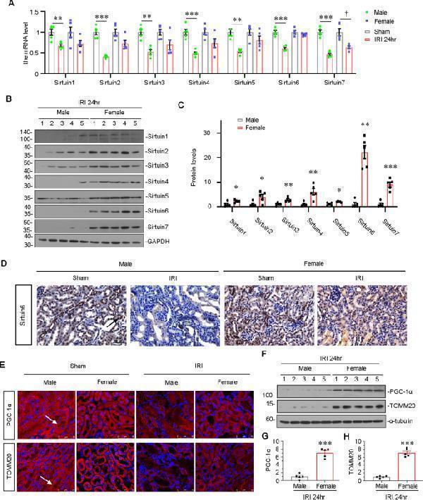
Sirtuin 6 is the possible contributor to gender differences upon IRI. A Graphical representations show the relative abundance of Sirtuin 1-7 mRNA in different groups. ** P < 0.01, *** P < 0.001 versus sham controls in male group ( n = 5); † P < 0.05 versus sham controls in female group ( n = 5). B , C Representative western blot ( B ) and graphical representations of ( C ) Sirtuin 1-7 protein expression levels are shown. * P < 0.05, ** P < 0.01, *** P < 0.001 versus male group ( n = 5). D Representative micrographs showing the expression of Sirtuin 6 in different groups, as indicated. Paraffin-embedded kidney sections were stained with an antibody against Sirtuin 6. Arrows indicate positive staining. Scale bar, 50 μm. E Representative micrographs showing the expression of PGC-1α and TOMM20 in different groups, as indicated. Frozen kidney sections were stained with an antibody against PGC-1α and TOMM20. Arrows indicate positive staining. Scale bar, 50 μm. F – H Representative western blot ( F ) and graphical representations of ( G ) PGC-1α and ( H ) TOMM20 protein expression levels are shown. *** P < 0.001 versus male group ( n = 5).
Index in PubMed under a CC BY license. PMID: 37185276

Click image to see more details
The expression of Sirtuin 6 was the key regulator for gender differences in rhabdomyolysis-induced AKI. A , B Representative western blot ( A ) and graphical representations of ( B ) Sirtuin 1-7 protein expression levels are shown. * P < 0.05, ** P < 0.01, *** P < 0.001 versus female group ( n = 5). C Representative micrographs show the expression of Sirtuin 6 in different groups, as indicated. Paraffin-embedded kidney sections were stained with an antibody against Sirtuin 6. Arrows indicate positive staining. Scale bar, 50 μm. D Representative micrographs showing the expression of PGC-1α and TOMM20 in different groups, as indicated. Frozen kidney sections were stained with an antibody against PGC-1α and TOMM20. Arrows indicate positive staining. Scale bar, 50 μm. E – G Representative western blot ( E ) and graphical representations of ( F ) PGC-1α and ( G ) TOMM20 protein expression levels are shown. * P < 0.05, *** P < 0.001 versus male group ( n = 5).
Index in PubMed under a CC BY license. PMID: 37185276

Click image to see more details
AR increases acetylation of PGC-1α by downregulating Sirtuin 6 expression. A – C Representative western blot ( A ) and graphical representations of ( B ) Sirtuin 1-7 and ( C ) PGC-1α protein expression levels are shown. * P < 0.05, ** P < 0.01 versus control group ( n = 3). HKC-8 cells were transfected with pcDNA3 or AR overexpression plasmid for 24 h. D Representative ChIP assay results showing the binding of AR to the Sirtuin 6 gene promoter region. HKC‐8 cells were incubated with DHT (10 μMol/L) or not for 24 h. Cell lysates were precipitated with an antibody against AR, histone H3, or nonimmune IgG, and the ChIP assay was performed for Sirtuin 6 gene promoters. Total diluted lysate was used as the total genomic input DNA. E Graphical representations show the relative abundance of Sirtuin 1-7 mRNA in different groups. ** P < 0.01 versus control group ( n = 3). F – I Representative western blot ( F ) and graphical representations of ( G ) Sirtuin 1-7, ( H ) PGC-1α and ( I ) AR protein expression levels are shown. HKC-8 cells were incubated with DHT (10 μMol/L) and transfected with AR-shRNA for 24 h. * P < 0.05, ** P < 0.01, *** P < 0.001 ( n = 3); † P < 0.05, †† P < 0.01, ††† P < 0.001 ( n = 3). J Representative graphs show the binding of PGC-1α with Sirtuin 6 or acetyl. HKC-8 cells were transfected with pcDNA3 or AR overexpression plasmid for 24 h. K Representative graphs show the binding of PGC-1α with acetyl, and the expression of PGC-1α in different groups, as indicated. HKC-8 cells were treated with DHT (10 μMol/L) and transfected with Sirtuin 6 overexpression plasmid for 24 h. L Representative graphs show the binding of PGC-1α with acetyl, and the protein levels of AR in nuclear fractions in different groups, as indicated. HKC-8 cells were treated with DHT (10 μMol/L) for 24 h. M Representative graphs show the binding of PGC-1α with acetyl, and the protein levels of AR in nuclear fractions in sham control and IRI group in male mice, as indicated.
Index in PubMed under a CC BY license. PMID: 37185276

Click image to see more details
Western blot analysis of SIRT1 using anti-SIRT1 antibody (PB9159).
Electrophoresis was performed on a 5-20% SDS-PAGE gel at 70V (Stacking gel) / 90V (Resolving gel) for 2-3 hours. The sample well of each lane was loaded with 30 ug of sample under reducing conditions.
Lane 1: human HepG2 whole cell lysates,
Lane 2: human MCF-7 whole cell lysates,
Lane 3: huamn SiHa whole cell lysates,
Lane 4: huamn Daudi whole cell lysates,
Lane 5: huamn K562 whole cell lysates.
After electrophoresis, proteins were transferred to a nitrocellulose membrane at 150 mA for 50-90 minutes. Blocked the membrane with 5% non-fat milk/TBS for 1.5 hour at RT. The membrane was incubated with rabbit anti-SIRT1 antigen affinity purified polyclonal antibody (Catalog # PB9159) at 0.5 μg/mL overnight at 4°C, then washed with TBS-0.1%Tween 3 times with 5 minutes each and probed with a goat anti-rabbit IgG-HRP secondary antibody at a dilution of 1:5000 for 1.5 hour at RT. The signal is developed using an Enhanced Chemiluminescent detection (ECL) kit (Catalog # EK1002) with Tanon 5200 system. A specific band was detected for SIRT1 at approximately 120 kDa. The expected band size for SIRT1 is at 82 kDa.

Click image to see more details
IF analysis of SIRT1 using anti-SIRT1 antibody (PB9159).
SIRT1 was detected in an immunocytochemical section of A431 cells. Enzyme antigen retrieval was performed using IHC enzyme antigen retrieval reagent (AR0022) for 15 mins. The cells were blocked with 10% goat serum. And then incubated with 5 μg/mL rabbit anti-SIRT1 Antibody (PB9159) overnight at 4°C. DyLight®488 Conjugated Goat Anti-Rabbit IgG (BA1127) was used as secondary antibody at 1:100 dilution and incubated for 30 minutes at 37°C. The section was counterstained with DAPI. Visualize using a fluorescence microscope and filter sets appropriate for the label used.
Specific Publications For Anti-SIRT1 Antibody Picoband® (PB9159)
Loading publications
Recommended Resources
Here are featured tools and databases that you might find useful.
- Boster's Pathways Library
- Protein Databases
- Bioscience Research Protocol Resources
- Data Processing & Analysis Software
- Photo Editing Software
- Scientific Literature Resources
- Research Paper Management Tools
- Molecular Biology Software
- Primer Design Tools
- Bioinformatics Tools
- Phylogenetic Tree Analysis
Customer Reviews
Have you used Anti-SIRT1 Antibody Picoband®?
Share your experimental results or join a short interview to earn up to $1,000 in product credits or other rewards.
0 Reviews For Anti-SIRT1 Antibody Picoband®
Customer Q&As
Have a question?
Find answers in Q&As, reviews.
Can't find your answer?
Submit your question
4 Customer Q&As for Anti-SIRT1 Antibody Picoband®
Question
We want using your anti-SIRT1 antibody for negative regulation of protein acetylation studies. Has this antibody been tested with western blotting on sw620 cell lysate? We would like to see some validation images before ordering.
Verified Customer
Verified customer
Asked: 2019-12-11
Answer
I appreciate your inquiry. This PB9159 anti-SIRT1 antibody is validated on hepg2 whole cell lysate, mcf whole cell lysate, sw620 cell lysate. It is guaranteed to work for WB in human. Our Boster guarantee will cover your intended experiment even if the sample type has not been be directly tested.
Boster Scientific Support
Answered: 2019-12-11
Question
We have seen staining in human prostate. What should we do? Is anti-SIRT1 antibody supposed to stain prostate positively?
P. Lewis
Verified customer
Asked: 2019-06-21
Answer
Based on literature prostate does express SIRT1. Based on Uniprot.org, SIRT1 is expressed in right adrenal gland, testis, prostate, cervix carcinoma, leukemic t-cell, cervix carcinoma erythroleukemia, liver, among other tissues. Regarding which tissues have SIRT1 expression, here are a few articles citing expression in various tissues:
Cervix carcinoma, Pubmed ID: 16964243, 18669648, 20068231
Cervix carcinoma, and Erythroleukemia, Pubmed ID: 23186163
Leukemic T-cell, Pubmed ID: 19690332
Liver, Pubmed ID: 24275569
Prostate, Pubmed ID: 15489334
Testis, Pubmed ID: 10381378
Boster Scientific Support
Answered: 2019-06-21
Question
We are currently using anti-SIRT1 antibody PB9159 for human tissue, and we are satisfied with the WB results. The species of reactivity given in the datasheet says human. Is it likely that the antibody can work on monkey tissues as well?
M. Baker
Verified customer
Asked: 2017-06-06
Answer
The anti-SIRT1 antibody (PB9159) has not been validated for cross reactivity specifically with monkey tissues, though there is a good chance of cross reactivity. We have an innovator award program that if you test this antibody and show it works in monkey you can get your next antibody for free. Please contact me if I can help you with anything.
Boster Scientific Support
Answered: 2017-06-06
Question
My team were satisfied with the WB result of your anti-SIRT1 antibody. However we have been able to see positive staining in right adrenal gland pml body using this antibody. Is that expected? Could you tell me where is SIRT1 supposed to be expressed?
R. Jackson
Verified customer
Asked: 2013-11-14
Answer
From what I have seen in literature, right adrenal gland does express SIRT1. Generally SIRT1 expresses in nucleus, pml body, sirtt1 75 kda fragment: cytoplasm. Regarding which tissues have SIRT1 expression, here are a few articles citing expression in various tissues:
Cervix carcinoma, Pubmed ID: 16964243, 18669648, 20068231
Cervix carcinoma, and Erythroleukemia, Pubmed ID: 23186163
Leukemic T-cell, Pubmed ID: 19690332
Liver, Pubmed ID: 24275569
Prostate, Pubmed ID: 15489334
Testis, Pubmed ID: 10381378
Boster Scientific Support
Answered: 2013-11-14
