This website uses cookies to ensure you get the best experience on our website.


- Table of Contents
This IHC guide uses membranous staining in hepatocytes, enterocytes and renal tubules as its tissue reference (HPA tissue IHC). It covers paraffin-section preparation and chromogenic detection using the catalog antibody (datasheet PB9436).

Expected localisation, validated protocols, controls and antibodies — the at-a-glance facts below, then the full design guide.
| Expected localisation | Cell membrane (UniProt); membranous tissue staining (HPA tissue IHC) | |
| Staining pattern | Membranous hepatocytes, enterocytes and renal tubules (HPA tissue IHC) | |
| Antigen retrieval | EDTA pH 8.0 HIER, heat-mediated (datasheet PB9436) | |
| Positive control | Duodenum+3 more · see all | |
| Negative control | Adipose tissue+4 more · see all |
| Fixation | Keep fixation consistent across paraffin sections (standard IHC practice; not target-specific) | |
| Caveat | Renal tubules stain less strongly than hepatocytes (HPA tissue IHC) | |
| Regulation | No staining-linked regulator specified (UniProt) | |
| Isoform / epitope | 2 isoforms; check whether the epitope is extracellular or cytoplasmic (UniProt) |
Compare the catalog antibody’s IHC-P protocol (datasheet: PB9436) with one published chromogenic SLC2A2 protocol using gastric tissue sections (PMC10773219).
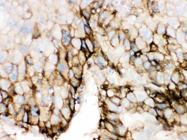
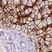
| Sample | Paraffin-embedded human liver cancer tissue; fixative not specified (datasheet PB9436) |
| Fixation | Image fixative and duration unreported (datasheet PB9436); verify before use. |
| Sectioning | 4–5 µm sections on charged slides (standard) |
| Deparaffinisation | Xylene, graded ethanol series to water (standard) |
| Antigen retrieval | Heat retrieval: EDTA pH 8.0 (datasheet PB9436); 20 min, 95–100 °C (standard) |
| Peroxidase block | 3% H2O2, 10 min, room temperature (standard) |
| Blocking | 10% goat serum (datasheet PB9436) |
| Primary antibody | Rabbit anti-SLC2A2, 0.5-1μg/ml (datasheet PB9436) |
| Primary incubation | Overnight at 4 °C (datasheet PB9436) |
| Detection | Streptavidin-biotin complex (SABC), DAB chromogen (datasheet PB9436) |
| Counterstain | Hematoxylin, blue, dehydrate and mount (standard) |
| Expected result | SLC2A2-positive staining in enterocytes of duodenum (HPA tissue IHC: High). HPA tissue profile: Selective membranous expression in hepatocytes, small intestine, duodenum and renal tubules. No signal in the no-primary control. |
SLC2A2 (GLUT2) is a cell-membrane transporter with 12 transmembrane segments (UniProt P11168 topology). In paraffin-section IHC, expect selective membranous staining in hepatocytes and enterocytes, with staining also reported in renal tubules (HPA tissue IHC: Enhanced reliability). HPA reports High staining in hepatocytes and duodenal and small-intestinal enterocytes, and Medium staining in kidney tubular cells (HPA tissue IHC).
| Crisp staining outlines hepatocytes or enterocytes; kidney tubular cells show a weaker outline. | This fits the reported membrane compartment and relative tissue levels: High in hepatocytes and enterocytes, Medium in tubular cells (UniProt P11168 subcellular location; HPA tissue IHC). Compare cells within each tissue; HPA levels are categorical observations, not a numeric staining threshold (HPA tissue IHC). |
| Signal is predominantly nuclear or fills the cytoplasm without a discernible membrane outline. | That distribution conflicts with the annotated cell-membrane location and HPA's selective membranous IHC profile (UniProt P11168 subcellular location; HPA tissue IHC). Treat it as an interpretation warning and assess whether background or nonspecific staining obscures a membrane signal (general IHC practice). |
| Strong staining appears in adipocytes or respiratory epithelial cells while expected cells are faint. | HPA reports SLC2A2 as Not detected in adipocytes of adipose tissue and respiratory epithelial cells of bronchus (HPA tissue IHC). Investigate antibody cross-reactivity or endogenous detection activity before calling those cells positive; an unexpected stain alone does not identify its cause (general IHC practice). |
| Color spreads across tissue and empty areas, making cell borders hard to distinguish. | Diffuse signal cannot be scored as the selective membrane pattern reported for SLC2A2 (HPA tissue IHC). Evaluate the detection background with an appropriate negative control and inspect whether blocking, washing, or chromogen development contributed (general IHC practice). |
| A liver section has no convincing hepatocyte membrane staining. | HPA reports High hepatocyte staining, so this is an informative failure of the current IHC run, though it does not prove that the specimen lacks SLC2A2 (HPA tissue IHC). Check tissue preservation, antibody performance, retrieval, and detection controls as general IHC practice; no SLC2A2-specific fixation sensitivity is established here. |
| Membrane topology | SLC2A2 has 12 transmembrane segments and is annotated at the cell membrane (UniProt P11168 topology; subcellular location). Judge IHC against a cell-border pattern; the supplied record does not identify an antibody epitope or establish which side of the membrane it recognizes. |
| Tissue and cell selection | Liver hepatocytes and duodenal or small-intestinal enterocytes provide reported High-staining examples; kidney tubular cells are Medium (HPA tissue IHC). Adipose-tissue adipocytes are reported Not detected and can help assess unexpected staining (HPA tissue IHC). |
| Strength of the tissue evidence | HPA rates the tissue IHC profile Enhanced, describing high consistency between antibody staining and RNA expression; three listed antibodies have IHC Enhanced status (HPA tissue IHC; HPA antibodies). This supports the reported pattern, without validating every antibody or specimen. |
| Isoforms and processing | UniProt lists 2 isoforms and a chain spanning residues 1–524, with no signal peptide or propeptide annotated (UniProt P11168 isoforms; processing). These annotations do not establish isoform-specific staining or a shed-product pattern. |
| Annotated modification | UniProt records a glycosylation site at residue 62 and a modified threonine at 523 (UniProt P11168 glycosylation; modified residues). The supplied sources do not show that either modification changes IHC antigen retrieval or staining intensity. |
| Situation | Likely cause | Next action |
|---|---|---|
| Expected membrane outlines are replaced by mostly nuclear or diffuse cytoplasmic color. | The result disagrees with the annotated cell-membrane location and selective membranous HPA profile; the cause is unresolved by appearance alone (UniProt P11168 subcellular location; HPA tissue IHC). | Review a negative control and the staining distribution at higher magnification; adjust blocking, washes, or detection conditions only if controls indicate background (general IHC practice). |
| An HPA-undetected cell type stains as strongly as hepatocytes. | Cross-reactivity or endogenous detection activity is possible; HPA reports High hepatocytes and Not detected adipose-tissue adipocytes (HPA tissue IHC; general IHC practice). | Check negative controls and compare the suspect cells with a liver positive control in the same run before assigning cell-specific positivity (HPA tissue IHC; general IHC practice). |
| The whole section has diffuse chromogen or staining outside identifiable cells. | Widespread background can obscure the selective membranous profile, but the image does not establish which reagent caused it (HPA tissue IHC; general IHC practice). | Inspect negative-control background, blocking, wash steps, and development time; reassess localization after the background is controlled (general IHC practice). |
| Known-positive liver lacks hepatocyte signal. | This contradicts HPA's High hepatocyte result, but the payload provides no target-specific fixation or retrieval effect to explain it (HPA tissue IHC). | Verify the liver control, antibody and detection steps, and the retrieval conditions used for that antibody; repeat with controls if the run failed (general IHC practice). |
| Kidney appears weaker than liver. | That ordering can match HPA's Medium tubular-cell level versus High hepatocyte level; intensity alone is insufficient to diagnose a failed stain (HPA tissue IHC). | Look for a convincing tubular-cell membrane outline and assess both tissues alongside run controls before changing conditions (HPA tissue IHC; general IHC practice). |
| Q: Can ICC-IF images define the expected paraffin-section IHC pattern? | HPA ICC-IF supports plasma-membrane localization and also reports mid-piece and principal-piece locations; those are subcellular observations, not paraffin-section tissue intensity scores (HPA subcellular ICC-IF; HPA tissue IHC). | A: Use the ICC-IF findings as localization context and interpret paraffin IHC against HPA's tissue-specific membranous profile; consult the separate IF/ICC guide for that application (HPA subcellular ICC-IF; HPA tissue IHC). |
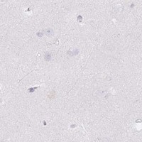
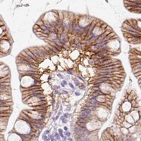
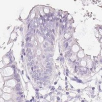


Comprehensive Human Protein Atlas IHC scoring per tissue (reliability: Enhanced — High consistency between antibody staining and RNA expression data.). Rows are taken directly from the HPA tissue chart — click any row's HPA link to view the source.
| Tissue | Cell type | Level | Evidence | Source |
|---|---|---|---|---|
| Duodenum | Enterocytes | High | Protein (IHC) | HPA → |
| Liver | Hepatocytes | High | Protein (IHC) | HPA → |
| Small intestine | Enterocytes | High | Protein (IHC) | HPA → |
| Kidney | Cells in tubules | Medium | Protein (IHC) | HPA → |
| Tissue | Cell type | Level | Evidence | Source |
|---|---|---|---|---|
| Adipose tissue | Adipocytes | Not detected | Protein (IHC) | HPA → |
| Adrenal gland | Glandular cells | Not detected | Protein (IHC) | HPA → |
| Appendix | Endocrine cells | Not detected | Protein (IHC) | HPA → |
| Bone marrow | Hematopoietic cells | Not detected | Protein (IHC) | HPA → |
| Breast | Adipocytes | Not detected | Protein (IHC) | HPA → |
Troubleshoot SLC2A2 staining by checking retrieval, membrane localisation, cell identity and controls before interpreting chromogenic signal.
The catalog lists human IHC antibodies for SLC2A2 and one paraffin-section image; it lists no IF/ICC data (catalog applications; PB9436 image caption; IF image alts).
PB9436 lists human IHC and shows staining in a paraffin-embedded human liver cancer section (PB9436 catalog applications/reactivity; PB9436 image caption). M02297-2 lists IHC reactivity for human, mouse, and rat, but provides no IHC image or specimen details (M02297-2 catalog applications/reactivity; IHC image alts).
Which to pick: For human paraffin-section IHC, choose PB9436: its own caption documents EDTA retrieval, 1 μg/ml primary antibody, and chromogenic detection; the fixative is unreported (PB9436 image caption). For cross-species IHC, consider the rabbit monoclonal M02297-2, whose catalog lists human, mouse, and rat reactivity but provides no tissue image or processing details (M02297-2 catalog). Neither SKU lists IF/ICC or provides an IF image, so neither has documented IF/ICC support here (catalog applications; IF image alts).