This website uses cookies to ensure you get the best experience on our website.


- Table of Contents
Plan chromogenic IHC for SLC30A8 in human pancreatic islets, where strong cytoplasmic staining is reported (HPA tissue IHC). Use the catalog antibody’s IHC-P conditions to guide staining (datasheet A01310-1), and compare endocrine-cell signal with appropriate controls (standard IHC practice).

Expected localisation, validated protocols, controls and antibodies — the at-a-glance facts below, then the full design guide.
| Expected localisation | Islet cytoplasm (HPA tissue IHC); vesicle membrane (UniProt) | |
| Staining pattern | Strong cytoplasmic staining in pancreatic endocrine cells (HPA tissue IHC) | |
| Antigen retrieval | EDTA pH 8.0 HIER, heat-mediated (datasheet A01310-1) | |
| Positive control | Pancreas | |
| Negative control | Adipose tissue+4 more · see all |
| Fixation | Matched tissue-IHC evidence does not establish the fixation claim. Validate the specimen-specific method before use. (selected-SKU IHC image A01310-1) | |
| Caveat | Fat expression does not establish adipocyte IHC staining (UniProt tissue specificity; HPA tissue IHC) | |
| Regulation | Lower subcutaneous fat expression in obesity (UniProt) | |
| Isoform / epitope | 2 isoforms; epitope coverage unknown; topology matters (UniProt) |
The catalog antibody’s IHC-P protocol is accompanied by one published SLC30A8 immunohistochemistry protocol for paraffin-embedded sections (PMC10328108).
| Sample | Paraffin-embedded human pancreas tissue; fixative not specified (datasheet A01310-1) |
| Fixation | Image fixative and duration unreported (datasheet A01310-1); verify before use. |
| Sectioning | 4–5 µm sections on charged slides (standard) |
| Deparaffinisation | Xylene, graded ethanol series to water (standard) |
| Antigen retrieval | Heat retrieval: EDTA pH 8.0 (datasheet A01310-1); 20 min, 95–100 °C (standard) |
| Peroxidase block | 3% H2O2, 10 min, room temperature (standard) |
| Blocking | 10% goat serum (datasheet A01310-1) |
| Primary antibody | Rabbit anti-SLC30A8, 2-5 μg/ml (datasheet A01310-1) |
| Primary incubation | Overnight at 4 °C (datasheet A01310-1) |
| Detection | HRP-conjugated secondary, DAB chromogen (datasheet A01310-1) |
| Counterstain | Hematoxylin, blue, dehydrate and mount (standard) |
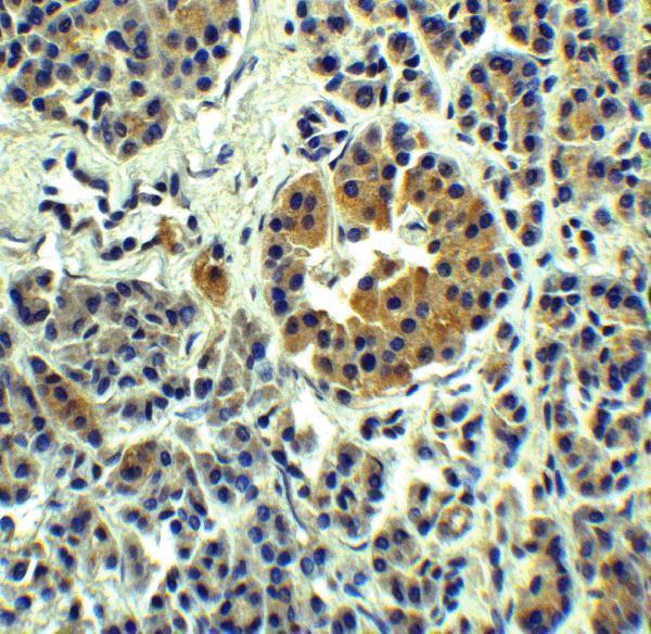
| Expected result | SLC30A8-positive staining in pancreatic endocrine cells of pancreas (HPA tissue IHC: High). HPA tissue profile: Strong cytoplasmic expression in Islets of Langerhans. No signal in the no-primary control. |
SLC30A8 is a six-pass membrane protein associated with secretory vesicles and the cell membrane (UniProt Q8IWU4 topology and subcellular location). In pancreatic sections, expect strong cytoplasmic staining in islets, principally in insulin-producing beta cells (HPA tissue IHC: High in pancreatic endocrine cells; UniProt Q8IWU4 tissue specificity). HPA rates the tissue IHC pattern Enhanced, citing agreement between staining and RNA expression (HPA tissue IHC: Enhanced).
| Strong, granular cytoplasmic staining is concentrated in pancreatic islets, with little staining in surrounding tissue. | This fits the reported strong islet pattern (HPA tissue IHC: High in pancreatic endocrine cells) and secretory-vesicle association (UniProt Q8IWU4 subcellular location). Interpret the granularity alongside islet morphology; chromogenic IHC alone does not identify individual granules (general IHC practice). |
| Predominantly nuclear staining appears in pancreatic endocrine cells. | A nuclear-dominant pattern conflicts with the reported vesicle and membrane locations (UniProt Q8IWU4 subcellular location; HPA subcellular ICC-IF). Check antibody and detection controls before scoring it as SLC30A8; nuclear colour can reflect nonspecific staining or detection artefact (general IHC practice). |
| Strong colour appears in adipocytes or other cells outside the expected pancreatic islet pattern. | HPA reports adipocytes as Not detected by tissue IHC, while its strongest reported signal is in pancreatic endocrine cells (HPA tissue IHC). Review morphology and controls for cross-reactivity or endogenous detection activity (general IHC practice). An unexpected result needs validation; it does not establish a new expression pattern. |
| Pale, diffuse colour covers positive and negative regions of the section. | A uniform haze does not match the concentrated islet staining reported by HPA (HPA tissue IHC). In chromogenic IHC, incomplete blocking, excess detection signal or inadequate washing can raise background (general IHC practice). Compare a primary-omission control and a negative tissue area before interpreting weak colour. |
| A well-preserved pancreatic section shows no endocrine-cell signal. | This conflicts with HPA's High pancreatic endocrine-cell staining (HPA tissue IHC). Check that islets are present, then review the IHC-validated antibody, detection reagents and a positive control (general IHC practice). Absence of colour alone cannot distinguish a technical failure from an unexpected specimen result. |
| Cell and tissue selection | Pancreatic endocrine cells provide the HPA High IHC reference; adipocytes are reported Not detected (HPA tissue IHC). UniProt specifies beta-cell expression within the endocrine pancreas (UniProt Q8IWU4 tissue specificity). Score cell populations separately rather than averaging colour across a whole section (general IHC practice). |
| Compartment and epitope interpretation | SLC30A8 has six transmembrane segments and cytoplasmic and vesicle-lumenal regions (UniProt Q8IWU4 topology). Its vesicle-membrane location supports cytoplasmic granularity, but the payload gives no antibody epitope; it cannot predict which molecular face a stain detects (UniProt Q8IWU4 topology; general IHC interpretation). |
| Antibody evidence | HPA rates tissue IHC Enhanced and lists HPA076165 as IHC Enhanced (HPA tissue IHC; HPA antibodies). HPA065953 is listed as ICC Supported, without an IHC status (HPA antibodies). Keep those assay validations distinct when selecting a reagent or interpreting a discrepant image. |
| Antigen retrieval | Retrieval conditions are an IHC-P workflow variable to verify against the chosen antibody instructions and controls (general IHC practice). Neither the supplied HPA pattern nor UniProt topology establishes an SLC30A8-specific retrieval condition or fixation effect (HPA tissue IHC; UniProt Q8IWU4 topology). |
| Situation | Likely cause | Next action |
|---|---|---|
| Pancreatic islets are present, but chromogenic signal is absent. | The result conflicts with HPA's High endocrine-cell IHC signal (HPA tissue IHC); a failed primary or detection step is possible (general IHC practice). | Confirm islet morphology and run a known-positive pancreatic control; review the chosen antibody's IHC-P instructions, retrieval, dilution and detection steps (general IHC practice). No target-specific dilution or retrieval setting is supplied here. |
| Colour is strong in nuclei but weak in islet cytoplasm. | Nuclear dominance is discordant with vesicle and membrane localization (UniProt Q8IWU4 subcellular location; HPA subcellular ICC-IF). Nonspecific staining remains possible (general IHC practice). | Compare a primary-omission control with the stained section; reassess localization using cell morphology and an IHC-validated antibody (general IHC practice; HPA antibodies: HPA076165 IHC Enhanced). |
| Adipocytes stain strongly alongside pancreatic endocrine cells. | HPA reports adipocytes as Not detected by tissue IHC (HPA tissue IHC). Endogenous detection activity or antibody cross-reactivity may produce unexpected colour (general IHC practice). | Check a primary-omission control and the detection system; score adipocytes and endocrine cells separately (general IHC practice). Treat persistent adipocyte staining as unconfirmed rather than extending the HPA IHC pattern. |
| Weak colour spreads across the entire section. | Diffuse background can arise from blocking, washing or detection conditions (general IHC practice); it obscures HPA's concentrated islet pattern (HPA tissue IHC). | Compare positive and negative regions and a primary-omission control; review blocking, washing and chromogen development under the chosen IHC-P workflow (general IHC practice). |
| Two antibodies give different tissue or compartment patterns. | HPA lists HPA076165 as IHC Enhanced but HPA065953 as ICC Supported, with no IHC status for the latter (HPA antibodies). Assay evidence therefore differs. | For paraffin IHC, interpret each reagent against its assay validation and pancreatic positive control; inspect staining by cell type and compartment (HPA antibodies; HPA tissue IHC; general IHC practice). |
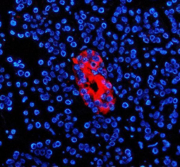
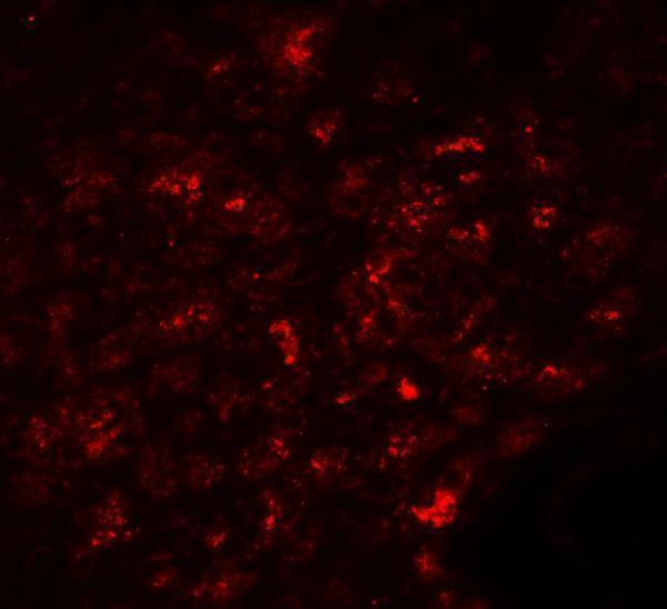
| IF/ICC: what localization should be expected? | HPA reports vesicles as Approved and plasma membrane as Supported in ICC-IF (HPA subcellular ICC-IF); UniProt also places SLC30A8 at secretory-vesicle and cell membranes (UniProt Q8IWU4 subcellular location). | Look for vesicular signal with possible plasma-membrane signal, using the separate IF/ICC guide for assay design (HPA subcellular ICC-IF). HPA065953 has ICC Supported status; its listing does not confer IHC validation (HPA antibodies). |
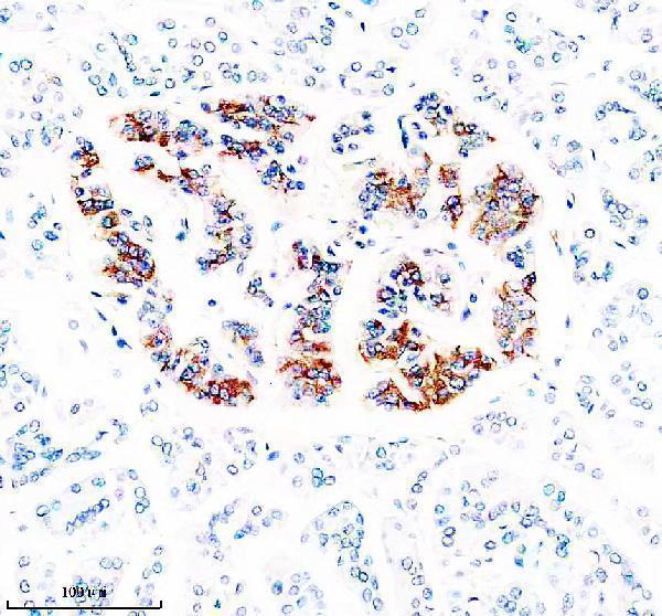


Comprehensive Human Protein Atlas IHC scoring per tissue (reliability: Enhanced — High consistency between antibody staining and RNA expression data.). Rows are taken directly from the HPA tissue chart — click any row's HPA link to view the source.
| Tissue | Cell type | Level | Evidence | Source |
|---|---|---|---|---|
| Pancreas | Pancreatic endocrine cells | High | Protein (IHC) | HPA → |
| Tissue | Cell type | Level | Evidence | Source |
|---|---|---|---|---|
| Adipose tissue | Adipocytes | Not detected | Protein (IHC) | HPA → |
| Adrenal gland | Glandular cells | Not detected | Protein (IHC) | HPA → |
| Appendix | Glandular cells | Not detected | Protein (IHC) | HPA → |
| Bone marrow | Hematopoietic cells | Not detected | Protein (IHC) | HPA → |
| Breast | Adipocytes | Not detected | Protein (IHC) | HPA → |
Troubleshoot SLC30A8 staining in paraffin sections by checking retrieval, cellular pattern, controls, and scoring against its secretory vesicle localisation.
Two human-reactive anti-SLC30A8 antibodies have IHC and IF images from human pancreas (catalog reactivity; IHC/IF image captions).
A01310-1 is listed for IHC and IF in human samples, with both images showing human pancreas; its IHC image documents a paraffin section (A01310-1 applications and image captions). A01310 is listed for IHC-P and IF in human samples, with both images showing human pancreas (A01310 applications and image captions).
Which to pick: For tissue IHC, choose A01310-1 when a documented paraffin-section workflow is useful: its caption specifies EDTA retrieval at pH 8.0, 2 μg/ml primary antibody and DAB detection; the fixative is unreported (A01310-1 IHC image caption). For IF, A01310-1 has a paraffin-section image at 5 μg/ml, while A01310 has a human-pancreas image at 20 μg/mL; ICC is not listed for either (IF image captions; catalog applications). Neither has listed reactivity beyond human, and clonality is unreported, so neither has a documented cross-species or clonality advantage (catalog reactivity and clone fields).