This website uses cookies to ensure you get the best experience on our website.


- Table of Contents
Plan SLC39A10 paraffin IHC using strongly stained ciliated cell bodies as reference tissue (HPA tissue IHC). This guide covers the observed cytoplasmic and membranous pattern (HPA tissue IHC), fixation, controls and the catalog antibody’s 2–5 μg/mL IHC range (datasheet A09043-1).

Expected localisation, validated protocols, controls and antibodies — the at-a-glance facts below, then the full design guide.
| Expected localisation | Cytoplasmic and membranous tissue staining (HPA tissue IHC) | |
| Staining pattern | Ciliated cell bodies: high; tissue signal cytoplasmic/membranous (HPA tissue IHC) | |
| Antigen retrieval | EDTA pH 8.0 HIER, heat-mediated (datasheet A09043-1) | |
| Positive control | Fallopian tube+4 more · see all | |
| Negative control | Adipose tissue+4 more · see all |
| Fixation | Matched tissue-IHC evidence does not establish the fixation claim. Validate the specimen-specific method before use. (selected-SKU IHC image A09043-1) | |
| Caveat | Medium staining–RNA consistency; verify the signal (HPA tissue IHC) | |
| Regulation | Expression regulation is not annotated (UniProt) | |
| Isoform / epitope | 2 isoforms; confirm extracellular vs cytoplasmic epitope (UniProt) |
The catalog antibody’s IHC-P protocol (datasheet A09043-1) is accompanied by one published SLC39A10 tissue IHC protocol (PMC8357404).
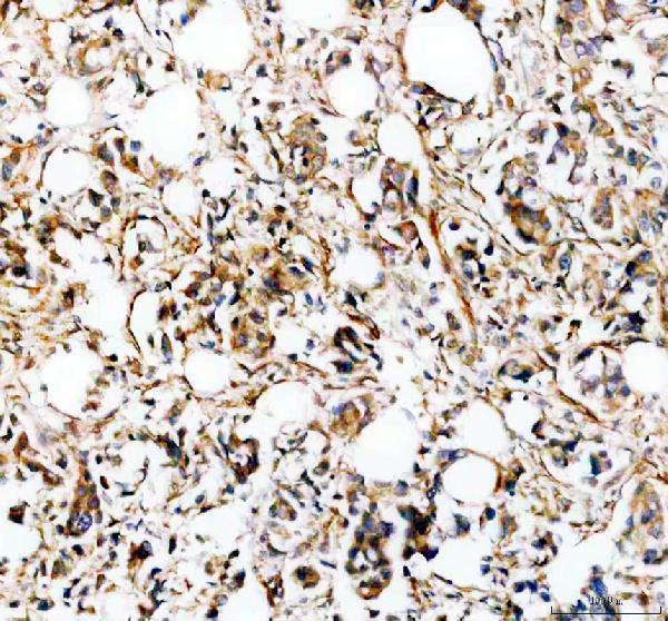
| Sample | Paraffin-embedded human brest cancer tissue; fixative not specified (datasheet A09043-1) |
| Fixation | Image fixative and duration unreported (datasheet A09043-1); verify before use. |
| Sectioning | 4–5 µm sections on charged slides (standard) |
| Deparaffinisation | Xylene, graded ethanol series to water (standard) |
| Antigen retrieval | Heat retrieval: EDTA pH 8.0 (datasheet A09043-1); 20 min, 95–100 °C (standard) |
| Peroxidase block | 3% H2O2, 10 min, room temperature (standard) |
| Blocking | 10% goat serum (datasheet A09043-1) |
| Primary antibody | Rabbit anti-SLC39A10, 2-5 μg/ml (datasheet A09043-1) |
| Primary incubation | Overnight at 4 °C (datasheet A09043-1) |
| Detection | HRP-conjugated secondary, DAB chromogen (datasheet A09043-1) |
| Counterstain | Hematoxylin, blue, dehydrate and mount (standard) |
| Expected result | SLC39A10-positive staining in ciliated cells (cell body) of fallopian tube (HPA tissue IHC: High). HPA tissue profile: Cytoplasmic and membranous expression in several tissues, including thyroid gland. No signal in the no-primary control. |
SLC39A10 is a seven-pass membrane protein with a reported apical location in kidney proximal tubules (UniProt Q9ULF5 topology and subcellular location). In paraffin IHC, expect cytoplasmic and membranous staining across several tissues, especially ciliated cells of the fallopian tube and nasopharynx (HPA tissue IHC). HPA rates the tissue profile Approved, with medium consistency against RNA data and external verification pending (HPA tissue IHC).
| Strong cell-body staining in fallopian-tube or nasopharyngeal ciliated cells (HPA tissue IHC: High). | This matches HPA’s strongest listed tissue IHC observations. Assess whether staining follows the expected cells and cytoplasmic or membranous distribution; intensity alone cannot establish specificity (HPA tissue IHC profile; general IHC practice). |
| Only extracellular or luminal chromogen is visible, without staining in the expected cells (HPA tissue IHC profile). | That distribution does not match the reported cellular pattern and may reflect deposited reagent or other artefact (HPA tissue IHC profile; general IHC practice). Inspect an omission control and repeat detection if the deposit persists. |
| Widespread staining appears in glomerular cells or adipocytes (HPA tissue IHC: Not detected in those cells). | This conflicts with the listed HPA observations and warrants a cross-reactivity or endogenous detection check (HPA tissue IHC; general IHC practice). The renal comparison is cell specific: UniProt places SLC39A10 at proximal-tubule apical membranes, not glomeruli (UniProt Q9ULF5 subcellular location). |
| A diffuse haze covers cells, stroma, and clear spaces, obscuring cell boundaries (general IHC practice). | Treat the slide as difficult to score: the haze can arise from nonspecific binding, incomplete washing, or detection background (general IHC practice). Compare an omission control before assigning any apparent SLC39A10-positive cell type. |
| No cell-body signal appears in adequately preserved fallopian-tube or nasopharyngeal ciliated cells (HPA tissue IHC: High). | A known-positive compartment is missing, so a negative study result is not yet interpretable (HPA tissue IHC; general IHC practice). Check section integrity, antibody and detection controls, then optimize routine IHC conditions without assuming a documented SLC39A10 fixation effect. |
| Which cells provide the clearest IHC reference? | Fallopian-tube and nasopharyngeal ciliated cells are listed as High; bronchial ciliated cells, endometrial glands, lung alveolar cells, placental trophoblasts, and prostate and seminal-vesicle glands are Medium (HPA tissue IHC). Use the same cell type when comparing sections. |
| What does membrane topology support? | UniProt annotates seven transmembrane segments and an apical proximal-tubule location (UniProt Q9ULF5 topology and subcellular location). These support a membrane component but do not specify the antibody epitope or predict chromogenic intensity in a particular tissue. |
| Do processing or isoforms alter this call? | UniProt lists a signal peptide at residues 1–25, a chain at 26–831, four glycosylation sites, and two isoforms (UniProt Q9ULF5 processing, glycosylation, isoforms). The supplied records do not map antibody epitopes or establish isoform-specific staining or shedding. |
| How should HPA reliability affect scoring? | HPA labels tissue staining Approved but reports medium RNA concordance and pending external verification; its listed antibodies have IHC Approved status rather than an IHC Enhanced designation (HPA tissue IHC and antibody validation). Score morphology and controls alongside intensity. |
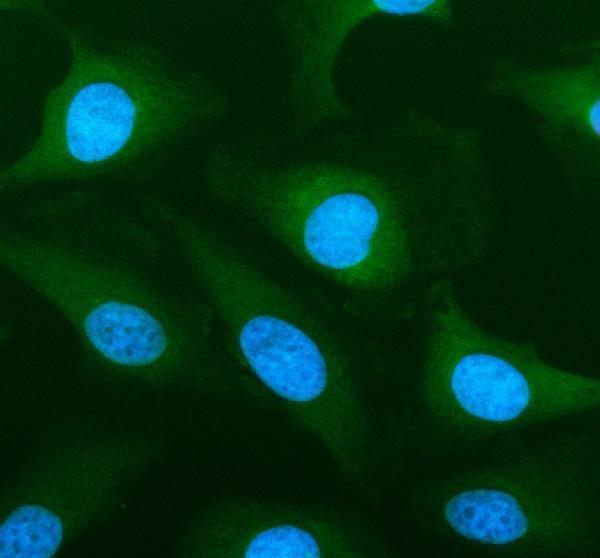
| What should IF/ICC show? | HPA ICC-IF reports mainly nucleoplasmic and cytosolic signal, with additional plasma-membrane signal; those locations are approved in that assay (HPA subcellular ICC-IF). A nuclear IF signal therefore needs its own interpretation and does not redefine the tissue IHC profile. |
| Situation | Likely cause | Next action |
|---|---|---|
| Positive-reference ciliated cells lack staining (HPA tissue IHC: High). | The run may have failed, or the section may lack intact target cells (general IHC practice). | Verify morphology and a working positive control; check primary antibody, detection reagents, and routine retrieval settings before interpreting negatives (general IHC practice). Target-specific fixation sensitivity is unreported in the supplied records. |
| Chromogen coats most of the section (general IHC practice). | Nonspecific binding or detection background may obscure the HPA cellular pattern (HPA tissue IHC profile; general IHC practice). | Compare a primary-omission control, review blocking and wash steps, and adjust antibody concentration using a known-positive section (general IHC practice). |
| Glomerular cells stain prominently (HPA tissue IHC: Not detected). | The signal conflicts with HPA’s glomerular result; endogenous activity or cross-reactivity is possible (HPA tissue IHC; general IHC practice). | Use omission and detection controls, then compare the glomerular signal with the expected positive cells (general IHC practice; HPA tissue IHC). Do not generalize this glomerular finding to proximal tubules (UniProt Q9ULF5 subcellular location). |
| Only nuclear staining is seen in tissue IHC (HPA tissue IHC profile). | It lacks the cytoplasmic and membranous tissue pattern, although HPA separately approves nucleoplasmic ICC-IF localization (HPA tissue IHC; HPA subcellular ICC-IF). | Check whether cellular boundaries and controls support the IHC signal; report the discordance instead of calling every nuclear signal artefactual (general IHC practice; HPA subcellular ICC-IF). |
| Signal is confined to lumen or extracellular material (HPA tissue IHC profile). | Acellular deposit does not match HPA’s reported cell-body or glandular staining (HPA tissue IHC); detection artefact is possible (general IHC practice). | Inspect an omission control and repeat washing or detection as needed; require staining within the expected cells for a positive call (general IHC practice; HPA tissue IHC). |
| A medium-level tissue looks weaker than the reference (HPA tissue IHC). | HPA lists several tissues at Medium, below its High ciliated-cell references (HPA tissue IHC). | Compare like cell types and section quality, then score signal above local background with controls; do not use one intensity threshold across all listed tissues (HPA tissue IHC; general IHC practice). |


Comprehensive Human Protein Atlas IHC scoring per tissue (reliability: Approved — Medium consistency between antibody staining and RNA expression data. Pending external verification.). Rows are taken directly from the HPA tissue chart — click any row's HPA link to view the source.
| Tissue | Cell type | Level | Evidence | Source |
|---|---|---|---|---|
| Fallopian tube | Ciliated cells (cell body) | High | Protein (IHC) | HPA → |
| Nasopharynx | Ciliated cells (cell body) | High | Protein (IHC) | HPA → |
| Bronchus | Ciliated cells (cell body) | Medium | Protein (IHC) | HPA → |
| Endometrium | Glandular cells | Medium | Protein (IHC) | HPA → |
| Lung | Alveolar cells | Medium | Protein (IHC) | HPA → |
| Tissue | Cell type | Level | Evidence | Source |
|---|---|---|---|---|
| Adipose tissue | Adipocytes | Not detected | Protein (IHC) | HPA → |
| Adrenal gland | Glandular cells | Not detected | Protein (IHC) | HPA → |
| Caudate | Glial cells | Not detected | Protein (IHC) | HPA → |
| Cerebellum | Cells in granular layer | Not detected | Protein (IHC) | HPA → |
| Duodenum | Glandular cells | Not detected | Protein (IHC) | HPA → |
Troubleshoot SLC39A10 staining in paraffin sections by checking retrieval, cell identity and subcellular pattern alongside tissue controls.
Both catalog antibodies have human tissue IHC images; A09043-1 also has mouse and rat tissue IHC images and IF images of HeLa cells and tissue sections (catalog image captions).
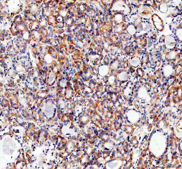
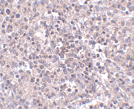
A09043 has IHC-P listed and an IHC image of human spleen at 2.5 μg/mL (catalog application list; A09043 image caption). A09043-1 has paraffin-section IHC images of human breast and thyroid cancer and mouse and rat brain, plus IF images of HeLa cells and human, mouse and rat tissue sections (A09043-1 image captions).
Which to pick: For paraffin-section tissue IHC, choose A09043-1 when a captioned retrieval and detection workflow is useful; its IHC captions specify EDTA retrieval, goat serum blocking and DAB detection, but do not report the fixative (A09043-1 IHC image captions). For IF/ICC, choose A09043-1 because those applications and corresponding images are listed for it (A09043-1 application list; IF image captions). For mouse or rat tissue IHC, A09043-1 has species-specific images; A09043 lists mouse and rat reactivity but its supplied IHC image is human spleen, and clonality is unreported for both (catalog reactivity; image captions; clone fields).