SLC40A1 / Ferroportin · IHC design guide

Design Immunohistochemistry for SLC40A1

Plan SLC40A1 IHC on paraffin sections using the catalog antibody's 2–5 μg/ml range (datasheet A01953-2). Compare membrane localisation expected from topology (UniProt) with low cytoplasmic staining in small intestine and bone marrow and distinct erythrocyte positivity (HPA tissue IHC).

Evidence assembled Oct 2026 · For research use; verify linked source records and product datasheet before use
Immunohistochemistry protocol sheet for SLC40A1 (IHC for SLC40A1): expected localisation Membrane expected (UniProt); low tissue cytoplasm observed (HPA tissue IHC), antibody A01953-2, validated IHC image, and IHC protocol steps
Printable SLC40A1 IHC protocol sheet — expected localisation Membrane expected (UniProt); low tissue cytoplasm observed (HPA tissue IHC), antibody A01953-2, controls and protocol steps. Open the full SLC40A1 IHC guide →

SLC40A1 Immunohistochemistry Experimental Design Guide

Expected localisation, validated protocols, controls and antibodies — the at-a-glance facts below, then the full design guide.

Must know before staining
Expected localisation Membrane expected (UniProt); low tissue cytoplasm observed (HPA tissue IHC)
Staining pattern Low cytoplasmic signal; distinct erythrocyte positivity (HPA tissue IHC)
Antigen retrieval EDTA pH 8.0 HIER, heat-mediated (datasheet A01953-2)
Positive control ⓘ Bone marrow
Negative control ⓘ Adipose tissue+4 more · see all
Important caveats
Reasons your staining may differ from the expected pattern.
Fixation Keep paraffin-section fixation consistent. (standard IHC practice; not target-specific)
Caveat Erythrocyte-rich areas need endogenous peroxidase control (HPA tissue IHC; standard IHC practice)
Regulation Hepcidin promotes ferroportin degradation (UniProt)
Isoform / epitope No isoforms annotated; epitope side matters across 12 helices (UniProt)
Section 1

Recommended SLC40A1 IHC & IF Protocols

The catalog antibody protocol uses EDTA pH 8.0 retrieval (datasheet). Two published chromogenic IHC protocols provide additional conditions for comparison (PMC5617709; PMC10247955).

Recommended immunohistochemistry (IHC-P) protocol parameters
SampleParaffin-embedded human larynx squamous cell carcinoma tissue; fixative not specified (datasheet A01953-2)
FixationImage fixative and duration unreported (datasheet A01953-2); verify before use.
Sectioning4–5 µm sections on charged slides (standard)
DeparaffinisationXylene, graded ethanol series to water (standard)
Antigen retrievalHeat retrieval: EDTA pH 8.0 (datasheet A01953-2); 20 min, 95–100 °C (standard)
Peroxidase block3% H2O2, 10 min, room temperature (standard)
Blocking10% goat serum (datasheet A01953-2)
Primary antibodyRabbit anti-SLC40A1, 2-5 μg/ml (datasheet A01953-2)
Primary incubationOvernight at 4 °C (datasheet A01953-2)
DetectionHRP-conjugated secondary, DAB chromogen (datasheet A01953-2)
CounterstainHematoxylin, blue, dehydrate and mount (standard)
Expected resultSLC40A1-positive staining in hematopoietic cells of bone marrow (HPA tissue IHC: Medium). HPA tissue profile: Low cytoplasmic expression in the small intestine and bone marrow with distinct positivity in erythrocytes. No signal in the no-primary control.
💡Decision noteStart with heat-mediated EDTA pH 8.0 for the catalog antibody (datasheet); consider pH 6.0 retrieval for mouse liver (PMC10247955).
Section 2

What Is the Expected SLC40A1 Staining Pattern?

SLC40A1 is a 12-pass cell-membrane protein; basolateral localization is reported in WIF-B cells (UniProt Q9NP59 topology and subcellular location). In paraffin IHC, expect distinct erythrocyte positivity, medium staining in bone-marrow hematopoietic cells, and low cytoplasmic staining in small-intestinal glandular cells (HPA tissue IHC, Approved). Interpret compartment and cell identity together.

What am I looking at on my slide?
Erythrocytes stain distinctly, marrow hematopoietic cells stain at medium intensity, and small-intestinal glandular cells show low cytoplasmic staining (HPA tissue IHC).This combination fits the reported IHC pattern (HPA tissue IHC, Approved). Compare each cell population separately: a strong intestinal signal is not required by the HPA profile, while a marrow result alone cannot establish the expected erythrocyte pattern.
Predominantly nuclear staining appears in cells whose membrane or cytoplasm is otherwise unstained.Treat an isolated nuclear IHC pattern cautiously: UniProt places SLC40A1 at the cell membrane (UniProt Q9NP59), while HPA calls nucleoplasmic ICC-IF localization uncertain (HPA subcellular). Check control slides and detection background before assigning that pattern to SLC40A1 (standard IHC practice).
Strong staining dominates adipocytes or bronchial respiratory epithelium, with little staining in the expected positive populations.HPA reports those sampled cell types as not detected (HPA tissue IHC). Such a distribution raises concern for cross-reactivity or endogenous detection activity; it does not prove either cause. Review cell identity, the no-primary control, and the positive tissue on the same run (standard IHC practice).
Color spreads across stroma, lumina, or most cell types without a clear cellular boundary.A diffuse deposit cannot be scored as the distinct erythrocyte or cell-associated pattern reported by HPA (HPA tissue IHC). Examine a no-primary control for detection background and reassess blocking, washes, and chromogen development (standard IHC practice).
Bone-marrow hematopoietic cells and erythrocytes are blank on an otherwise evaluable slide.This conflicts with HPA's medium marrow-cell staining and distinct erythrocyte positivity (HPA tissue IHC). First verify that the expected cells are present; then check antibody dilution, antigen retrieval, detection reagents, and the run control (standard IHC practice).
💡Expected SLC40A1 appearanceCall a positive result when distinct erythrocyte staining accompanies medium marrow hematopoietic-cell staining or low small-intestinal glandular cytoplasmic staining (HPA tissue IHC, Approved); isolated strong nuclear staining or diffuse color across unrelated structures warrants investigation (UniProt Q9NP59 subcellular location; HPA subcellular; standard IHC practice).
How each factor affects the staining
Membrane topology and compartmentSLC40A1 has 12 transmembrane segments and is assigned to the cell membrane (UniProt Q9NP59 topology and subcellular location). HPA nevertheless describes low cytoplasmic staining in intestinal IHC (HPA tissue IHC). Score the observed paraffin-section pattern without requiring a sharp membrane outline in every positive cell.
Choice of tissue and cell populationHPA reports medium staining in bone-marrow hematopoietic cells, low staining in duodenal and small-intestinal glandular cells, and distinct erythrocyte positivity (HPA tissue IHC). Its not-detected calls apply to the specified sampled cell populations; they are not blanket claims that every cell in those organs lacks SLC40A1.
Antibody evidenceThe listed rabbit polyclonal antibody HPA065634 is Approved for IHC; the tissue profile has Approved reliability, with external characterization supporting staining (HPA antibodies; HPA tissue IHC). Approval supports use of the reported pattern, but unexpected staining still needs controls and cell-level interpretation (standard IHC practice).
ICC-IF Q&A: Should its image pattern dictate this IHC call?No. HPA reports supported plasma-membrane ICC-IF localization, uncertain cytosolic localization, and uncertain additional nucleoplasmic localization (HPA subcellular; HPA antibodies: ICC Supported). Use the tissue IHC profile for paraffin-section expectations (HPA tissue IHC); ICC-IF belongs to its separate guide.
Why is my staining missing, weak or wrong?
SituationLikely causeNext action
No staining in marrow cells or erythrocytesThe result misses the reported positive populations (HPA tissue IHC); inadequate retrieval, antibody application, or detection is possible (standard IHC practice).Confirm those cells are present, compare a known-positive section in the same run, and review retrieval conditions, antibody dilution, reagent activity, and incubation records (standard IHC practice).
Erythrocytes stain in the no-primary controlEndogenous detection activity or deposited chromogen may contribute to apparent erythrocyte staining (standard chromogenic IHC practice).Evaluate endogenous-enzyme blocking and detection controls before calling the erythrocyte signal specific (standard IHC practice). Distinct erythrocyte positivity is expected in the HPA profile, but that expectation does not replace a control (HPA tissue IHC).
Strong staining in a sampled HPA-negative cell typeFor example, adipocytes and bronchial respiratory epithelial cells were not detected in the HPA tissue profile (HPA tissue IHC). Cross-reactivity, background, or cell misidentification are possible (standard IHC practice).Check the annotated cell type against morphology, inspect the no-primary control, and compare expected positive cells before interpreting the new distribution (standard IHC practice; HPA tissue IHC).
Broad haze obscures cellular localizationDiffuse background can reflect blocking, washing, antibody concentration, or chromogen-development conditions (standard IHC practice).Inspect the no-primary slide; optimize blocking and washes, then reassess dilution and development time while retaining a positive control (standard IHC practice). Score only interpretable cell-associated staining against the HPA pattern (HPA tissue IHC).
Only strong nuclear staining is visibleUniProt assigns SLC40A1 to the cell membrane, and HPA rates nucleoplasmic ICC-IF localization uncertain (UniProt Q9NP59; HPA subcellular). The IHC finding therefore needs verification.Check detection background and tissue morphology, then seek the reported erythrocyte, marrow-cell, or low intestinal pattern on a controlled run (standard IHC practice; HPA tissue IHC).

Sample controls for SLC40A1 IHC & IF

🧪Run bone marrow first and assess staining in its hematopoietic cells (HPA: Medium in bone marrow hematopoietic cells). Use adipose tissue adipocytes as the negative comparison (HPA: Not detected in adipocytes); on the bone marrow slide, cells without specific membrane staining should remain at background levels relative to the no-primary control (UniProt Q9NP59: cell membrane localization; standard IHC practice).
Positive control tissue: Bone marrow (Hematopoietic cells, HPA Medium)
Negative control tissue: Adipose tissue (HPA Not detected)
ICC-IF cell lines (HPA subcellular resource): HPA ICC-IF images show SLC40A1 in Hep-G2, RT-4, U2OS, with annotated localisation: Plasma membrane (supported), Cytosol (uncertain) (HPA subcellular).
Technical controls: Include a no-primary, secondary-only control, a concentration-matched rabbit IgG control matched to the primary antibody’s clonality, and SLC40A1 knockout tissue or a validated peptide-block control (A01953-2 tissue-IHC caption: rabbit primary antibody; standard IHC practice). Block endogenous peroxidase and check for pigment-related background in bone marrow before interpreting DAB staining (standard IHC practice).
⚠️Feasibility: A target-specific fixation window and fixation effect are unreported in the supplied evidence; the selected paraffin-section caption does not state the fixative (A01953-2 tissue-IHC caption). The reported IHC procedure uses heat-mediated retrieval in EDTA at pH 8.0, but whether that retrieval is required across specimens is unreported (A01953-2 tissue-IHC caption). Whether frozen sections or IF/ICC are easier for this antibody is unreported; bone marrow requires attention to endogenous peroxidase and pigment background during chromogenic scoring (standard IHC practice).

HPA tissue IHC evidence for SLC40A1

Comprehensive Human Protein Atlas IHC scoring per tissue (reliability: Approved — External characterization data supports antibody staining. Antibody staining in cells/structures not annotated, view images.). Rows are taken directly from the HPA tissue chart — click any row's HPA link to view the source.

Positive expression · recommended positive controls

TissueCell typeLevelEvidenceSource
Bone marrow Hematopoietic cells Medium Protein (IHC) HPA →

Undetected expression · recommended negative controls

TissueCell typeLevelEvidenceSource
Adipose tissue Adipocytes Not detected Protein (IHC) HPA →
Adrenal gland Glandular cells Not detected Protein (IHC) HPA →
Appendix Glandular cells Not detected Protein (IHC) HPA →
Breast Adipocytes Not detected Protein (IHC) HPA →
Bronchus Respiratory epithelial cells Not detected Protein (IHC) HPA →
Section 3

Advanced SLC40A1 IHC Tips

Troubleshoot SLC40A1 chromogenic staining in paraffin sections using the catalog antibody’s tissue image, reported localisation, and matched controls.

What retrieval should I try first when SLC40A1 staining is weak?
Start with heat-mediated retrieval in EDTA at pH 8.0 for paraffin sections (datasheet A01953-2). The selected tissue image used that condition, followed by 2 μg/ml primary antibody overnight at 4°C (datasheet A01953-2). If staining remains weak, compare a second retrieval condition on adjacent sections while keeping antibody concentration and detection constant (standard IHC practice). Include a positive tissue control in each run so a failed retrieval step is distinguishable from a low-expressing specimen (standard IHC practice). Record heating time and temperature because the caption does not specify them (datasheet A01953-2).
Could fixation explain inconsistent staining between paraffin blocks?
Target-specific fixation sensitivity is unknown: the selected paraffin-section caption does not report its fixative (datasheet A01953-2). Compare blocks with documented fixation histories using the same EDTA pH 8.0 retrieval and detection run (datasheet A01953-2; standard IHC practice). Include a known positive section and a no-primary control to separate specimen variation from staining failure or nonspecific signal (standard IHC practice). Record fixative, duration, processing schedule, and section age before attributing a weak result to SLC40A1 biology (standard IHC practice). Do not infer a target-specific fixation effect from expression patterns or membrane topology alone (HPA tissue IHC; UniProt Q9NP59 topology).
Should ferroportin staining be membranous or cytoplasmic?
Prioritise a cell-boundary pattern when assessing ferroportin, which is annotated at the cell membrane (UniProt Q9NP59 subcellular location). The subcellular atlas supports plasma-membrane localisation, while its cytosolic and nucleoplasmic assignments are uncertain (HPA subcellular). Tissue IHC also reports low cytoplasmic expression in small intestine and bone marrow, so cytoplasmic signal warrants evaluation rather than automatic rejection (HPA tissue IHC). Compare staining with an appropriate cell-boundary reference and inspect intact cells at high magnification (standard IHC practice). Score membrane and cytoplasmic compartments separately, especially where cell borders are difficult to resolve in 1 section (standard IHC practice).
How does epitope position affect my interpretation of SLC40A1 staining?
Confirm the catalog antibody’s immunogen or mapped epitope before interpreting access to either side of the membrane; its position is not supplied here (datasheet A01953-2). Ferroportin has 12 annotated transmembrane segments and cytoplasmic and extracellular loops, making epitope accessibility a practical concern (UniProt Q9NP59 topology). A glycosylation site is annotated at residue 434, within an extracellular region, but its effect on this antibody is unknown (UniProt Q9NP59 topology and glycosylation). No alternative isoforms are annotated in the supplied record; this does not establish epitope recognition experimentally (UniProt Q9NP59 isoforms). Check staining after the documented EDTA pH 8.0 retrieval before changing conditions (datasheet A01953-2).
How can I assess SLC40A1 by IF alongside a cell-type marker?
Treat IF as a separate optimisation: the selected antibody evidence is chromogenic staining of a paraffin section, with no IF conditions supplied (datasheet A01953-2). Pair SLC40A1 with a validated marker for the cell population under study and inspect whether their signals occur in the same cells (standard IF practice). Select a fluorophore channel with low specimen autofluorescence, then image unstained and single-stain controls using identical acquisition settings (standard IF practice). Confirm the epitope’s membrane-facing side before choosing permeabilisation, since SLC40A1 has both cytoplasmic and extracellular regions (UniProt Q9NP59 topology). Evaluate cell-boundary signal separately from diffuse cytoplasmic fluorescence (UniProt Q9NP59 subcellular location; standard IF practice).
What should I change when DAB obscures specific staining?
First inspect a no-primary section for signal caused by the detection system or endogenous peroxidase (standard IHC practice). Apply a peroxidase block before HRP detection and limit DAB development to the point at which positive cells remain distinguishable (standard IHC practice). The selected image used 10% goat serum blocking, 2 μg/ml primary antibody overnight at 4°C, and an HRP/DAB workflow (datasheet A01953-2). If background persists, titrate primary antibody around that documented concentration and compare matched positive and negative sections (standard IHC practice). Distinguish diffuse deposit, tissue-edge staining, and pigmented material from staining that follows intact cell boundaries (standard IHC practice; UniProt Q9NP59 subcellular location).
How should I quantify SLC40A1 staining across tissue samples? ⚠ ANSWER MARKED FOR VERIFICATION
Define the cell population and compartment before scoring, because SLC40A1 is a membrane protein and tissue staining may also appear cytoplasmic (UniProt Q9NP59 subcellular location; HPA tissue IHC). Report the percentage of positive cells and an intensity-weighted H-score, or positive-cell density per mm² when tissue area is the relevant denominator (standard IHC practice). Normalise cell-based scores to the number of evaluable cells of the same type, and area-based scores to viable tissue area (standard IHC practice). Use fixed thresholds, matched imaging conditions, and positive and no-primary controls across runs (standard IHC practice). Keep membrane and cytoplasmic results separate so differences in localisation remain visible (standard IHC practice).
How do I distinguish true SLC40A1 positivity from artefact?
Look for reproducible staining in intact cells, with particular attention to cell boundaries because ferroportin is annotated at the cell membrane (UniProt Q9NP59 subcellular location). Bone-marrow hematopoietic cells show medium staining, while several listed cell populations have no detected staining; use such patterns as context, not universal controls (HPA tissue IHC). Treat isolated nuclear staining cautiously because the atlas labels nucleoplasmic localisation uncertain (HPA subcellular). Exclude necrotic regions and tissue edges from scoring, and check a no-primary section for endogenous enzyme or nonspecific DAB signal (standard IHC practice). A compartment-matched signal that recurs across sections supports interpretation more strongly than diffuse stain alone (standard IHC practice).
Boster reagents

Best SLC40A1 / Ferroportin IHC Antibodies

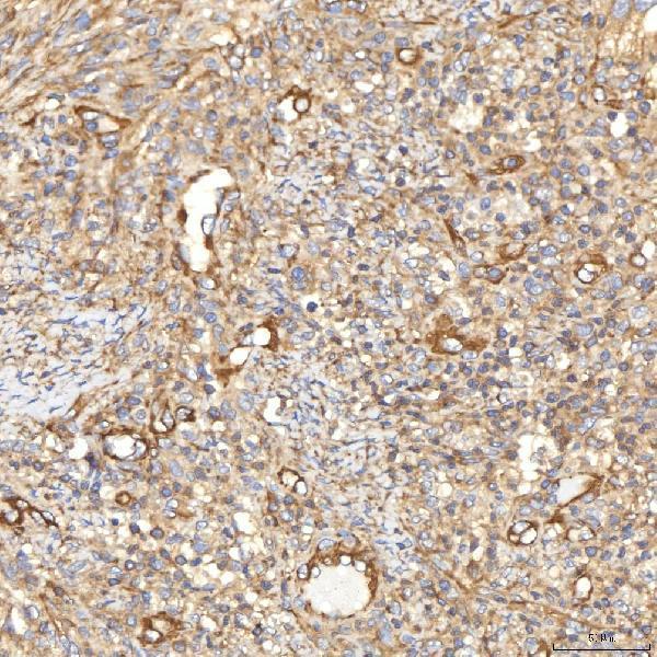
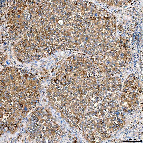
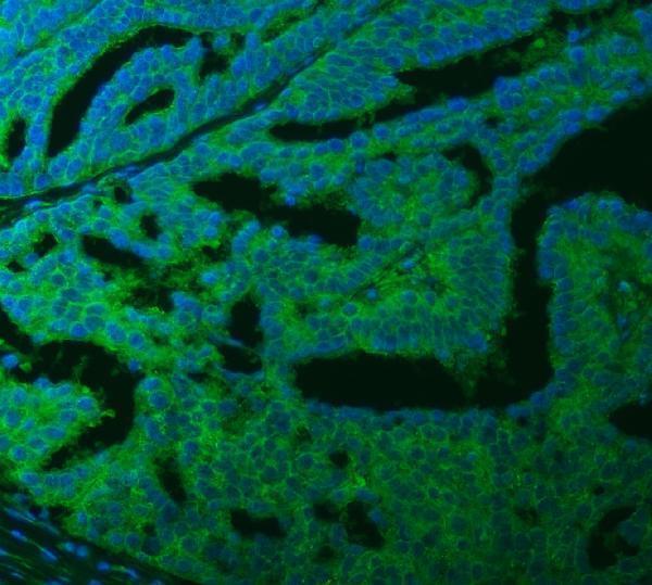
A01953-2 has real IHC data from human paraffin-embedded tumor sections (catalog IHC captions) and IF data from a human ovarian cancer paraffin section (catalog IF caption).

Real IHC data IHC analysis of SLC40A1 using anti-SLC40A1 antibody (A01953-2). SLC40A1 was detected in a paraffin-embedded section of human larynx squamous cell carcinoma tissue. Heat mediated antigen retrieval was performed in EDTA buffer (pH 8.0, epitope retrieval solution). The tissue section was blocked with 10% goat serum. The tissue section was then incubated with 2 μg/ml rabbit anti-SLC40A1 Antibody (A01953-2) overnight at 4°C. Peroxidase Conjugated Goat Anti-rabbit IgG was used as secondary antibody and incubated for 30 minutes at 37°C. The tissue section was developed using HRP Conjugated Rabbit IgG Super Vision Assay Kit (Catalog # SV0002) with DAB as the chromogen.
Anti-SLC40A1 Antibody ®
Cat # A01953-2

A01953-2 is listed for IHC and IF, with Human, Mouse, and Rat reactivity (catalog applications and reactivity). Its images document chromogenic IHC on human paraffin-embedded tumor sections and IF on a human ovarian cancer paraffin section (A01953-2 image captions).

Which to pick: For chromogenic tissue IHC, choose A01953-2: its IHC captions document human paraffin sections, EDTA retrieval at pH 8.0, and 2 μg/ml primary antibody; the fixative is unreported (A01953-2 IHC captions). For IF, A01953-2 has a paraffin-section image at 5 μg/ml; ICC validation is unreported (A01953-2 IF caption; catalog applications). For cross-species work, A01953-2 lists Human, Mouse, and Rat reactivity, but its illustrated IHC and IF data are human only; clonality is unreported (catalog reactivity and clone field; A01953-2 image captions).

Each figure is that product's own IHC / IF validation image from its datasheet.

References

  1. UniProt Consortium. UniProt entry Q9NP59 (S40A1_HUMAN, Ferroportin).
  2. Human Protein Atlas. SLC40A1 tissue IHC expression (reliability: Approved).
  3. Human Protein Atlas. SLC40A1 subcellular location (ICC-IF): Mainly localized to the plasma membrane and cytosol. In addition localized to the nucleoplasm..
  4. Human Protein Atlas. SLC40A1 antibody validation summary (1 antibodies).
  5. Type 4B hereditary hemochromatosis associated with a novel mutation in the SLC40A1 gene: A case report and a review of the literature. Medicine 2017 — PMC5617709.
  6. SLC40A1-mediated positive feedback loop with M1 macrophages suppresses epithelial ovarian cancer progression. Frontiers in immunology 2025 — PMC12847247.
  7. Hepatocyte-specific damage in acute toxicity of sodium ferrous citrate: Presentation of a human autopsy case and experimental results in mice. Toxicology reports 2023 — PMC10247955.
  8. Biomembrane-coated Nanoparticles Targeting circHIF1α Suppress Ovarian Cancer Metastasis and Cisplatin Resistance by Mediating System Xc⁻ Inactivation via SLC7A11/SLC3A2 to Induce Ferroptosis in Cancer Stem Cells. International journal of biological sciences 2026 — PMC13215457.
  9. PubMed PMID:10747949 — UniProt-cited evidence.
  10. PubMed PMID:10882071 — UniProt-cited evidence.
  11. PubMed PMID:10693807 — UniProt-cited evidence.