Click image to see more details
-

-
-
-
-
+6
Product Info Summary
| SKU: | A01953-2 |
|---|---|
| Size: | 100 μg/vial |
| Reactive Species: | Human, Mouse, Rat |
| Host: | Rabbit |
| Application: | ELISA, Flow Cytometry, IF, IHC, WB |
Customers Who Bought This Also Bought

Product info
Product Name
Anti-SLC40A1 Antibody Picoband®
SKU/Catalog Number
A01953-2
Size
100 μg/vial
Form
Lyophilized
Description
Boster Bio Anti-SLC40A1 Antibody Picoband® catalog # A01953-2. Tested in ELISA, Flow Cytometry, IF, IHC, WB applications. This antibody reacts with Human, Mouse, Rat. The brand Picoband indicates this is a premium antibody that guarantees superior quality, high affinity, and strong signals with minimal background in Western blot applications. Only our best-performing antibodies are designated as Picoband, ensuring unmatched performance.
Storage & Handling
At -20°C for one year from date of receipt. After reconstitution, at 4°C for one month. It can also be aliquotted and stored frozen at -20°C for six months. Avoid repeated freezing and thawing.
Cite This Product
Anti-SLC40A1 Antibody Picoband® (Boster Biological Technology, Pleasanton CA, USA, Catalog # A01953-2)
Host
Rabbit
Contents
Each vial contains 4 mg Trehalose, 0.9 mg NaCl, 0.2 mg Na2HPO4.
Clonality
Polyclonal
Isotype
Rabbit IgG
Immunogen
E.coli-derived human SLC40A1 recombinant protein (Position: M1-V571).
Cross-reactivity
No cross-reactivity with other proteins.
Reactive Species
A01953-2 is reactive to SLC40A1 in Human, Mouse, Rat
Observed Molecular Weight
70 kDa
Calculated molecular weight
62.5 kDa
Background of SLC40A1
Ferroportin-1, also known as solute carrier family 40 member 1 (SLC40A1) or iron-regulated transporter 1 (IREG1), is a protein that in humans is encoded by the SLC40A1 gene, and is part of the Ferroportin (Fpn) Family.
Antibody Validation
Boster validates all antibodies on WB, IHC, ICC, Immunofluorescence, and ELISA with known positive control and negative samples to ensure specificity and high affinity, including thorough antibody incubations.
Application & Images
Applications
A01953-2 is guaranteed for ELISA, Flow Cytometry, IF, IHC, WB Boster Guarantee
Assay Dilutions Recommendation
The recommendations below provide a starting point for assay optimization. The actual working concentration varies and should be decided by the user.
Western blot, 0.25-0.5 μg/ml, Human, Mouse, Rat
Immunohistochemistry(Paraffin-embedded Section), 2-5 μg/ml, Human
Immunofluorescence, 5 μg/ml, Human
Flow Cytometry (Fixed), 1-3 μg/1x106 cells, Human
ELISA, 0.1-0.5 μg/ml, -
Positive Control
WB: human placenta tissue, rat liver tissue, mouse spleen tissue, mouse liver tissue
IHC: human larynx squamous cell carcinoma tissue, human liver cancer tissue, human ovarian serous adenocarcinoma tissue, human prostate adenocarcinoma tissue, human rectum adenocarcinoma tissue
IF: human ovarian cancer tissue
FCM: U20S cell
Validation Images & Assay Conditions
Click image to see more details
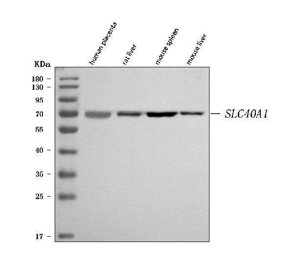
Western blot analysis of SLC40A1 using anti-SLC40A1 antibody (A01953-2).
Electrophoresis was performed on a 5-20% SDS-PAGE gel at 70V (Stacking gel) / 90V (Resolving gel) for 2-3 hours. The sample well of each lane was loaded with 30 ug of sample unde
r reducing conditions.
Lane 1: human placenta tissue lysates,
Lane 2: rat liver tissue lysates,
Lane 3: mouse spleen tissue lysates,
Lane 4: mouse liver tissue lysates.
After electrophoresis, proteins were transferred to a nitrocellulose membrane at 150 mA for 50-90 minutes. Blocked the membrane with 5% non-fat milk/TBS for 1.5 hour at RT. The membrane was incubated with rabbit anti-SLC40A1 antigen affinity purified polyclonal antibody (Catalog # A01953-2) at 0.5 μg/mL overnight at 4°C, then washed with TBS-0.1%Tween 3 times with 5 minutes each and probed with a goat anti-rabbit IgG-HRP secondary antibody at a dilution of 1:5000 for 1.5 hour at RT. The signal is developed using an Enhanced Chemiluminescent detection (ECL) kit (Catalog # EK1002) with Tanon 5200 system. A specific band was detected for SLC40A1 at approximately 70 kDa. The expected band size for SLC40A1 is at 63 kDa.

Click image to see more details
FSGS rats showed iron metabolism disorder. ( A ) Prussian blue staining (scale bar: 100 μm). ( B ) Fe 2+ results. ( C ) Western blot analysis of hepcidin, ferroportin and TFR. ( D ) Relative expression of hepcidin. ( E ) Relative expression of ferroportin. ( F ) Relative expression of TFR. * P < 0.05.
Index in PubMed under a CC BY license. PMID: 38097813

Click image to see more details
IHC analysis of SLC40A1 using anti-SLC40A1 antibody (A01953-2).
SLC40A1 was detected in a paraffin-embedded section of human larynx squamous cell carcinoma tissue. Heat mediated antigen retrieval was performed in EDTA buffer (pH 8.0, epitope retrieval solution). The tissue section was blocked with 10% goat serum. The tissue section was then incubated with 2 μg/ml rabbit anti-SLC40A1 Antibody (A01953-2) overnight at 4°C. Peroxidase Conjugated Goat Anti-rabbit IgG was used as secondary antibody and incubated for 30 minutes at 37°C. The tissue section was developed using HRP Conjugated Rabbit IgG Super Vision Assay Kit (Catalog # SV0002) with DAB as the chromogen.

Click image to see more details
IHC analysis of SLC40A1 using anti-SLC40A1 antibody (A01953-2).
SLC40A1 was detected in a paraffin-embedded section of human liver cancer tissue. Heat mediated antigen retrieval was performed in EDTA buffer (pH 8.0, epitope retrieval solution). The tissue section was blocked with 10% goat serum. The tissue section was then incubated with 2 μg/ml rabbit anti-SLC40A1 Antibody (A01953-2) overnight at 4°C. Peroxidase Conjugated Goat Anti-rabbit IgG was used as secondary antibody and incubated for 30 minutes at 37°C. The tissue section was developed using HRP Conjugated Rabbit IgG Super Vision Assay Kit (Catalog # SV0002) with DAB as the chromogen.

Click image to see more details
IHC analysis of SLC40A1 using anti-SLC40A1 antibody (A01953-2).
SLC40A1 was detected in a paraffin-embedded section of human ovarian serous adenocarcinoma tissue. Heat mediated antigen retrieval was performed in EDTA buffer (pH 8.0, epitope retrieval solution). The tissue section was blocked with 10% goat serum. The tissue section was then incubated with 2 μg/ml rabbit anti-SLC40A1 Antibody (A01953-2) overnight at 4°C. Peroxidase Conjugated Goat Anti-rabbit IgG was used as secondary antibody and incubated for 30 minutes at 37°C. The tissue section was developed using HRP Conjugated Rabbit IgG Super Vision Assay Kit (Catalog # SV0002) with DAB as the chromogen.

Click image to see more details
IHC analysis of SLC40A1 using anti-SLC40A1 antibody (A01953-2).
SLC40A1 was detected in a paraffin-embedded section of human prostate adenocarcinoma tissue. Heat mediated antigen retrieval was performed in EDTA buffer (pH 8.0, epitope retrieval solution). The tissue section was blocked with 10% goat serum. The tissue section was then incubated with 2 μg/ml rabbit anti-SLC40A1 Antibody (A01953-2) overnight at 4°C. Peroxidase Conjugated Goat Anti-rabbit IgG was used as secondary antibody and incubated for 30 minutes at 37°C. The tissue section was developed using HRP Conjugated Rabbit IgG Super Vision Assay Kit (Catalog # SV0002) with DAB as the chromogen.

Click image to see more details
IHC analysis of SLC40A1 using anti-SLC40A1 antibody (A01953-2).
SLC40A1 was detected in a paraffin-embedded section of human rectum adenocarcinoma tissue. Heat mediated antigen retrieval was performed in EDTA buffer (pH 8.0, epitope retrieval solution). The tissue section was blocked with 10% goat serum. The tissue section was then incubated with 2 μg/ml rabbit anti-SLC40A1 Antibody (A01953-2) overnight at 4°C. Peroxidase Conjugated Goat Anti-rabbit IgG was used as secondary antibody and incubated for 30 minutes at 37°C. The tissue section was developed using HRP Conjugated Rabbit IgG Super Vision Assay Kit (Catalog # SV0002) with DAB as the chromogen.

Click image to see more details
IF analysis of SLC40A1 using anti-SLC40A1 antibody (A01953-2).
SLC40A1 was detected in a paraffin-embedded section of human ovarian cancer tissue. Heat mediated antigen retrieval was performed in EDTA buffer (pH 8.0, epitope retrieval solution). The tissue section was blocked with 10% goat serum. The tissue section was then incubated with 5 μg/mL rabbit anti-SLC40A1 Antibody (A01953-2) overnight at 4°C. FITC Conjugated Goat Anti-Rabbit IgG (BA1105) was used as secondary antibody at 1:500 dilution and incubated for 30 minutes at 37°C. The section was counterstained with DAPI. Visualize using a fluorescence microscope and filter sets appropriate for the label used.

Click image to see more details
Flow Cytometry analysis of U20S cells using anti-SLC40A1 antibody (A01953-2).
Overlay histogram showing U20S cells stained with A01953-2 (Blue line). To facilitate intracellular staining, cells were fixed with 4% paraformaldehyde and permeabilized with permeabilization buffer. The cells were blocked with 10% normal goat serum. And then incubated with rabbit anti-SLC40A1 Antibody (A01953-2, 1 μg/1x106 cells) for 30 min at 20°C. DyLight®488 conjugated goat anti-rabbit IgG (BA1127, 5-10 μg/1x106 cells) was used as secondary antibody for 30 minutes at 20°C. Isotype control antibody (Green line) was rabbit IgG (1 μg/1x106) used under the same conditions. Unlabelled sample without incubation with primary antibody and secondary antibody (Red line) was used as a blank control.

Click image to see more details
Western blot analysis of SLC40A1 using anti-SLC40A1 antibody (A04887-1).
Electrophoresis was performed on a 5-20% SDS-PAGE gel at 80V (Stacking gel) / 120V (Resolving gel) for 2 hours. The sample well of each lane was loaded with 30 ug of sample under reducing conditions.
Lane 1-5: model group-human uterine tissue lysates,
Lane 6-10: young group-human uterine tissue lysates.
After electrophoresis, proteins were transferred to a nitrocellulose membrane at 150 mA for 50-90 minutes. Blocked the membrane with 5% non-fat milk/TBS for 1.5 hour at RT. The membrane was incubated with rabbit anti-SLC40A1 antigen affinity purified polyclonal antibody (A04887-1) at 1:1000 overnight at 4°C, then washed with TBS-0.1%Tween 3 times with 5 minutes each and probed with a goat anti-rabbit IgG-HRP secondary antibody at a dilution of 1:5000 for 1 hour at RT. The signal is developed using an ECL Plus Western Blotting Substrate with Tanon 5200 system. A specific band was detected for SLC40A1 at approximately 60-70 kDa. The expected band size for SLC40A1 is at 70 kDa.
Specific Publications For Anti-SLC40A1 Antibody Picoband® (A01953-2)
Loading publications
Recommended Resources
Here are featured tools and databases that you might find useful.
- Boster's Pathways Library
- Protein Databases
- Bioscience Research Protocol Resources
- Data Processing & Analysis Software
- Photo Editing Software
- Scientific Literature Resources
- Research Paper Management Tools
- Molecular Biology Software
- Primer Design Tools
- Bioinformatics Tools
- Phylogenetic Tree Analysis
Customer Reviews
Have you used Anti-SLC40A1 Antibody Picoband®?
Share your experimental results or join a short interview to earn up to $1,000 in product credits or other rewards.
1 Reviews For Anti-SLC40A1 Antibody Picoband®
The SLC40A1 antibody was used to detect the expression of the target protein in human uterine tissue. Although two bands were observed, the differences in expression levels were still clearly discernible.
Excellent

| SKU | A01953-2 |
|---|---|
| Application | Western Blot |
| Sample | human uterine tissue |
| Sample Processing Description | The tissue was minced and further disrupted by sonication, then lysed on ice for 1 hour using RIPA buffer. After centrifugation, the supernatant was collected and quantified using the BCA method. The appropriate amount of loading buffer was added, and the samples were boiled in a water bath to denature the proteins. Finally, 15 μL of each protein sample was loaded into each lane of the SDS-PAGE gel. |
| Other Reagents | 5% Non-fat milk |
| Primary Antibody | Anti-SLC40A1 Antibody Picoband® |
| Primary Incubation | overnight at 4 ℃ |
| Secondary Antibody | HRP-conjugated Anti-Rabbit IgG Secondary Antibody |
| Secondary Incubation | 1 hour in room temperature |
| Detection | Substrate: Ultra-sensitive ECL luminescent reagent (Cat# AR1191), Imaging system:Tanon |
| Results Summary | The SLC40A1 antibody was used to detect the expression of the target protein in human uterine tissue. Although two bands were detected, the differences in expression levels were still clearly observable and did not affect the analysis of the experimental results. |
Anfeng Ning, Peking University Third Hospital
Verified customer
Submitted 2025-11-11
Customer Q&As
Have a question?
Find answers in Q&As, reviews.
Can't find your answer?
Submit your question
