This website uses cookies to ensure you get the best experience on our website.
- Table of Contents
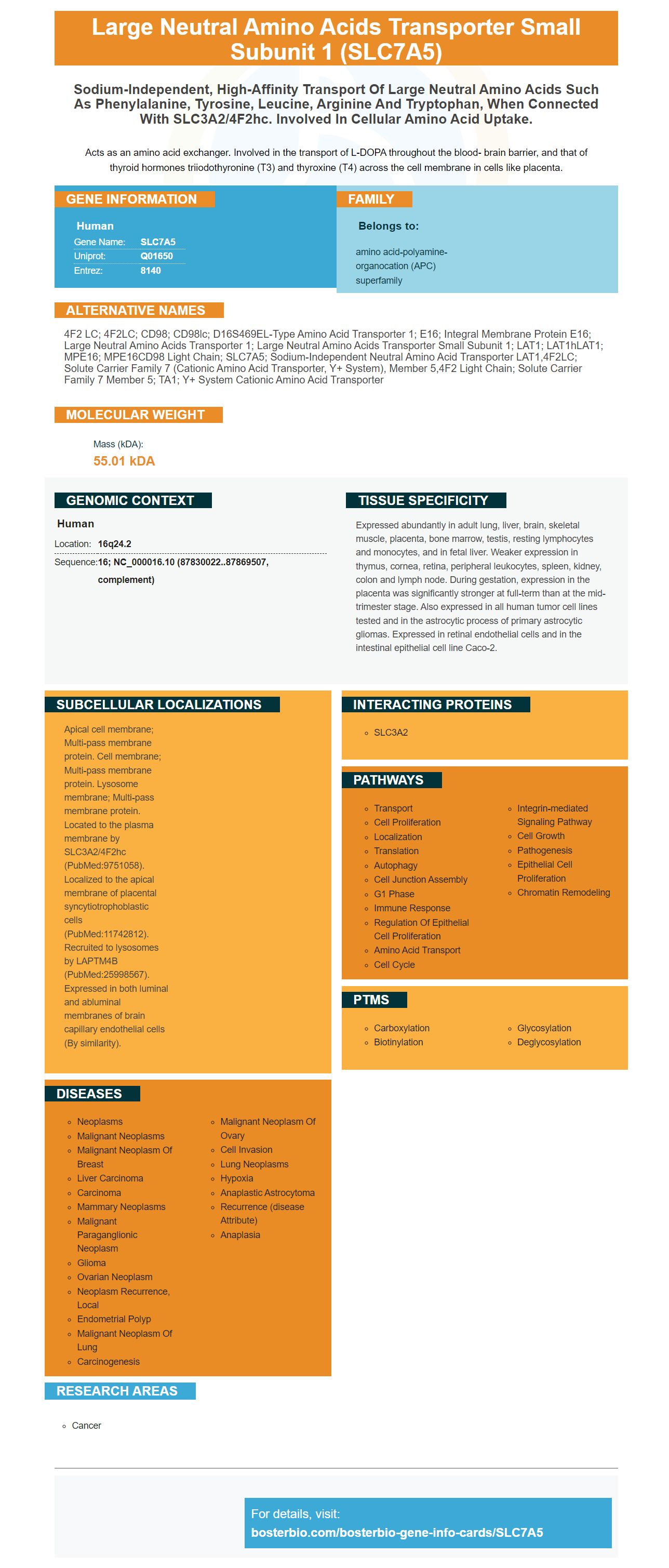
Facts about Large neutral amino acids transporter small subunit 1.
Acts as an amino acid exchanger. Involved in the transport of L-DOPA throughout the blood- brain barrier, and that of thyroid hormones triiodothyronine (T3) and thyroxine (T4) across the cell membrane in cells like placenta.
Human | |
---|---|
Gene Name: | SLC7A5 |
Uniprot: | Q01650 |
Entrez: | 8140 |
Belongs to: |
---|
amino acid-polyamine-organocation (APC) superfamily |
4F2 LC; 4F2LC; CD98; CD98lc; D16S469EL-type amino acid transporter 1; E16; Integral membrane protein E16; large neutral amino acids transporter 1; large neutral amino acids transporter small subunit 1; LAT1; LAT1hLAT1; MPE16; MPE16CD98 light chain; SLC7A5; sodium-independent neutral amino acid transporter LAT1,4F2LC; solute carrier family 7 (cationic amino acid transporter, y+ system), member 5,4F2 light chain; Solute carrier family 7 member 5; TA1; y+ system cationic amino acid transporter
Mass (kDA):
55.01 kDA
Human | |
---|---|
Location: | 16q24.2 |
Sequence: | 16; NC_000016.10 (87830022..87869507, complement) |
Expressed abundantly in adult lung, liver, brain, skeletal muscle, placenta, bone marrow, testis, resting lymphocytes and monocytes, and in fetal liver. Weaker expression in thymus, cornea, retina, peripheral leukocytes, spleen, kidney, colon and lymph node. During gestation, expression in the placenta was significantly stronger at full-term than at the mid-trimester stage. Also expressed in all human tumor cell lines tested and in the astrocytic process of primary astrocytic gliomas. Expressed in retinal endothelial cells and in the intestinal epithelial cell line Caco-2.
Apical cell membrane; Multi-pass membrane protein. Cell membrane; Multi-pass membrane protein. Lysosome membrane; Multi-pass membrane protein. Located to the plasma membrane by SLC3A2/4F2hc (PubMed:9751058). Localized to the apical membrane of placental syncytiotrophoblastic cells (PubMed:11742812). Recruited to lysosomes by LAPTM4B (PubMed:25998567). Expressed in both luminal and abluminal membranes of brain capillary endothelial cells (By similarity).
Researchers are always looking for reliable antibodies. Make sure you choose a company which validates antibodies on multiple platforms and has known positive samples. Boster validates antibodies to ensure high specificity and high affinity. Boster also offers product credits to early reviewers, which is a great way for scientists around the world to be rewarded. Learn more about Boster’s quality inspection process for antibodies.
This study examined the relationship between SLC7A5 protein expression and clinical outcome in patients with different types of human cancers. It used two large validation and discovery cohorts to find that high levels of SLC7A5 proteins were associated with poor clinical outcome, higher Ki67 expression and lower survival rates in low-proliferation tumors. The mean rank value for SLC7A5 was lower than that of other histological subtypes.
SLC7A5 transports large, neutral ammonia. It is sodium-independent. It is responsible for supplying amino acid to cancer cells and maintaining intracellular Leucine, which is the master regulator in the mTORC1 pathway. The protein is needed for homodimerization of SLC3A2, a member in the heavy chain family of SLC3.
SLC7A5 was amplified, respectively, in 0.3% of BC cases and 68% of BC cases in the METABRIC, TCGA datasets. The mutation frequency was low, with one case reported in a TCGA dataset with a missense mutation. SLC7A5 lies on chromosome 16. (16q24.2) Annotated gene pairs were selected to investigate whether SLC7A5 in low-proliferation carcinomas is associated the high copy number of these genes.
Recent research shows that PTC patients who have high levels of LAT1 or SLC7A5 gene expression are associated with poor prognosis. These results suggest SLC7A3/SLC7A5 gene expression may be useful biomarkers in diagnosing and treating PTC. These results indicate that LAT1 and lung carcinoma are not supported by any specific clinical trials. However, these findings suggest that they may play a significant role in tumorigenesis.
CRC cells express SLC6A14, which promotes tumor growth. CRC cells were able to grow in vivo by overexpressing SLC6A14 and this led to an increase of tumor size and mass. Immunohistochemistry also revealed a positive association between activated STAT3 and upregulated SLC6A14 in CRC cells.
Researchers concluded that SLC7A5 in lobular tumors is associated with tumour size and quality, as well as poor Nottingham Prognostic Index. SLC7A5 protein expression was not associated with metastases to other organs, such as the liver or bone. It is worth noting that the gene expression was related to a shorter OS.
Multiple tumor types and cell lines show high levels of SLC7A5 protein expression. These studies used large TCGA populations to identify associations between SLC7A5 expression and clinical outcomes. A correlation was also found between higher Ki-67 expression and high SLC7A5 expression. These findings are consistent with previous studies that showed a link between SLC7A5 and Ki-67 expression.
The authors also found SLC7A5 gene variation was associated to tumor grade and subtype. SLC7A5 copies were more prevalent in basal-like tumors than those of luminal. SLC7A5 copies lost and gained were also significantly associated with luminal A subtypes. Although these associations may not be conclusive they suggest that SLC7A5 variants in copy number could play a role as lobular carcinoma progress.
SLC7A5 works as a sodium-independent, amino acid transporter. It acts as an amino acid transporter, importing large quantities of neutral amino acids into the cells. It is vital in supplying amino acids for cancer cells. It also plays a key role in maintaining intracellular leucine, a master regulator in the mTORC1 signalling pathway. The gene is positively correlated to SLC3A2 during lobular carcinoma.
Although SLC7A5 expression has been shown to be significantly associated with enzymes involved for glutamine conversion, it is not clear how this will impact patient outcomes. The researchers found that SLC7A5 expression was correlated with p53 protein, a marker for TP53 mutations. SLC7A5 is also highly correlated to the expression of Ecadherin in lung, ovarian and other cancers.
SLC7A5 gene expression in lobular cancer cells was significantly linked to tumour grade and nodal metastaticsis. The gene was also associated to poor patient outcome and shorter DMFS for luminal B malignancies. SLC7A5 expression is associated with poorer BCSS scores. The researchers also discovered a relationship between SLC7A5 gene expression and pmTORC1 transcription.
SLC7A5 has a high expression level in many cancer cells and human cancers. This marker was recently used in a study to determine if there were any associations between SLC7A5 and clinical outcomes in TCGA populations. Previous studies have shown SLC7A5 to be associated with higher levels (a marker of proliferation) of Ki67 in certain types of cancer.
The SLC7A5 gene is expressed in cancer cells of the luminal A subtype. SLC7A5 expression was highly predictive for luminal A subtype. It was able distinguish luminal A cells, basal-like cells, and between the two types. About 50% of all luminal cells in the human body are affected by the SLC7A5 gene.
The study also demonstrated that high levels of SLC7A5 protein were associated with poorer DMFS in ER+ patients with breast cancer. In addition, higher SLC7A5 mRNA levels were associated with higher OS in DMFS. These results support the prognostic value of SLC7A5 mRNA expression in luminal cancers.
A large number of cancers express SLC7A5. These cancers often express both SLC7A5 & mTORC1, proteins that control protein expression and apoptosis. SLC7A5 expression was found in almost all cancers. It was also co-expressed along with SLC1A5, an enzyme that has been linked with the progression of tumours. This means that a positive correlation between SLC7A5 and mTOR is an important risk factor for cancer patients.
SLC7A5, an amino-acid transporter, is associated both with poor prognosis as well as low survival rates in breast tumors. It may be a therapeutic target for cancer patients, but further functional assessment is required to determine its role in the highly proliferative ER+ subtype of breast cancer. The SLC7A5 genes are also known to be increased in HER2+ tumors.
Boster Bio has developed a highly sensitive antibody with high affinity. Its sensitivity is up to picograms, and it has been validated against over 250 tissues. Boster's antibodies have been validated for WB, IHC, ELISA, and Flow Cytometry. The antibody can also be purchased through Sanbio, the BeNeLux distributor.
Many studies have examined the clinical relevance of SLC7A5 mRNA expression in breast cancer patients. SLC7A5 expression was associated with several clinicopathological variables including tumor stage and ER status. SLC7A5 mRNA expression was correlated more with genes involved in cancer development than ERpositive tumors. Future research is needed to determine if SLC7A5 expression can be used to distinguish between different types of breast cancer.
Histopathological grade has been shown in ER+ breast cancer patients with SLC7A5 expression. This suggests the importance of the amino acids transporter in the biology of breast disease. SLC7A5 proteins were not associated with any other prognostic or patient outcome factors. More research is needed to confirm the correlation. However, SLC7A5 expression has been associated with endocrine resistance, and it may represent a novel target for treatment in breast cancer.
SLC7A5 gene expression has been linked to a lower Nottingham Prognostic Index, although it is not directly related to tumor size. It is also associated with distant metastases in lung and brain, but it is not associated with bone or liver. SLC7A5 is associated with a higher level of tumour grade. Low levels of SLC7A5 expressed in breast cancer do not necessarily indicate poor survival.
One study showed that SLC7A5 mRNA expression was correlated ERBB2, cyclin D1, BHLH, and cyclin D1 transcription factors. It was also associated the genes regulating hypoxia and cell metabolism of breast cancer cells. This suggests that SLC7A5 could play a role the biology of breast cancer that is endocrinologically driven. A recent study found that SLC7A5 might play a role when it comes to breast cancer development.
SLC7A5 expression is associated to increased mTORC1 activation. This pathway is crucial for the proliferation and prevention apoptosis of tumor cells. Also, high levels SLC7A5 transcription are associated to increased levels of carbonic anihydrase. This regulates cellular pH. This suggests that SLC7A5 may be involved in metabolic reprogramming in breast carcinoma cells.
PMID: 9751058 by Mastroberardino L., et al. Amino-acid transport by heterodimers of 4F2hc/CD98 and members of a permease family.
PMID: 10049700 by Prasad P.D., et al. Human LAT1, a subunit of system L amino acid transporter: molecular cloning and transport function.