This website uses cookies to ensure you get the best experience on our website.


- Table of Contents
Plan SMAD1 staining in paraffin sections using the documented IHC workflow (datasheet M00728-3). This guide helps assess cytoplasmic and nuclear staining and choose high-staining glandular tissue references (HPA tissue IHC).

Expected localisation, validated protocols, controls and antibodies — the at-a-glance facts below, then the full design guide.
| Expected localisation | Cytoplasmic and nuclear staining in most tissues (HPA tissue IHC) | |
| Staining pattern | Glandular cells show cytoplasmic and nuclear staining (HPA tissue IHC) | |
| Antigen retrieval | Citrate pH 6 HIER, heat-mediated (datasheet M00728-3) | |
| Positive control | Epididymis+4 more · see all | |
| Negative control | Adrenal gland+4 more · see all |
| Fixation | Formaldehyde-fixed paraffin sections are documented (selected-SKU IHC image M00728-3) | |
| Caveat | Antibody staining has low concordance with RNA data (HPA tissue IHC) | |
| Regulation | BMP signaling promotes nuclear entry (UniProt) | |
| Isoform / epitope | Two isoforms; epitope coverage is unspecified (UniProt) |
The catalog antibody protocol is followed by published paraffin-section IHC methods for total SMAD1 and, separately, phospho-SMAD1/5/8 (PMC6566125; PMC4440936; PMC12316925).
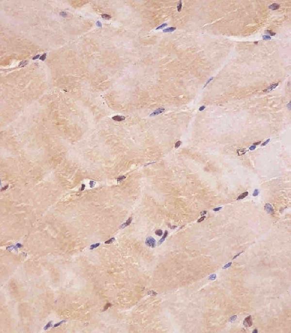
| Sample | Formaldehyde-fixed, paraffin-embedded human skeletal muscle tissue (datasheet M00728-3) |
| Fixation | Image formalin-fixed; duration unreported (datasheet M00728-3); verify before use. |
| Sectioning | 4–5 µm sections on charged slides (standard) |
| Deparaffinisation | Xylene, graded ethanol series to water (standard) |
| Antigen retrieval | Heat retrieval: Citrate pH 6 (datasheet M00728-3); 20 min, 95–100 °C (standard) |
| Peroxidase block | 3% H2O2, 10 min, room temperature (standard) |
| Blocking | 10% normal serum of the secondary host, 30 min, room temperature (standard) |
| Primary antibody | Mouse monoclonal (clone 1356CT119.18.55) anti-SMAD1, 1:25 (datasheet M00728-3) |
| Primary incubation | 1 hours at 37°C (datasheet M00728-3) |
| Detection | HRP-polymer secondary, DAB chromogen 5–10 min (standard) |
| Counterstain | Hematoxylin, blue, dehydrate and mount (standard) |
| Expected result | SMAD1-positive staining in glandular cells of epididymis (HPA tissue IHC: High). HPA tissue profile: Cytoplasmic and nuclear expression in most tissues. No signal in the no-primary control. |
SMAD1 is found in cytoplasm and nucleus; it can move into the nucleus during BMP signaling (UniProt Q15797). In tissue IHC, expect cytoplasmic and nuclear staining in most tissues, including high staining in epididymal and stomach glandular cells (HPA tissue IHC). HPA rates the tissue profile Approved but reports low consistency between antibody staining and RNA expression data (HPA tissue IHC). SMAD1 has no transmembrane segment (UniProt Q15797 topology).
| Cytoplasmic staining, with or without nuclear staining, in epididymal or stomach glandular cells. | This fits the reported high staining in those cells and the cytoplasmic and nuclear tissue profile (HPA tissue IHC). Nuclear enrichment is compatible with SMAD1 signaling, but total SMAD1 staining alone cannot establish BMP activation (UniProt Q15797 function and subcellular location). |
| Predominantly membrane outlining, with little cytoplasmic or nuclear staining. | A membrane-only pattern falls outside the reported cytoplasmic and nuclear locations (HPA tissue IHC; UniProt Q15797 subcellular location). SMAD1 has no transmembrane segment (UniProt Q15797 topology). Review morphology and detection controls before assigning the stain to SMAD1 (general IHC practice). |
| Strong staining confined to a cell population reported as not detected, such as adrenal glandular cells. | That conflicts with the reported absence of staining in those cells (HPA tissue IHC). Cross-reactivity or endogenous detection activity is possible; a discrepant stain alone does not establish either cause (general IHC practice). Compare cell identity and run appropriate controls. |
| Diffuse color across cells, stroma and empty areas, without clear cell boundaries. | A noncellular haze is hard to reconcile with the reported cellular pattern (HPA tissue IHC). Nonspecific reagent binding, endogenous detection activity or inadequate washing may contribute (general IHC practice). Judge the slide against a negative control and tissue morphology. |
| No discernible staining in epididymal or stomach glandular cells. | Those cells are reported as high staining references (HPA tissue IHC), so a blank result calls for a technical review. It does not prove SMAD1 is absent: the HPA profile is Approved with low antibody-staining versus RNA consistency (HPA tissue IHC). |
| Compartment and signaling state | SMAD1 is cytoplasmic without ligand and migrates to the nucleus in a SMAD4 complex (UniProt Q15797 subcellular location). A mixture of cytoplasmic and nuclear staining can therefore be plausible; total SMAD1 IHC does not by itself measure phosphorylation (UniProt Q15797 function). |
| Tissue and cell choice | Epididymal and stomach glandular cells are reported High; appendix and breast glandular cells are Medium (HPA tissue IHC). Adrenal glandular cells and liver cholangiocytes are reported Not detected (HPA tissue IHC). Score the specified cell population, since a tissue name alone does not identify its expected signal. |
| Evidence limits | The tissue profile is Approved, with low consistency between antibody staining and RNA expression data (HPA tissue IHC). CAB005389 is IHC Approved rather than IHC Enhanced in the supplied validation record (HPA antibodies). Treat a matching pattern as supportive, especially when interpreting a weak or unexpected stain. |
| Protein structure and variants | SMAD1 has two listed isoforms, no signal peptide or propeptide, and no transmembrane segment (UniProt Q15797 isoforms, processing and topology). The record gives no antibody epitope, so it cannot establish whether this antibody detects both isoforms or whether a particular domain changes staining. |
| IF/ICC comparison | HPA reports supported nucleoplasm and cytosol localization in ICC-IF and lists MCF-7 and U2OS image sets (HPA subcellular). CAB005389 has ICC Supported status (HPA antibodies). This agrees with the broad compartment expectation, but the cell images do not set intensity thresholds for paraffin tissue IHC. |
| Situation | Likely cause | Next action |
|---|---|---|
| Known high-reference glandular cells show no stain. | An unsuccessful IHC run or an unsuitable staining condition is possible; high staining is reported in epididymal and stomach glandular cells (HPA tissue IHC). | Confirm tissue identity and inspect the run control, then review the catalog antibody's IHC-P conditions, retrieval, dilution and detection steps (general IHC practice). Do not infer target-specific retrieval or fixation sensitivity from the HPA profile. |
| Stain is weak throughout a section. | This may reflect the staining workflow, or a sample dominated by cells with lower reported expression; endometrial glandular cells are Low (HPA tissue IHC). | Compare the specified cell population with a high-reference tissue, and check reagent performance and detection exposure using the same run (HPA tissue IHC; general IHC practice). Interpret weak signal cautiously given the reported low staining versus RNA consistency (HPA tissue IHC). |
| A reported negative cell type stains strongly. | Adrenal glandular cells, liver cholangiocytes and salivary glandular cells are reported Not detected (HPA tissue IHC). Misidentified cells, cross-reactivity or endogenous detection activity may explain a discrepancy (general IHC practice). | Recheck morphology and cell identity; compare negative-reagent and detection-system controls, then assess whether the signal reproduces with an independent approach (general IHC practice). |
| Color is diffuse across cells and background. | Nonspecific binding, endogenous detection activity or incomplete washing can obscure cell localization (general IHC practice). The expected tissue distribution is cellular and cytoplasmic or nuclear (HPA tissue IHC). | Inspect a negative control and blank areas; check blocking, washes and detection chemistry, then score only clearly localized cellular signal (general IHC practice). |
| Only cell borders or membranes are stained. | The location conflicts with the reported cytoplasmic and nuclear pattern and with SMAD1 lacking a transmembrane segment (HPA tissue IHC; UniProt Q15797 topology). | Check whether border color follows tissue edges or detection artifacts; review controls and cell morphology before treating the signal as SMAD1 (general IHC practice). |
| Nuclear staining varies between fields or specimens. | SMAD1 can move from cytoplasm to nucleus during BMP signaling (UniProt Q15797 function and subcellular location). Variation can also reflect sampling or staining differences (general IHC practice). | Score nuclear and cytoplasmic compartments separately in comparable cell types and review run controls; avoid calling pathway activation from total SMAD1 localization alone (UniProt Q15797 function; general IHC practice). |


Comprehensive Human Protein Atlas IHC scoring per tissue (reliability: Approved — Low consistency between antibody staining and RNA expression data.). Rows are taken directly from the HPA tissue chart — click any row's HPA link to view the source.
| Tissue | Cell type | Level | Evidence | Source |
|---|---|---|---|---|
| Epididymis | Glandular cells | High | Protein (IHC) | HPA → |
| Stomach | Glandular cells | High | Protein (IHC) | HPA → |
| Appendix | Glandular cells | Medium | Protein (IHC) | HPA → |
| Breast | Glandular cells | Medium | Protein (IHC) | HPA → |
| Bronchus | Respiratory epithelial cells | Medium | Protein (IHC) | HPA → |
| Tissue | Cell type | Level | Evidence | Source |
|---|---|---|---|---|
| Adrenal gland | Glandular cells | Not detected | Protein (IHC) | HPA → |
| Esophagus | Squamous epithelial cells | Not detected | Protein (IHC) | HPA → |
| Liver | Cholangiocytes | Not detected | Protein (IHC) | HPA → |
| Ovary | Ovarian stroma cells | Not detected | Protein (IHC) | HPA → |
| Parathyroid gland | Glandular cells | Not detected | Protein (IHC) | HPA → |
Troubleshoot SMAD1 staining in paraffin sections by checking retrieval, compartment-specific signal, controls and scoring before interpreting BMP activity.
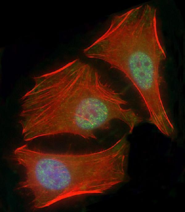
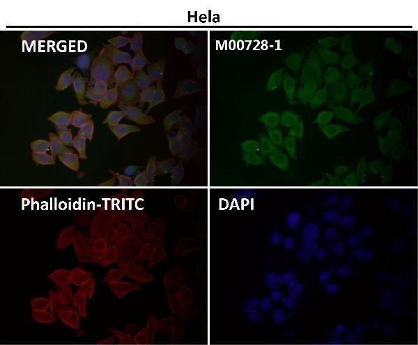
Anti-SMAD1 options include human paraffin-section IHC images (M00728, M00728-3, M00728-1 image captions) and human cell IF data (M00728-3 IF caption); M00728-1 also lists mouse ICC/IF reactivity (catalog).
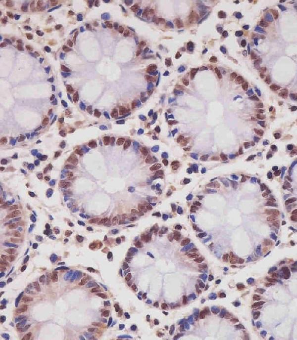
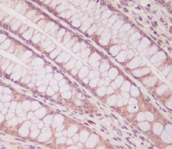
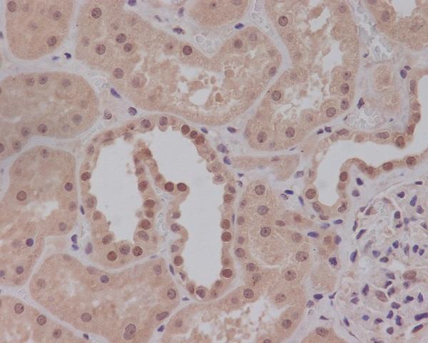
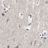
M00728-3 shows IHC staining of formaldehyde-fixed human skeletal muscle sections (IHC image caption); M00728 shows paraffin-embedded human colon IHC, with fixative unreported (IHC image caption). M00728-1 has a paraffin-embedded human kidney IHC image caption, with fixative unreported, while its listed applications are ICC/IF rather than IHC (IHC image caption; catalog applications).
Which to pick: For tissue IHC, choose mouse monoclonal M00728-3 when the documented formaldehyde-fixed paraffin-section workflow fits your sample (catalog; M00728-3 IHC image caption), or rabbit monoclonal M00728 for paraffin-embedded human colon; its caption does not report a fixative (catalog; M00728 IHC image caption). For IF/ICC, M00728-3 has a human HeLa IF image, while rabbit monoclonal M00728 lists both applications without an IF image in this payload (M00728-3 IF image caption; M00728 catalog). For human and mouse ICC/IF, choose rabbit monoclonal M00728-1 based on its listed reactivity and applications; its kidney IHC caption does not establish IHC as a listed application (M00728-1 catalog; M00728-1 IHC image caption).