This website uses cookies to ensure you get the best experience on our website.


- Table of Contents
Plan paraffin SMURF1 IHC around cytoplasmic and membranous staining in glandular cells (HPA tissue IHC). This guide covers tissue controls and the catalog antibody’s 0.5–1 μg/mL IHC range (datasheet PB9892).

Expected localisation, validated protocols, controls and antibodies — the at-a-glance facts below, then the full design guide.
| Expected localisation | Cytoplasmic and membranous tissue staining (HPA tissue IHC) | |
| Staining pattern | Glandular cells show cytoplasmic and membranous staining (HPA tissue IHC) | |
| Antigen retrieval | EDTA pH 8.0 HIER, heat-mediated (datasheet PB9892) | |
| Positive control | Breast+4 more · see all | |
| Negative control | Adipose tissue+4 more · see all |
| Fixation | Keep fixation consistent across sections (standard IHC practice; not target-specific) | |
| Caveat | Staining shows low consistency with RNA expression (HPA tissue IHC) | |
| Regulation | Expression regulation is not established (UniProt) | |
| Isoform / epitope | 2 isoforms, Long and Short; epitope coverage is unknown (UniProt) |
The catalog antibody uses heat-mediated EDTA retrieval at pH 8.0 (datasheet PB9892). Published SMURF1 IHC methods describe automated staining (PMC12941280) and an FFPE workflow (PMC5377408).
| Sample | Paraffin-embedded human intestinal cancer tissue; fixative not specified (datasheet PB9892) |
| Fixation | Image fixative and duration unreported (datasheet PB9892); verify before use. |
| Sectioning | 4–5 µm sections on charged slides (standard) |
| Deparaffinisation | Xylene, graded ethanol series to water (standard) |
| Antigen retrieval | Heat retrieval: EDTA pH 8.0 (datasheet PB9892); 20 min, 95–100 °C (standard) |
| Peroxidase block | 3% H2O2, 10 min, room temperature (standard) |
| Blocking | 10% goat serum (datasheet PB9892) |
| Primary antibody | Rabbit anti-SMURF1, 0.5-1μg/ml (datasheet PB9892) |
| Primary incubation | Overnight at 4 °C (datasheet PB9892) |
| Detection | Streptavidin-biotin complex (SABC), DAB chromogen (datasheet PB9892) |
| Counterstain | Hematoxylin, blue, dehydrate and mount (standard) |
| Expected result | SMURF1-positive staining in glandular cells of breast (HPA tissue IHC: High). HPA tissue profile: Cytoplasmic and membranous expression in several different tissue types. No signal in the no-primary control. |
SMURF1 is cytoplasmic and cell membrane associated, with no transmembrane segment (UniProt Q9HCE7 localization and topology). In paraffin IHC, expect cytoplasmic and membranous staining in several tissue types, including high staining in breast glandular cells, lung alveolar cells and bladder urothelial cells (HPA tissue IHC). HPA rates the tissue staining Approved but reports low consistency between antibody staining and RNA expression; interpret tissue comparisons cautiously (HPA tissue IHC).
| Cytoplasmic staining with some membrane accent in glandular cells. | This matches the reported pattern. Breast, cervix, fallopian tube, gallbladder and stomach glandular cells are each scored High (HPA tissue IHC). Compare compartments and cell types as well as intensity; HPA reports low consistency with RNA expression (HPA tissue IHC). |
| Predominantly nuclear staining, with little cytoplasmic or membranous signal. | This is discordant with the tissue IHC profile and warrants a specificity check (HPA tissue IHC; general IHC practice). Nucleoplasmic signal is listed as uncertain in ICC-IF, so an isolated nuclear signal should not establish the expected paraffin IHC pattern (HPA subcellular ICC-IF). |
| Strong color in cells outside the expected positive population. | First identify the stained cells: HPA scores alveolar cells High in lung and trophoblastic cells High in placenta (HPA tissue IHC). Unexpected staining may reflect cross-reactivity or endogenous detection activity; compare a no-primary control and the expected cell population (general IHC practice). |
| Diffuse color covers cells and extracellular spaces without clear boundaries. | An uninterpretable background can arise from nonspecific binding or detection activity (general IHC practice). It cannot confirm the cytoplasmic and membranous distribution or the cell-specific HPA pattern; assess a no-primary control before scoring cells (HPA tissue IHC; general IHC practice). |
| No signal in a section expected to contain high-staining cells. | Check that the relevant cells are present before calling the stain negative: breast glandular, lung alveolar and bladder urothelial cells are scored High (HPA tissue IHC). If they are present, review the staining run and a positive control (general IHC practice). |
| Compartment and topology | UniProt places SMURF1 in cytoplasm and at the cell membrane, despite no transmembrane segment (UniProt Q9HCE7 localization and topology). Membrane accent can therefore fit the record; a transmembrane staining requirement would overstate it (UniProt Q9HCE7 topology). |
| Choice of comparison cells | HPA scores breast and stomach glandular cells, lung alveolar cells, placental trophoblastic cells and bladder urothelial cells High (HPA tissue IHC). Adipocytes and skeletal muscle myocytes are scored Not detected; use them as pattern comparisons, not proof of absolute absence (HPA tissue IHC). |
| Tissue evidence and antibody status | The tissue profile is Approved, with low antibody-staining versus RNA consistency and unannotated endothelial staining (HPA tissue IHC). HPA055245 is IHC Approved; HPA019671 is ICC Approved with no IHC status listed (HPA antibodies). Do not transfer one antibody’s application status to the other. |
| Isoforms and target region | UniProt lists Long and Short isoforms and C2, two WW and HECT domains (UniProt Q9HCE7 isoforms and domains). No antibody epitope is supplied, so these features cannot predict which isoform or domain a particular stain detects. |
| IF/ICC comparison | Q: Should an ICC-IF image define the paraffin IHC result? A: No. Vesicles are the approved main ICC-IF location; nucleoplasm is uncertain (HPA subcellular ICC-IF). Use the cytoplasmic and membranous tissue profile to interpret paraffin IHC (HPA tissue IHC). |
| Situation | Likely cause | Next action |
|---|---|---|
| A high-scoring comparison tissue is blank. | The expected cells may be absent from the section, or the staining run may have failed (HPA tissue IHC; general IHC practice). | Verify the relevant cells on a counterstained section, then review the positive control and general retrieval, antibody and detection steps (general IHC practice). No SMURF1-specific retrieval condition is supplied. |
| Every compartment has weak, hazy color. | Nonspecific binding or excessive detection background can obscure the reported pattern (general IHC practice; HPA tissue IHC). | Compare a no-primary control, then adjust general blocking, washing or detection conditions as indicated by that control (general IHC practice). Score only interpretable cell-associated signal. |
| Color persists when the primary antibody is omitted. | Endogenous detection activity or reagent background may contribute to the signal (general IHC practice). | Check the detection-system controls and address the implicated activity using the system’s standard controls and blocking steps (general IHC practice). Do not count that color as SMURF1. |
| Strong nuclear staining dominates the section. | This differs from the cytoplasmic and membranous tissue profile; the additional nucleoplasmic ICC-IF location is uncertain (HPA tissue IHC; HPA subcellular ICC-IF). | Inspect no-primary and positive controls and confirm whether cytoplasmic or membrane signal is present in expected cells (general IHC practice; HPA tissue IHC). Avoid scoring nuclear color alone. |
| Endothelial staining is prominent. | HPA flags endothelial antibody staining as unannotated and reports low staining-versus-RNA consistency (HPA tissue IHC). | Record endothelial signal separately from the expected cell populations and compare controls and morphology (general IHC practice; HPA tissue IHC). Avoid treating it as validated SMURF1 localization. |
| An ICC-IF pattern appears different from paraffin IHC. | ICC-IF lists approved vesicles and uncertain nucleoplasm, while tissue IHC describes cytoplasmic and membranous expression (HPA subcellular ICC-IF; HPA tissue IHC). | Interpret each application against its own HPA profile and antibody status: HPA055245 is IHC Approved; HPA019671 is ICC Approved (HPA antibodies). Do not use the ICC-IF observation as an IHC protocol option. |




Comprehensive Human Protein Atlas IHC scoring per tissue (reliability: Approved — Low consistency between antibody staining and RNA expression data. Antibody staining in endothelial cells not annotated, view images.). Rows are taken directly from the HPA tissue chart — click any row's HPA link to view the source.
| Tissue | Cell type | Level | Evidence | Source |
|---|---|---|---|---|
| Breast | Glandular cells | High | Protein (IHC) | HPA → |
| Cervix | Glandular cells | High | Protein (IHC) | HPA → |
| Fallopian tube | Glandular cells | High | Protein (IHC) | HPA → |
| Gallbladder | Glandular cells | High | Protein (IHC) | HPA → |
| Lung | Alveolar cells | High | Protein (IHC) | HPA → |
| Tissue | Cell type | Level | Evidence | Source |
|---|---|---|---|---|
| Adipose tissue | Adipocytes | Not detected | Protein (IHC) | HPA → |
| Adrenal gland | Glandular cells | Not detected | Protein (IHC) | HPA → |
| Heart muscle | Cardiomyocytes | Not detected | Protein (IHC) | HPA → |
| Lymph node | Germinal center cells | Not detected | Protein (IHC) | HPA → |
| Oral mucosa | Squamous epithelial cells | Not detected | Protein (IHC) | HPA → |
SMURF1 IHC troubleshooting focuses on retrieval, compartment-aware scoring, and control tissues because reported staining is cytoplasmic and membranous (HPA tissue IHC).
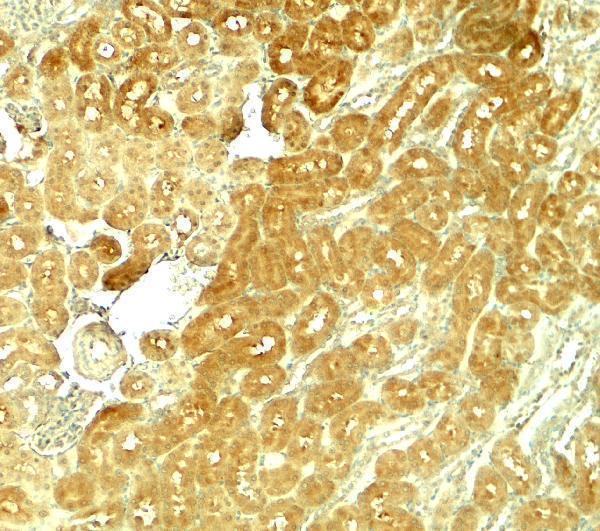
Two anti-SMURF1 antibodies have tissue IHC images: human paraffin sections and rat kidney tissue; one also has IF data in HeLa cells (catalog images: PB9892, A02823).
PB9892 shows IHC in paraffin sections of human intestinal and breast cancer tissue and IF in HeLa cells (PB9892 image captions). A02823 shows IHC in rat kidney tissue; its listed reactivity includes human, mouse and rat (A02823 image caption and catalog).
Which to pick: For human paraffin-section IHC, choose PB9892: its IHC image captions show that processing, while the fixative is unreported (PB9892 image captions). For IF/ICC, choose PB9892, which lists both applications and shows IF in HeLa cells (PB9892 catalog and IF image caption). For a mouse-reactive option, consider A02823, but its pictured IHC evidence is from rat kidney; clone type is unreported for both antibodies (A02823 catalog and image caption; PB9892 catalog).