This website uses cookies to ensure you get the best experience on our website.


- Table of Contents
Plan chromogenic SOAT1 IHC on paraffin sections using adrenal zona fasciculata cells as a high-staining reference (HPA tissue IHC). The guide covers fixation consistency, staining controls and interpretation of cytoplasmic signal (HPA tissue IHC).

Expected localisation, validated protocols, controls and antibodies — the at-a-glance facts below, then the full design guide.
| Expected localisation | Cytoplasmic staining in select cells (HPA tissue IHC) | |
| Staining pattern | Adrenal zona fasciculata and salivary gland cells: cytoplasmic (HPA tissue IHC) | |
| Antigen retrieval | EDTA pH 8.0 HIER, heat-mediated (datasheet A05096-1) | |
| Positive control | Adrenal gland+4 more · see all | |
| Negative control | Adipose tissue+4 more · see all |
| Fixation | Keep paraffin-section fixation consistent across samples. (standard IHC practice; not target-specific) | |
| Caveat | Staining varies by cell type within a tissue (HPA tissue IHC) | |
| Regulation | No staining-linked regulator annotated (UniProt) | |
| Isoform / epitope | Three isoforms; confirm epitope coverage (UniProt) |
The catalog antibody’s IHC-P protocol (datasheet: A05096-1) is followed by four published SOAT1 IHC protocols (PMC8152151; PMC7006231; PMC5811932; PMC9385470).
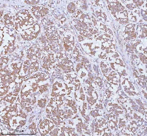
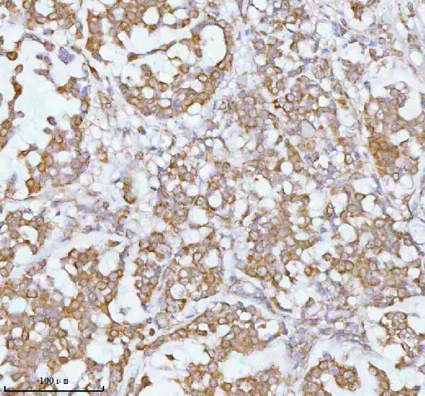
| Sample | Paraffin-embedded human breast cancer tissue; fixative not specified (datasheet A05096-1) |
| Fixation | Image fixative and duration unreported (datasheet A05096-1); verify before use. |
| Sectioning | 4–5 µm sections on charged slides (standard) |
| Deparaffinisation | Xylene, graded ethanol series to water (standard) |
| Antigen retrieval | Heat retrieval: EDTA pH 8.0 (datasheet A05096-1); 20 min, 95–100 °C (standard) |
| Peroxidase block | 3% H2O2, 10 min, room temperature (standard) |
| Blocking | 10% goat serum (datasheet A05096-1) |
| Primary antibody | Rabbit anti-SOAT1, 2-5 μg/ml (datasheet A05096-1) |
| Primary incubation | Overnight at 4 °C (datasheet A05096-1) |
| Detection | HRP-conjugated secondary, DAB chromogen (datasheet A05096-1) |
| Counterstain | Hematoxylin, blue, dehydrate and mount (standard) |
| Expected result | SOAT1-positive staining in cells in zona fasciculata of adrenal gland (HPA tissue IHC: High). HPA tissue profile: Cytoplasmic expression in a subset of tissues including adrenal gland, salivary gland, pancreatic ducts, Leydig cells and decidual cells. No signal in the no-primary control. |
SOAT1 is an endoplasmic reticulum membrane protein with 9 transmembrane segments (UniProt P35610 topology). In paraffin-section IHC, expect cytoplasmic staining in selected cells, especially adrenal zona fasciculata cells, salivary glandular cells, sebaceous glands, decidual cells and intestinal endocrine cells (HPA tissue IHC). HPA rates its tissue profile Enhanced, citing high consistency between staining and RNA expression (HPA tissue IHC).
| Cytoplasmic stain in adrenal zona fasciculata cells, with nearby cells distinguishable by morphology (HPA: High in zona fasciculata). | This fits the reported high-expression positive control and the expected intracellular compartment (HPA tissue IHC; UniProt P35610 subcellular location). Judge staining within identified cells rather than treating the whole section as uniformly positive (general IHC practice). |
| Predominantly nuclear, surface-restricted or extracellular signal in cells scored as positive (UniProt P35610 subcellular location). | Those compartments conflict with the annotated endoplasmic reticulum location and HPA cytoplasmic tissue profile (UniProt P35610; HPA tissue IHC). Check morphology, counterstain and antibody-dependent background before assigning SOAT1 positivity (general IHC practice). |
| Strong stain in adipocytes while the intended positive cells are absent or weak (HPA: adipocytes Not detected). | The adipocyte result conflicts with HPA tissue IHC and raises concern for nonspecific binding or detection-related signal (HPA tissue IHC; general IHC practice). A single discordant cell type does not identify which mechanism is responsible; inspect controls (general IHC practice). |
| Color spreads across stroma, lumina or most cells without clear cellular boundaries (general IHC practice). | A diffuse field cannot be scored confidently as the selective cytoplasmic pattern reported by HPA (HPA tissue IHC). Review the no-primary control, blocking, washes and chromogen development for background (general IHC practice). |
| No stain in adrenal zona fasciculata cells on a section with preserved morphology (HPA: High in zona fasciculata). | A negative known-positive control makes a biological absence call unreliable (HPA tissue IHC; general IHC practice). Check the antibody's IHC-P validation, reagent sequence, retrieval conditions and detection controls before scoring the sample (general IHC practice). |
| Cell and tissue choice (HPA tissue IHC) | High staining is reported in adrenal zona fasciculata, duodenal and small-intestinal endocrine cells, decidual cells, salivary glandular cells and sebaceous glands; adipocytes are reported Not detected (HPA tissue IHC). Compare like cell types when selecting controls (general IHC practice). |
| Compartment and epitope position (UniProt P35610 topology) | SOAT1 has 9 membrane-spanning segments and both cytoplasmic and lumenal regions (UniProt P35610 topology). The supplied record does not locate the antibody epitope; topology alone cannot predict how a given IHC reagent will stain after retrieval (UniProt P35610 topology; general IHC practice). |
| Antibody validation (HPA antibodies) | HPA lists 2 rabbit polyclonal antibodies, HPA047171 and CAB009533, with Enhanced IHC validation (HPA antibodies). That supports the reported tissue pattern, but agreement still requires matching the stained cell type and compartment in the section being assessed (HPA tissue IHC; general IHC practice). |
| Isoforms and processing (UniProt P35610) | UniProt lists 3 isoforms and a chain spanning residues 1–550, with no annotated signal peptide or propeptide (UniProt P35610). The supplied evidence gives no isoform-specific antibody epitope, so an IHC intensity difference cannot be assigned to a particular isoform (UniProt P35610; general IHC practice). |
| Chromogenic detection background (general IHC practice) | Endogenous enzyme activity can add color when the detection system uses that enzyme (general IHC practice). A no-primary control and the appropriate enzyme-blocking step help distinguish detection signal from antibody-dependent staining (general IHC practice); neither establishes SOAT1-specific reactivity by itself (general IHC practice). |
| Situation | Likely cause | Next action |
|---|---|---|
| High background obscures cell boundaries (general IHC practice). | Incomplete blocking or washing, excessive antibody concentration, or overdevelopment can obscure selective staining (general IHC practice). | Inspect the no-primary control; adjust blocking, washes, antibody concentration and development within the chosen detection workflow (general IHC practice). |
| Adrenal zona fasciculata is blank (HPA: High in zona fasciculata). | A failed staining run or an antibody/retrieval combination that performs poorly in this workflow is possible (general IHC practice). | Target-specific fixation effects are not established by the supplied assay evidence. Verify with a matched IHC source before attributing a result to fixation. |
| Adipocytes stain strongly (HPA: adipocytes Not detected). | The pattern is discordant with the HPA reference; nonspecific antibody or detection signal are possibilities (HPA tissue IHC; general IHC practice). | Compare with a no-primary control and a known-positive cell population; reassess cell identification and antibody dilution before interpreting the signal (general IHC practice). |
| Only nuclei appear positive (UniProt P35610 subcellular location). | Nuclear localization conflicts with the annotated endoplasmic reticulum and HPA cytoplasmic profile (UniProt P35610; HPA tissue IHC). | Recheck counterstain and brightfield color separation, then compare the same run's positive tissue and no-primary control (general IHC practice). |
| A weak section is being called SOAT1-negative (general IHC practice). | HPA reports medium staining in kidney distal tubules and fallopian-tube non-ciliated cells, so intensity varies by cell type (HPA tissue IHC). | Score the named cell population against an appropriate positive control and record intensity and distribution separately (general IHC practice); avoid applying the adrenal high-intensity expectation to every tissue (HPA tissue IHC). |
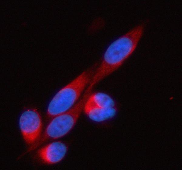
| IF/ICC: where should signal appear? (HPA subcellular ICC-IF). | HPA reports enhanced endoplasmic reticulum localization by ICC-IF, consistent with UniProt membrane annotation (HPA subcellular ICC-IF; UniProt P35610). | Expect intracellular ER-associated fluorescence rather than a nuclear-only pattern (HPA subcellular ICC-IF; UniProt P35610). Use the separate IF/ICC guide for assay setup (general IHC/IF practice). |


Comprehensive Human Protein Atlas IHC scoring per tissue (reliability: Enhanced — High consistency between antibody staining and RNA expression data.). Rows are taken directly from the HPA tissue chart — click any row's HPA link to view the source.
| Tissue | Cell type | Level | Evidence | Source |
|---|---|---|---|---|
| Adrenal gland | Cells in zona fasciculata | High | Protein (IHC) | HPA → |
| Duodenum | Endocrine cells | High | Protein (IHC) | HPA → |
| Placenta | Decidual cells | High | Protein (IHC) | HPA → |
| Salivary gland | Glandular cells | High | Protein (IHC) | HPA → |
| Skin | Sebaceous glands | High | Protein (IHC) | HPA → |
| Tissue | Cell type | Level | Evidence | Source |
|---|---|---|---|---|
| Adipose tissue | Adipocytes | Not detected | Protein (IHC) | HPA → |
| Bone marrow | Hematopoietic cells | Not detected | Protein (IHC) | HPA → |
| Breast | Adipocytes | Not detected | Protein (IHC) | HPA → |
| Caudate | Glial cells | Not detected | Protein (IHC) | HPA → |
| Cerebellum | Cells in granular layer | Not detected | Protein (IHC) | HPA → |
Troubleshoot SOAT1 staining in paraffin-section IHC by checking retrieval, cellular context and controls before comparing staining intensity across samples.
A05096-1 has real IHC data from human paraffin-embedded breast and prostatic cancer sections and IF data from PC-3 cells (catalog image captions). Its listed reactivity is Human, Mouse and Rat (catalog applications/reactivity).
The rendered card is A05096-1, with IHC images from human paraffin-embedded breast and prostatic cancer sections (catalog IHC captions). Its IF image shows PC-3 cells (catalog IF caption).
Which to pick: Choose A05096-1 for paraffin-section IHC: its caption documents EDTA retrieval at pH 8.0 and 2 μg/ml primary antibody, but does not report the fixative (A05096-1 IHC caption). For IF/ICC, A05096-1 has a PC-3 cell image at 5 μg/ml; A05096 lists IF at 1:50–1:200 but has no supplied IF image (catalog IF captions/dilutions). Both list Human, Mouse and Rat reactivity, while A05096-1 lists paraffin IHC for Human and Rat and IF/ICC for Human; verify performance in other species and applications before relying on it (catalog applications/reactivity).