SPG11 / Spatacsin · IHC design guide

Design Immunohistochemistry for SPG11

Start paraffin IHC with the catalog antibody at 2.5 μg/mL; its reported IHC validation used mouse brain (datasheet). Assess cytoplasmic staining in neuronal and glandular cells, using adipocytes as a low-signal comparator (HPA tissue IHC).

Evidence assembled Oct 2026 · For research use; verify linked source records and product datasheet before use
Immunohistochemistry protocol sheet for SPG11 (IHC for SPG11): expected localisation General cytoplasmic staining (HPA tissue IHC), antibody A03005, validated IHC image, and IHC protocol steps
Printable SPG11 IHC protocol sheet — expected localisation General cytoplasmic staining (HPA tissue IHC), antibody A03005, controls and protocol steps. Open the full SPG11 IHC guide →

SPG11 Immunohistochemistry Experimental Design Guide

Expected localisation, validated protocols, controls and antibodies — the at-a-glance facts below, then the full design guide.

Must know before staining
Expected localisation General cytoplasmic staining (HPA tissue IHC)
Staining pattern Cytoplasmic staining in glandular and neuronal cells (HPA tissue IHC)
Antigen retrieval Tris-EDTA pH 9.0 HIER, 95–98 °C, 20 min (rule: nuclear antigen)
Positive control ⓘ Adrenal gland+4 more · see all
Negative control ⓘ Adipose tissue
Important caveats
Reasons your staining may differ from the expected pattern.
Fixation Matched tissue-IHC evidence does not establish the fixation claim. Validate the specimen-specific method before use. (selected-SKU IHC image A03005)
Caveat Cerebellar IHC is low despite reported high expression (HPA tissue IHC; UniProt)
Regulation No specific staining regulator reported (UniProt)
Isoform / epitope 3 isoforms; antibody epitope coverage is unknown (UniProt; datasheet)
Section 1

Recommended SPG11 IHC & IF Protocols

The catalog antibody’s IHC-P protocol is accompanied by published SPG11 immunohistochemistry conditions for brain and spinal cord sections (PMC12005084; PMC5839621).

Recommended immunohistochemistry (IHC-P) protocol parameters
SampleTissue sections; selected-image fixative not specified (standard IHC workflow)
FixationImage fixative and duration unreported (datasheet A03005); verify before use.
Sectioning4–5 µm sections on charged slides (standard)
DeparaffinisationXylene, graded ethanol series to water (standard)
Antigen retrievalHeat-induced epitope retrieval in Tris-EDTA buffer, pH 9.0, 20 min at 95–98 °C (standard rule: nuclear antigen)
Peroxidase block3% H2O2, 10 min, room temperature (standard)
Blocking10% normal serum of the secondary host, 30 min, room temperature (standard)
Primary antibodyRabbit anti-SPG11, 2.5 μg/mL (datasheet A03005)
Primary incubationOvernight at 4 °C (standard)
DetectionHRP-polymer secondary, DAB chromogen 5–10 min (standard)
CounterstainHematoxylin, blue, dehydrate and mount (standard)
Expected resultSPG11-positive staining in glandular cells of adrenal gland (HPA tissue IHC: Medium). HPA tissue profile: General cytoplasmic expression. No signal in the no-primary control.
💡Decision noteStart with Tris-EDTA pH 9.0 retrieval at 95–98 °C for 20 min (page retrieval). Interpret staining cautiously: one study suspected off-target signal (PMC12005084).
Section 2

What Is the Expected SPG11 Staining Pattern?

SPG11 (spatacsin) is mainly cytoplasmic, with reported nuclear and neuronal projection localisation and no transmembrane segment (UniProt Q96JI7). In paraffin-section IHC, expect predominantly cytoplasmic staining in neuronal cells of the cerebral cortex or caudate and in several glandular epithelia (HPA: medium staining). HPA describes a general cytoplasmic pattern, but rates its tissue IHC evidence Approved, with medium consistency against RNA data and external verification pending (HPA: tissue IHC).

What am I looking at on my slide?
Cytoplasmic staining in cortical or caudate neuronal cells, with identifiable cell outlines and restrained background.This fits the expected IHC distribution (HPA: medium neuronal staining; general cytoplasmic expression). Neuronal projections may also stain (UniProt Q96JI7: axon and dendrite localisation). Judge the cell pattern alongside the negative control rather than intensity alone (general IHC practice).
Signal is predominantly nuclear, extracellular, or confined to a sharply defined compartment without convincing cytoplasmic staining.Treat this as discordant with the main tissue pattern (HPA: general cytoplasmic expression; UniProt Q96JI7: mainly cytoplasmic). Some nuclear localisation is reported by UniProt, so nuclear signal alone does not prove an artefact. Review controls and morphology before assigning specificity (general IHC practice).
Strong staining appears in adipocytes or another cell population outside the expected pattern.Adipocytes were not detected in the supplied HPA tissue IHC record (HPA: adipose tissue, adipocytes). Consider cross-reactivity or endogenous detection activity, especially if the negative control also stains (general IHC practice). One discrepant tissue does not establish the underlying mechanism.
Brown signal spreads across stroma, empty spaces, and multiple cell types without clear cellular boundaries.A diffuse field is difficult to interpret against the reported cytoplasmic cell pattern (HPA: tissue IHC). Check the negative control for background and inspect whether pigment or precipitate follows tissue structure (general chromogenic IHC practice). Do not score diffuse colour as SPG11 positive solely because it is present.
No convincing staining appears in a control section containing cortical or caudate neuronal cells.These neuronal populations show medium staining in the supplied HPA observations (HPA: cerebral cortex and caudate). First assess tissue preservation, reagent performance, and the chosen IHC workflow (general IHC practice). A negative result alone cannot distinguish absent antigen from assay failure.
💡Expected SPG11 appearanceCall a positive result when neuronal or glandular cells show interpretable, chiefly cytoplasmic staining around medium intensity (HPA: tissue IHC); isolated adipocyte staining or diffuse colour outside cells warrants control review (HPA: adipocytes not detected; general IHC practice).
How each factor affects the staining
Tissue and cell selectionCortex and caudate neuronal cells, plus listed glandular epithelia, showed medium IHC staining (HPA: tissue IHC). Adipocytes were not detected (HPA: adipose tissue); use this comparison cautiously because HPA rates the tissue pattern Approved with medium RNA concordance.
Compartment and topologySPG11 is mainly cytoplasmic and has no annotated transmembrane segment (UniProt Q96JI7). This supports cytoplasmic IHC interpretation; the supported plasma-membrane signal comes from ICC-IF images and should not be imposed as a required paraffin-section pattern (HPA: subcellular ICC-IF).
Alternative isoforms and processingUniProt lists 3 isoforms and a single annotated spatacsin chain spanning residues 1–2443, with no signal peptide or propeptide (UniProt Q96JI7). The supplied evidence does not map either IHC antibody's epitope, so it cannot establish which isoforms an antibody detects.
Phosphorylation and retrievalA phosphoserine at residue 1955 is annotated (UniProt Q96JI7), but no evidence here links that modification to staining. Antigen retrieval is a standard paraffin IHC variable (general IHC practice); no SPG11-specific retrieval condition or fixation sensitivity is established by the supplied sources.
Antibody evidenceBoth listed antibodies have Approved IHC status; one also has Supported ICC status (HPA: HPA040412 and HPA040947). These classifications guide confidence in an observed pattern but do not prove that every stain in every tissue is specific (HPA: tissue IHC reliability description).
Why is my staining missing, weak or wrong?
SituationLikely causeNext action
Expected neuronal control is blank.The section or detection run may have failed; the supplied sources do not establish a SPG11-specific fixation or retrieval failure mode.Check tissue morphology and assay controls, then review antibody dilution, retrieval, and detection against the validated IHC workflow (general IHC practice). Use a listed HPA medium-staining tissue for comparison (HPA: cortex or caudate).
Adipocytes stain strongly.This conflicts with the supplied adipocyte observation (HPA: not detected) and may reflect background, cross-reactivity, or endogenous detection activity (general IHC practice).Compare with a negative control on the same tissue, inspect cell identity, and reassess the blocking and detection steps (general IHC practice). Avoid calling SPG11 positivity from adipocyte colour alone.
Signal is nuclear or membrane dominant.The main tissue pattern is cytoplasmic (HPA: tissue IHC). UniProt also reports nuclear localisation, while HPA supports plasma-membrane localisation in ICC-IF; neither makes an exclusively nuclear or membrane IHC pattern diagnostic.Check whether cytoplasmic staining accompanies the signal, inspect controls, and report the observed compartment explicitly (general IHC practice). Treat isolated compartment staining as uncertain.
The entire section has diffuse brown background.Diffuse colour can arise from nonspecific binding, endogenous detection activity, or chromogen deposits (general chromogenic IHC practice); it obscures the reported cellular pattern (HPA: general cytoplasmic expression).Compare a negative control, examine clean tissue areas, and review blocking, washes, and chromogen development (general IHC practice). Score only interpretable cellular staining.
Cerebellum is weaker than cortex despite UniProt's brain-expression statement.UniProt reports high cerebellar expression, while the supplied HPA IHC observation is low in granular-layer cells and medium in cortical neuronal cells (UniProt Q96JI7; HPA: tissue IHC). RNA or expression summaries need not predict identical IHC intensity.Record the tissue and cell layer separately. Use the observed HPA IHC levels as the staining comparison, with its Approved, medium-consistency caveat (HPA: tissue IHC).
Q: How should an ICC-IF membrane or nucleolar signal affect this IHC call?HPA supports cytosol and plasma membrane as main ICC-IF locations and nucleoli as an additional location (HPA: subcellular ICC-IF). These are observations from a different application.A: Keep the paraffin-section decision anchored to the chiefly cytoplasmic tissue pattern (HPA: tissue IHC). Evaluate ICC-IF with its own controls and guide; it does not provide an IHC protocol option here.

Sample controls for SPG11 IHC & IF

🧪Run caudate first and assess staining in neuronal cells (HPA: Medium in caudate neuronal cells). Run adipose tissue as the negative tissue (HPA: Not detected in adipocytes); on the caudate slide, cells without staining should show only background chromogen, but their target-negative status must be validated before treating them as internal negatives (standard IHC practice).
Positive control tissue: Adrenal gland (Glandular cells, HPA Medium)
Negative control tissue: Adipose tissue (HPA Not detected)
ICC-IF cell lines (HPA subcellular resource): HPA ICC-IF images show SPG11 in A-431, U-251MG, U2OS, with annotated localisation: Plasma membrane (supported), Cytosol (supported) (HPA subcellular).
Technical controls: Include a no-primary, secondary-only control; an irrelevant immunoglobulin matched to the primary antibody’s host species, class, and clonality; and SPG11 knockout tissue, if available, as a biological negative (standard IHC practice). Check for endogenous peroxidase signal in chromogenic IHC and autofluorescence if assessing brain tissue by IF (standard IHC/IF practice).
⚠️Feasibility: A target-specific fixation window or fixation effect is unreported, and the selected A03005 mouse-brain IHC caption does not state a fixative (selected A03005 tissue-IHC caption). Whether SPG11 staining depends on antigen retrieval is unreported; optimize retrieval on paraffin sections with the controls above (standard IHC practice). The supplied evidence does not establish that frozen sections or IF are easier; brain autofluorescence can complicate IF interpretation (standard IF practice).

HPA tissue IHC evidence for SPG11

Comprehensive Human Protein Atlas IHC scoring per tissue (reliability: Approved — Medium consistency between antibody staining and RNA expression data. Pending external verification.). Rows are taken directly from the HPA tissue chart — click any row's HPA link to view the source.

Positive expression · recommended positive controls

TissueCell typeLevelEvidenceSource
Adrenal gland Glandular cells Medium Protein (IHC) HPA →
Appendix Glandular cells Medium Protein (IHC) HPA →
Breast Glandular cells Medium Protein (IHC) HPA →
Bronchus Respiratory epithelial cells Medium Protein (IHC) HPA →
Caudate Neuronal cells Medium Protein (IHC) HPA →

Undetected expression · recommended negative controls

TissueCell typeLevelEvidenceSource
Adipose tissue Adipocytes Not detected Protein (IHC) HPA →
Section 3

Advanced SPG11 IHC Tips

Troubleshoot SPG11 staining in paraffin sections by checking retrieval, controls, cellular localisation and scoring before interpreting a chromogenic signal.

What retrieval conditions should I start with for SPG11 in paraffin sections?
Start with Tris-EDTA at pH 9.0 for heat-induced epitope retrieval at 95–98 °C for 20 min (page retrieval setting). Allow sections to cool in the retrieval solution, then keep washing and detection conditions identical across the test and control sections (standard IHC practice). If staining is weak, compare a milder retrieval condition on adjacent sections while holding antibody concentration and development time constant; excessive heating can damage tissue morphology (standard IHC practice). Judge the result against neuronal staining in cerebral cortex or caudate, where medium staining is reported, while recognizing that the HPA tissue profile has medium consistency with RNA data (HPA: cerebral cortex and caudate neuronal cells; HPA: Approved, pending external verification).
How should I troubleshoot fixation when SPG11 staining is weak?
Target-specific fixation sensitivity is unknown from the supplied evidence, and the selected A03005 mouse brain caption does not state a fixative (selected A03005 tissue-IHC caption). Record the actual fixative, fixation interval, tissue thickness and processing history for each block before changing retrieval or antibody concentration (standard IHC practice). Compare similarly processed sections in one staining run, using a consistent control section to detect batch effects (standard IHC practice). If an archived block stains poorly, test a freshly processed section under the same retrieval and detection conditions before attributing the difference to fixation; do not infer a SPG11-specific fixation effect from tissue staining patterns or protein annotations (standard IHC practice).
Which cellular pattern is credible for SPG11 chromogenic staining?
Look first for predominantly cytoplasmic staining: the tissue-IHC profile describes general cytoplasmic expression, and UniProt calls SPG11 mainly cytoplasmic (HPA: tissue IHC; UniProt Q96JI7: subcellular location). Cytosolic staining is also consistent with supported cell-imaging localisation, although that evidence additionally reports plasma membrane and nucleolar signal (HPA: subcellular localisation). Score nuclear-only or sharply membranous staining separately from the expected cytoplasmic pattern rather than automatically calling it negative or specific (standard IHC interpretation; HPA: subcellular localisation). In brain sections, compare identified neuronal cells with surrounding cells and tissue structure; cortical projection-neuron expression is reported, while the selected A03005 caption documents mouse brain staining without naming a cell type (UniProt Q96JI7: tissue specificity; selected A03005 tissue-IHC caption).
Could isoform choice or epitope masking explain discordant SPG11 staining?
SPG11 has 3 annotated isoforms, but the supplied evidence does not map the catalog antibody’s epitope to any isoform (UniProt Q96JI7: isoforms; supplied antibody evidence). Check the antibody’s documented immunogen or epitope against the isoform sequences before treating a negative section as evidence that all SPG11 forms are absent (standard antibody-validation practice; UniProt Q96JI7: isoforms). The protein has no annotated transmembrane segment or glycosylation sites, and a phosphoserine is annotated at residue 1955; none establishes what this antibody detects after processing (UniProt Q96JI7: topology, glycosylation and modified residues). If an independent antibody with a documented different epitope is available, compare patterns on adjacent sections using matched retrieval and scoring (standard IHC validation practice).
How can IF help assess a disputed SPG11 IHC pattern?
Use IF as a separate localisation check, multiplexing SPG11 with a validated neuronal marker when examining the reported cortical projection-neuron population (UniProt Q96JI7: tissue specificity; standard IF practice). Choose spectrally separated fluorophores after checking tissue autofluorescence in an unstained control, and include single-colour controls to assess bleed-through (standard IF practice). SPG11 has no annotated transmembrane segment and is mainly cytoplasmic, so use a mild permeabilisation condition when testing access to an intracellular epitope; the supplied evidence does not identify this antibody’s epitope (UniProt Q96JI7: topology and subcellular location; standard IF practice; supplied antibody evidence). Compare cytosolic and any nucleolar signal with the reported cell-imaging pattern, without assuming that an IF result validates chromogenic staining in paraffin sections (HPA: subcellular localisation; standard assay-validation practice).
What should I change when SPG11 staining is diffuse or dirty?
Run a section without primary antibody to assess detection-system background, and inspect tissue edges and damaged regions before interpreting diffuse brown signal (standard chromogenic IHC practice). Include an appropriate peroxidase block before DAB development and use a protein block plus thorough washes to reduce nonspecific deposition (standard chromogenic IHC practice). If background persists, titrate the primary antibody and shorten DAB development while keeping retrieval and section thickness constant across comparisons (standard IHC practice). The selected A03005 mouse brain image caption reports 2.5 µg/mL antibody, but does not establish that concentration as optimal for every paraffin specimen or detection system (selected A03005 tissue-IHC caption; standard IHC optimisation practice).
How should I quantify SPG11 staining across paraffin sections? ⚠ ANSWER MARKED FOR VERIFICATION
Define the cell population and region before scoring, then record cytoplasmic intensity and the percentage of positive cells using an H-score or a stated percentage-positive threshold (standard IHC scoring practice; HPA: general cytoplasmic expression). For spatial comparisons, positive-cell density per mm² is useful when the sampled areas differ; report the analysed area and number of evaluable cells (standard image-analysis practice). Normalise comparisons to the same cell type or compartment, section area, staining run and background threshold, and exclude folds, edges and necrotic regions by a prespecified rule (standard IHC quantification practice). Report neuronal and non-neuronal compartments separately in brain analyses because neuronal staining is reported in cerebral cortex and caudate (HPA: cerebral cortex and caudate neuronal cells; standard IHC quantification practice).
When is a brown SPG11 signal convincing rather than artefactual?
A convincing pattern should be reproducible in intact cells, predominantly cytoplasmic, and compatible with the sampled cell population (HPA: general cytoplasmic expression; UniProt Q96JI7: mainly cytoplasmic; standard IHC interpretation). In cerebral cortex or caudate, medium neuronal staining has been reported, but the HPA tissue-IHC reliability remains Approved with medium consistency and pending external verification (HPA: tissue IHC). Treat signal restricted to section edges, necrotic areas or the no-primary control as suspect, and check the peroxidase block when DAB appears independent of primary antibody (standard chromogenic IHC practice). Investigate an exclusively nuclear or unrelated-cell pattern with adjacent sections and an independent reagent before making a biological claim; nucleolar localisation is reported in cell imaging but does not by itself establish a tissue-IHC pattern (HPA: subcellular localisation; standard IHC validation practice).
Boster reagents

Best SPG11 / Spatacsin IHC Antibodies

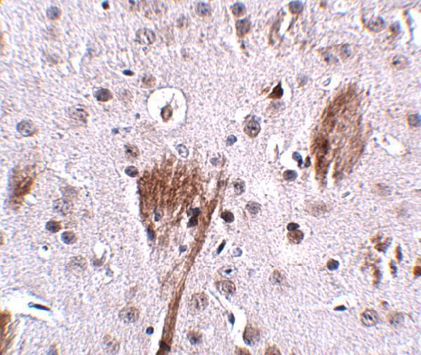
A03005 has real IHC data from mouse brain at 2.5 μg/mL (A03005 image caption). No IF data are supplied (catalog: IF images).

Real IHC data Immunohistochemistry of SPG11 in mouse brain tissue with SPG11 antibody at 2.5 μg/mL.
Anti-Spatacsin SPG11 Antibody
Cat # A03005

A03005 is listed for IHC-P and human, mouse, and rat reactivity (catalog: applications and reactivity). Its IHC figure shows mouse brain at 2.5 μg/mL (A03005 image caption); no IF figure is supplied (catalog: IF images).

Which to pick: For paraffin-section tissue IHC, choose A03005: it is a rabbit antibody listed for IHC-P, with mouse brain staining shown at 2.5 μg/mL; the fixative is unreported (catalog: host and applications; A03005 image caption). No IF/ICC option is supported because A03005 has no IF/ICC application or IF figure (catalog: applications and IF images). For human or rat IHC, A03005 lists reactivity with both species, but its IHC figure documents mouse tissue only (catalog: reactivity; A03005 image caption).

Each figure is that product's own IHC / IF validation image from its datasheet.

References

  1. UniProt Consortium. UniProt entry Q96JI7 (SPTCS_HUMAN, Spatacsin).
  2. Human Protein Atlas. SPG11 tissue IHC expression (reliability: Approved).
  3. Human Protein Atlas. SPG11 subcellular location (ICC-IF): Mainly localized to the plasma membrane and cytosol. In addition localized to the nucleoli..
  4. Human Protein Atlas. SPG11 antibody validation summary (2 antibodies).
  5. Hereditary spastic paraplegia with thin corpus callosum and SPG11 mutation: A neuropathological evaluation. Neuropathology : official journal of the Japanese Society of Neuropathology 2025 — PMC12005084.
  6. Inhibition of Lysosome Membrane Recycling Causes Accumulation of Gangliosides that Contribute to Neurodegeneration. Cell reports 2018 — PMC6045775.
  7. A role for mutations in AK9 and other genes affecting ependymal cells in idiopathic normal pressure hydrocephalus. Proceedings of the National Academy of Sciences of the United States of America 2023 — PMC10743366.
  8. Motor neuron degeneration in spastic paraplegia 11 mimics amyotrophic lateral sclerosis lesions. Brain : a journal of neurology 2016 — PMC5839621.
  9. PubMed PMID:11347906 — UniProt-cited evidence.
  10. PubMed PMID:12168954 — UniProt-cited evidence.
  11. PubMed PMID:16572171 — UniProt-cited evidence.