This website uses cookies to ensure you get the best experience on our website.


- Table of Contents
Plan SPIB chromogenic IHC on paraffin sections with the catalog antibody, starting at 2.5 μg/mL (datasheet). Compare the observed cytoplasmic tissue staining (HPA tissue IHC) with SPIB’s annotated nuclear and cytoplasmic locations (UniProt), while accounting for low staining–RNA consistency (HPA tissue IHC).

Expected localisation, validated protocols, controls and antibodies — the at-a-glance facts below, then the full design guide.
| Expected localisation | Cytoplasmic staining in several tissues (HPA tissue IHC) | |
| Staining pattern | Cytoplasmic staining in immune and other tissue cells (HPA tissue IHC) | |
| Antigen retrieval | Tris-EDTA pH 9.0 HIER, 95–98 °C, 20 min (rule: nuclear antigen) | |
| Positive control | Spleen+4 more · see all | |
| Negative control | Esophagus+4 more · see all |
| Fixation | Keep fixation consistent across sections. (standard IHC practice; not target-specific) | |
| Caveat | Staining and RNA show low consistency (HPA tissue IHC) | |
| Regulation | Expressed in pDCs and B cells (UniProt) | |
| Isoform / epitope | 3 isoforms; epitope effects are unknown (UniProt) |
The catalog antibody’s IHC-P protocol accompanies three published SPIB IHC methods using lymphoma, colorectal, and gastric specimens (PMC5021043; PMC8176860; PMC13383869).
| Sample | Tissue sections; selected-image fixative not specified (standard IHC workflow) |
| Fixation | Image fixative and duration unreported (datasheet A02805-1); verify before use. |
| Sectioning | 4–5 µm sections on charged slides (standard) |
| Deparaffinisation | Xylene, graded ethanol series to water (standard) |
| Antigen retrieval | Heat-induced epitope retrieval in Tris-EDTA buffer, pH 9.0, 20 min at 95–98 °C (standard rule: nuclear antigen) |
| Peroxidase block | 3% H2O2, 10 min, room temperature (standard) |
| Blocking | 10% normal serum of the secondary host, 30 min, room temperature (standard) |
| Primary antibody | Rabbit anti-SPIB, 2.5 μg/mL (datasheet A02805-1) |
| Primary incubation | Overnight at 4 °C (standard) |
| Detection | HRP-polymer secondary, DAB chromogen 5–10 min (standard) |
| Counterstain | Hematoxylin, blue, dehydrate and mount (standard) |
| Expected result | SPIB-positive staining in cells in red pulp of spleen (HPA tissue IHC: High). HPA tissue profile: Cytoplasmic expression in several tissues, including immune cells. No signal in the no-primary control. |
SPIB is a nuclear and cytoplasmic transcription factor with no transmembrane segment (UniProt Q01892). Expression is reported in B cells and plasmacytoid dendritic cells, but not T cells or granulocytes (UniProt Q01892). HPA reports cytoplasmic staining across several tissues; its tissue IHC reliability is Uncertain because staining and RNA data agree poorly, and it flags antibody cross-target and transcript discrepancies (HPA: tissue IHC).
| Discrete nuclear staining in a subset of immune cells, with or without cytoplasmic signal. | This is compatible with SPIB localisation and its reported B cell and plasmacytoid dendritic cell expression (UniProt Q01892). Morphology alone cannot identify those subsets. Score nuclear and cytoplasmic staining separately, then confirm cell identity with appropriate markers (general IHC practice). HPA has no ICC-IF images establishing a preferred compartment (HPA: subcellular). |
| Predominantly cytoplasmic staining in spleen red pulp cells or tonsil squamous epithelial cells. | HPA reports High staining in both populations and describes a broader cytoplasmic tissue pattern (HPA: tissue IHC). Red pulp staining does not identify the positive cell subtype. Tonsil epithelial staining should be interpreted cautiously alongside UniProt’s immune cell expression profile and HPA’s Uncertain tissue IHC reliability (UniProt Q01892; HPA: tissue IHC). |
| Sharp membrane outlining or extracellular deposits presented as SPIB positive cells. | These are discordant with the reported nuclear and cytoplasmic localisation and lack of a transmembrane segment (UniProt Q01892). Review tissue morphology and detection controls before calling them specific staining (general IHC practice). Cytoplasmic signal by itself is not a wrong compartment: both UniProt and HPA allow it (UniProt Q01892; HPA: tissue IHC). |
| Strong staining assigned to T cells or granulocytes, or broad staining across unrelated cell types. | T cells and granulocytes are reported as SPIB negative (UniProt Q01892). Confirm the stained cell identities before inferring cross-reactivity or endogenous detection activity (general IHC practice). Broad positivity also warrants caution because HPA flags cross-target staining and poor agreement with RNA expression (HPA: tissue IHC). |
| Uniform haze, or no signal in a section containing an expected positive population. | Haze that obscures cell boundaries prevents compartment scoring (general IHC practice). An absent signal may reflect the sampled cells, detection performance or antibody performance; it does not establish biological absence (general IHC practice). Compare with spleen red pulp cells, which HPA scores High, while retaining HPA’s Uncertain reliability caveat (HPA: tissue IHC). |
| Compartment and tissue evidence | UniProt places SPIB in nucleus and cytoplasm; HPA describes cytoplasmic tissue staining (UniProt Q01892; HPA: tissue IHC). Record the compartments independently. HPA lists no ICC-IF image or main subcellular location, so tissue IHC observations cannot establish a validated IF distribution (HPA: subcellular). |
| Cell selection and comparator tissue | UniProt reports expression in B cells and plasmacytoid dendritic cells, and absence in T cells and granulocytes (UniProt Q01892). HPA scores spleen red pulp cells High and skeletal muscle myocytes Not detected (HPA: tissue IHC). Use cell identity and compartment when comparing sections; a tissue label alone is insufficient (general IHC practice). |
| Antibody and transcript uncertainty | The listed HPA antibody, HPA018523, has Uncertain IHC validation; HPA flags possible staining of protein from more than one gene and a splice or transcript discrepancy (HPA: antibodies; HPA: tissue IHC). UniProt lists three SPIB isoforms (UniProt Q01892). These records do not identify which isoforms an antibody detects or resolve discordant cell staining. |
| IF/ICC question: is the expected pattern validated in cells? | No ICC-IF images or main subcellular location are available in HPA’s SPIB subcellular record, and ICC validation is unlisted for HPA018523 (HPA: subcellular; HPA: antibodies). Nuclear and cytoplasmic localisation is the available biological expectation (UniProt Q01892). Assess IF/ICC methods in its own guide; this IHC section offers no IF protocol. |
| Situation | Likely cause | Next action |
|---|---|---|
| No staining in spleen red pulp cells. | Positive cells may be absent from the sampled field, or the IHC detection may have failed (general IHC practice). | Locate intact red pulp cells, inspect a matched positive control, and check the catalog antibody’s IHC-P protocol (general IHC practice); HPA rates these cells High but tissue IHC reliability Uncertain (HPA: tissue IHC). |
| Every cell shows diffuse brown haze. | Background may arise from nonspecific binding, endogenous enzyme activity or detection reagents (general IHC practice). | Compare a primary-antibody omission control; review blocking, washes and chromogen development using general IHC practice. Do not score haze as SPIB, even where HPA reports cytoplasmic tissue expression (HPA: tissue IHC). |
| Only membrane rims or extracellular material stains. | That distribution conflicts with SPIB’s reported nucleus/cytoplasm localisation and lack of a transmembrane segment (UniProt Q01892). | Check morphology and controls, then repeat with the validated IHC conditions if needed (general IHC practice). Require cellular nuclear or cytoplasmic signal before assigning SPIB localisation (UniProt Q01892). |
| Staining appears in cells identified as T cells or granulocytes. | The cell assignment may be wrong, or the signal may be nonspecific; UniProt reports neither population expresses SPIB (UniProt Q01892). | Verify cell identity with appropriate markers and compare antibody-omission controls (general IHC practice). Treat persistent signal cautiously because HPA flags possible cross-target staining (HPA: tissue IHC). |
| Tonsil epithelium is strongly positive while expected immune cells are weak. | HPA scores tonsil squamous epithelial cells High, but its tissue IHC reliability is Uncertain (HPA: tissue IHC). | Document each cell population and compartment separately; verify cell identity and compare an independent IHC-validated antibody where available (general IHC practice). Do not use epithelial positivity alone to infer SPIB specificity. |
| IF/ICC shows a different distribution from the IHC section. | HPA supplies no SPIB ICC-IF images or assigned main subcellular location for comparison (HPA: subcellular). | Interpret IF/ICC using its own guide and controls. Keep the IHC tissue observation distinct from UniProt’s nuclear and cytoplasmic localisation annotation (HPA: tissue IHC; UniProt Q01892). |
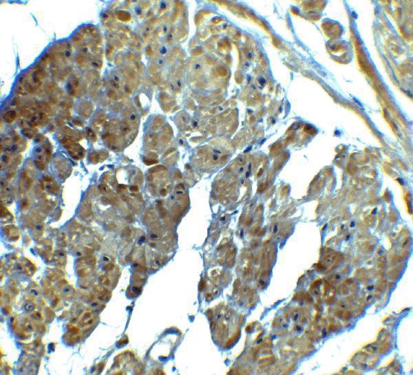


Comprehensive Human Protein Atlas IHC scoring per tissue (reliability: Uncertain — Low consistency between antibody staining and RNA expression data. Caution, targets protein from more than one gene. Caution, Splice and/or transcript discrepancy exists.). Rows are taken directly from the HPA tissue chart — click any row's HPA link to view the source.
| Tissue | Cell type | Level | Evidence | Source |
|---|---|---|---|---|
| Spleen | Cells in red pulp | High | Protein (IHC) | HPA → |
| Tonsil | Squamous epithelial cells | High | Protein (IHC) | HPA → |
| Adipose tissue | Adipocytes | Medium | Protein (IHC) | HPA → |
| Adrenal gland | Glandular cells | Medium | Protein (IHC) | HPA → |
| Appendix | Glandular cells | Medium | Protein (IHC) | HPA → |
| Tissue | Cell type | Level | Evidence | Source |
|---|---|---|---|---|
| Esophagus | Squamous epithelial cells | Not detected | Protein (IHC) | HPA → |
| Liver | Cholangiocytes | Not detected | Protein (IHC) | HPA → |
| Skeletal muscle | Myocytes | Not detected | Protein (IHC) | HPA → |
| Smooth muscle | Smooth muscle cells | Not detected | Protein (IHC) | HPA → |
| Vagina | Squamous epithelial cells | Not detected | Protein (IHC) | HPA → |
Troubleshoot SPIB staining by checking retrieval, compartment, cell identity and controls before comparing signal across sections.
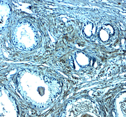
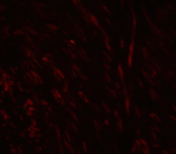
A02805-1 has IHC and IF figures from human prostate tissue (IHC/IF image captions). Its listed reactivity covers human, mouse, and rat (catalog reactivity).
A02805-1 will render with human prostate tissue IHC at 2.5 μg/mL (IHC image caption). Its IF figure shows human prostate tissue at 20 μg/mL (IF image caption).
Which to pick: Choose A02805-1 for paraffin-section tissue IHC (catalog applications: IHC-P); its own IHC caption shows human prostate tissue, but does not report the fixative (IHC image caption). For IF, A02805-1 has a human prostate tissue figure; ICC is not separately documented (IF image caption; catalog applications). For mouse or rat samples, A02805-1 lists reactivity with both species, while the supplied IHC and IF figures show human tissue (catalog reactivity; IHC/IF image captions).