SPIN1 / Spindlin-1 · IHC design guide

Design Immunohistochemistry for SPIN1

Plan chromogenic SPIN1 IHC in paraffin sections using ovarian stromal cells or epididymal glandular cells as positive references (HPA tissue IHC). Assess nuclear and cytoplasmic staining (HPA tissue IHC), and use a peptide-preabsorbed antibody control to check specificity (datasheet A07281).

Evidence assembled Oct 2026 · For research use; verify linked source records and product datasheet before use
Immunohistochemistry protocol sheet for SPIN1 (IHC for SPIN1): expected localisation Nuclear and cytoplasmic staining (HPA tissue IHC), antibody A07281, validated IHC image, and IHC protocol steps
Printable SPIN1 IHC protocol sheet — expected localisation Nuclear and cytoplasmic staining (HPA tissue IHC), antibody A07281, controls and protocol steps. Open the full SPIN1 IHC guide →

SPIN1 Immunohistochemistry Experimental Design Guide

Expected localisation, validated protocols, controls and antibodies — the at-a-glance facts below, then the full design guide.

Must know before staining
Expected localisation Nuclear and cytoplasmic staining (HPA tissue IHC)
Staining pattern Most tissues show nuclear and cytoplasmic staining (HPA tissue IHC)
Antigen retrieval Tris-EDTA pH 8.0 HIER, heat-mediated (datasheet A07281)
Positive control ⓘ Epididymis+4 more · see all
Negative control ⓘ Adrenal gland+3 more · see all
Important caveats
Reasons your staining may differ from the expected pattern.
Fixation Keep paraffin-section fixation consistent across samples. (standard IHC practice; not target-specific)
Caveat HPA antibody may recognize more than one gene (HPA tissue IHC)
Regulation High expression in ovarian cancer tissue (UniProt)
Isoform / epitope No annotated isoforms or cleavage; one 1–262 chain (UniProt)
Section 1

Recommended SPIN1 IHC & IF Protocols

The catalog antibody has a datasheet IHC-P protocol (datasheet A07281); four published SPIN1 IHC methods provide additional tissue protocols (PMC4467114; PMC8810024; PMC6171160; PMC11568276).

Recommended immunohistochemistry (IHC-P) protocol parameters
SampleParaffin-embedded Human breast cancer tissue; fixative not specified (datasheet A07281)
FixationImage fixative and duration unreported (datasheet A07281); verify before use.
Sectioning4–5 µm sections on charged slides (standard)
DeparaffinisationXylene, graded ethanol series to water (standard)
Antigen retrievalHeat retrieval: Tris-EDTA pH 8.0 (datasheet A07281); 20 min, 95–100 °C (standard)
Peroxidase block3% H2O2, 10 min, room temperature (standard)
Blocking10% normal serum of the secondary host, 30 min, room temperature (standard)
Primary antibodyRabbit anti-SPIN1, 1:100-1:300 (datasheet A07281)
Primary incubationOvernight at 4 °C (standard)
DetectionHRP-polymer secondary, DAB chromogen 5–10 min (standard)
CounterstainHematoxylin, blue, dehydrate and mount (standard)
Expected resultSPIN1-positive staining in glandular cells of epididymis (HPA tissue IHC: High). HPA tissue profile: Nuclear and cytoplasmic expression in most tissues. No signal in the no-primary control.
💡Decision noteStart with heat-mediated Tris-EDTA pH 8.0 (datasheet A07281). Published alternatives include EDTA pH 9.0 (PMC8810024), sodium citrate (PMC11568276), and trypsin (PMC6171160).
Section 2

What Is the Expected SPIN1 Staining Pattern?

SPIN1 is a nuclear protein with reported nucleolar localization (UniProt Q9Y657: subcellular location) and no transmembrane segment (UniProt Q9Y657: topology). In paraffin sections, look for nuclear staining in epididymal and seminal vesicle glandular cells and ovarian stromal cells (HPA: High). HPA also reports cytoplasmic staining across tissues, but rates its tissue IHC profile Uncertain because antibody staining and RNA show medium consistency and the antibody may recognize protein from more than one gene (HPA: tissue IHC).

What am I looking at on my slide?
Clear nuclear staining in epididymal or seminal vesicle glandular cells, or ovarian stromal cells (HPA: High).This matches the strongest listed tissue patterns (HPA: tissue IHC) and SPIN1's nuclear localization (UniProt Q9Y657: subcellular location). Record the stained cell type and nuclear intensity; morphology alone cannot establish antibody specificity because tissue IHC reliability is Uncertain (HPA: tissue IHC).
Predominantly membrane or extracellular staining, with little nuclear signal.That distribution conflicts with the nuclear localization reported for SPIN1 (UniProt Q9Y657: subcellular location) and its lack of a transmembrane segment (UniProt Q9Y657: topology). Treat it as suspect and assess controls before assigning it to SPIN1 (general IHC practice).
Strong staining in an unexpected cell population, such as germinal center cells in lymph node or tonsil (HPA: Not detected).The contrast with HPA's reference pattern raises possible cross-reactivity or endogenous detection activity; it does not prove either cause (HPA: tissue IHC; general IHC practice). Check cell identity, antibody specificity and a no-primary control (general IHC practice).
Diffuse color across cells and surrounding tissue, without a discernible nuclear pattern.This is difficult to score as SPIN1 despite the broad nuclear and cytoplasmic expression reported in tissue IHC (HPA: tissue IHC). Review no-primary staining, blocking and detection background before interpreting weak cellular color (general IHC practice).
No staining in epididymal glandular cells or ovarian stromal cells (HPA: High).A negative result conflicts with these HPA reference patterns, but does not alone establish that SPIN1 is absent (HPA: tissue IHC). Confirm the expected cells are present, then review retrieval, antibody dilution and detection controls (general IHC practice).
💡Expected SPIN1 appearanceA convincing positive shows distinct nuclear staining in the listed High cell populations (HPA: tissue IHC; UniProt Q9Y657: subcellular location), while dominant membrane or extracellular color is suspect (UniProt Q9Y657: topology; general IHC practice).
How each factor affects the staining
Tissue reference and cell identityEpididymal and seminal vesicle glandular cells and ovarian stromal cells are High references (HPA: tissue IHC); glandular cells in adrenal gland and fallopian tube are Not detected references (HPA: tissue IHC). Compare the named cells, rather than whole-section color (general IHC practice).
Antibody validationThe listed tissue IHC antibody HPA000162 is rated Uncertain, and HPA cautions that the tissue antibody may recognize protein from more than one gene (HPA: antibodies; HPA: tissue IHC). A plausible distribution therefore needs control-based interpretation (general IHC practice).
Cellular compartmentUniProt places SPIN1 in the nucleus and nucleolus, while HPA tissue IHC reports nuclear and cytoplasmic expression in most tissues (UniProt Q9Y657: subcellular location; HPA: tissue IHC). Cytoplasmic color alone cannot settle specificity; compare it with nuclear staining and controls (general IHC practice).
Topology and processingSPIN1 has no transmembrane segment, signal peptide or propeptide, and its annotated chain spans residues 1–262 (UniProt Q9Y657: topology and processing). These annotations do not predict antigen retrieval conditions or fixation sensitivity (UniProt Q9Y657: annotations).
IF/ICC Q: What pattern should a separate IF/ICC assessment compare?A: Mainly nucleoplasmic staining is the supported HPA subcellular result; HPA068784 has Supported ICC validation (HPA: subcellular; HPA: antibodies). That validation belongs to a different antibody from the listed tissue IHC antibody HPA000162, whose IHC rating is Uncertain (HPA: antibodies).
Why is my staining missing, weak or wrong?
SituationLikely causeNext action
Known-positive cells show no chromogenic signal (HPA: High).The expected cells may be absent from the section, or a staining step may have failed (general IHC practice).Verify cell identity and section quality; check retrieval, antibody dilution and detection using a suitable positive control (general IHC practice).
Nuclear signal is weak beside diffuse tissue color.Background can obscure a cellular pattern and make weak staining difficult to score (general IHC practice).Compare a no-primary control, then review blocking, washes and detection development; score only identifiable cells (general IHC practice).
Color appears in a no-primary control.Primary-antibody-independent signal can arise from endogenous detection activity or the detection system (general IHC practice).Address the control signal through appropriate endogenous-activity blocking and detection controls before interpreting tissue staining (general IHC practice).
Membranes stain more strongly than nuclei.A dominant membrane pattern disagrees with SPIN1's nuclear location and lack of a transmembrane segment (UniProt Q9Y657: subcellular location and topology).Review controls and cellular boundaries; do not score membrane-only color as a convincing SPIN1 positive (general IHC practice).
Unexpected cells stain strongly while listed High cells do not (HPA: tissue IHC).The pattern could reflect cell identification error or antibody cross-reactivity; HPA rates tissue IHC Uncertain (HPA: tissue IHC; general IHC practice).Recheck morphology and positive and no-primary controls; report the discrepancy rather than assigning the unexpected staining to SPIN1 (general IHC practice).
Cytoplasmic staining accompanies nuclear staining.HPA reports both compartments in most tissues, although its tissue IHC reliability is Uncertain (HPA: tissue IHC).Record nuclear and cytoplasmic staining separately by cell type, and interpret the cytoplasmic component against controls (general IHC practice).

Sample controls for SPIN1 IHC & IF

🧪Run ovary first and look for nuclear staining in ovarian stroma cells (HPA: High in ovarian stroma cells; UniProt: nucleus and nucleolus). Run fallopian tube as the negative tissue, focusing on glandular cells (HPA: Not detected in fallopian tube glandular cells); on the ovary slide, use cells without nuclear chromogen as an internal background reference if present, without assigning an unlisted cell type as negative.
Positive control tissue: Epididymis (Glandular cells, HPA High)
Negative control tissue: Adrenal gland (HPA Not detected)
ICC-IF cell lines (HPA subcellular resource): HPA ICC-IF images show SPIN1 in HaCaT, HeLa, U2OS, with annotated localisation: Nucleoplasm (supported) (HPA subcellular).
Technical controls: Include a no-primary (secondary-only) control, an isotype control matched to the primary antibody’s host species and monoclonal or polyclonal format, and a knockout sample or immunogen-peptide block as a biological specificity control (selected-SKU IHC caption: peptide-preabsorbed negative control). For chromogenic HRP detection, quench endogenous peroxidase in the ovary section; block endogenous biotin if using avidin–biotin detection (standard IHC practice).
⚠️Feasibility: A target-specific fixation window or fixation effect is unreported, and the fixative is unreported in the selected-SKU paraffin-section caption (selected-SKU tissue-IHC caption). That caption uses high-pressure, heated Tris-EDTA retrieval at pH 8.0 in breast cancer tissue, but does not establish that SPIN1 staining depends on retrieval in ovary (selected-SKU tissue-IHC caption). Relative ease of frozen-section staining or IF is unreported; for IF, assess nuclear signal against tissue autofluorescence, while ICC-IF images support nucleoplasmic localization (HPA: nucleoplasm supported; standard IF practice).

HPA tissue IHC evidence for SPIN1

Comprehensive Human Protein Atlas IHC scoring per tissue (reliability: Uncertain — Medium consistency between antibody staining and RNA expression data. Caution, targets protein from more than one gene.). Rows are taken directly from the HPA tissue chart — click any row's HPA link to view the source.

Positive expression · recommended positive controls

TissueCell typeLevelEvidenceSource
Epididymis Glandular cells High Protein (IHC) HPA →
Ovary Ovarian stroma cells High Protein (IHC) HPA →
Seminal vesicle Glandular cells High Protein (IHC) HPA →
Adipose tissue Adipocytes Medium Protein (IHC) HPA →
Bone marrow Hematopoietic cells Medium Protein (IHC) HPA →

Undetected expression · recommended negative controls

TissueCell typeLevelEvidenceSource
Adrenal gland Glandular cells Not detected Protein (IHC) HPA →
Fallopian tube Glandular cells Not detected Protein (IHC) HPA →
Lymph node Germinal center cells Not detected Protein (IHC) HPA →
Tonsil Germinal center cells Not detected Protein (IHC) HPA →
Section 3

Advanced SPIN1 IHC Tips

Troubleshoot SPIN1 staining by checking retrieval, nuclear localisation, cell type and controls before comparing signal across paraffin sections.

What retrieval should I try first if SPIN1 staining is weak?
Use heat-mediated Tris-EDTA at pH 8.0 for antigen retrieval in paraffin sections (datasheet A07281). The selected tissue image used high pressure and temperature retrieval with that buffer, followed by antibody at 1:100 overnight at 4°C (caption A07281). If staining remains weak, compare shorter and longer heating intervals on adjacent sections while holding antibody dilution and detection constant (standard IHC practice). Examine tissue preservation and negative controls alongside nuclear signal; stronger staining after harsher retrieval is not sufficient evidence of improved specificity (standard IHC practice; UniProt Q9Y657 localisation).
How should I troubleshoot inconsistent SPIN1 staining after fixation?
The selected paraffin-section caption does not state a fixative, so SPIN1-specific fixation sensitivity is unknown (caption A07281). Record the fixative, fixation duration and processing history for every specimen before comparing staining intensity (standard IHC practice). On comparable sections, hold retrieval at Tris-EDTA pH 8.0 and antibody incubation at the reported 1:100, overnight at 4°C, while assessing morphology and signal (datasheet A07281; caption A07281). If batches differ, stain a shared reference section in each run and interpret residual differences cautiously; neither tissue staining patterns nor protein annotations establish a fixation effect (standard IHC practice).
Should SPIN1 staining be nuclear, nucleolar or cytoplasmic?
Prioritise nuclear staining when judging SPIN1 in chromogenic sections: UniProt places it in the nucleus and nucleolus, and subcellular imaging supports a mainly nucleoplasmic location (UniProt Q9Y657 localisation; HPA subcellular). HPA tissue IHC also describes nuclear and cytoplasmic staining in most tissues, but rates that tissue evidence uncertain (HPA tissue IHC). Score nuclear and cytoplasmic compartments separately rather than combining them into one intensity value (standard IHC practice). If only diffuse cytoplasmic colour appears, check the no-primary control, tissue edges and cell morphology before assigning it to SPIN1 (standard IHC practice; UniProt Q9Y657 localisation).
Could an isoform or modified epitope explain variable staining?
The supplied UniProt record lists 0 annotated isoforms and a 1–262 protein chain, but it does not locate this antibody’s epitope (UniProt Q9Y657 processing and isoforms; caption A07281). SPIN1 has annotated acetylation and phosphorylation sites, including phosphorylation at residues 109, 124 and 199; their effects on this antibody’s binding are unknown (UniProt Q9Y657 modified residues). Compare staining after the reported retrieval and dilution across matched sections before attributing a difference to epitope masking (datasheet A07281; caption A07281; standard IHC practice). The caption’s peptide preabsorption control supports testing reagent-dependent staining, but does not by itself resolve cross-reactivity (caption A07281; standard IHC practice).
How can I assess SPIN1 by IF alongside a cell-type marker?
For the separate IF/ICC workflow, pair SPIN1 with a validated marker for the cell population being examined; HPA reports high tissue staining in ovarian stromal cells and in epididymal glandular cells (HPA tissue IHC). Choose well-separated fluorophores and place a weak SPIN1 signal in a far-red channel if shorter wavelengths show tissue autofluorescence (standard IF practice). Because SPIN1 is mainly nucleoplasmic and has no annotated transmembrane segment, use a nuclear-access permeabilisation step and verify that cell morphology remains intact (HPA subcellular; UniProt Q9Y657 topology; standard IF practice). Include single-colour and secondary-only controls to check bleed-through and background; the paraffin-section caption supplies no IF fixation conditions (standard IF practice; caption A07281).
How do I separate SPIN1 signal from chromogenic background?
Inspect a no-primary control and a section with the primary antibody before interpreting brown nuclear or cytoplasmic deposits (standard IHC practice). The selected caption reports a peptide-preabsorbed negative control for its human breast cancer paraffin section at 1:100; treat that as an antibody-dependent staining check, not proof that every positive cell contains SPIN1 (caption A07281; standard IHC practice). Apply a peroxidase block before DAB detection and compare counterstain intensity across sections (standard chromogenic IHC practice). If diffuse background persists, adjust blocking, washing or antibody concentration while retaining the reported retrieval as the starting condition (standard IHC practice; datasheet A07281).
How should I score SPIN1 across paraffin sections? ⚠ ANSWER MARKED FOR VERIFICATION
Define the cell population and nuclear compartment before scoring, because SPIN1 is mainly nucleoplasmic while HPA tissue IHC also reports cytoplasmic staining (HPA subcellular; HPA tissue IHC). Report the percentage of positive nuclei and an intensity-based H-score from 0–300, using the same thresholds and counterstain conditions across cases (standard IHC scoring practice). Normalise counts to the number of eligible cells, or report positive-cell density per mm² of viable analysed tissue (standard IHC quantification practice). Exclude folds, necrosis and section edges, and retain a shared reference section across runs to assess staining drift (standard IHC practice).
When is apparent SPIN1 positivity most likely an artefact?
Give greatest weight to reproducible nuclear staining in intact cells: UniProt places SPIN1 in the nucleus and nucleolus, while HPA subcellular imaging supports the nucleoplasm (UniProt Q9Y657 localisation; HPA subcellular). Check whether the stained cell population fits the specimen; HPA reports high staining in ovarian stromal cells but no detected staining in fallopian-tube glandular cells (HPA tissue IHC). Treat isolated edge staining, necrotic deposits or colour present in a no-primary control as possible processing or detection artefacts (standard IHC practice). Interpret diffuse cytoplasmic staining cautiously because HPA rates its tissue IHC evidence uncertain and warns that its antibody may detect proteins from more than one gene (HPA tissue IHC).
Boster reagents

Best SPIN1 / Spindlin-1 IHC Antibodies

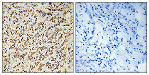
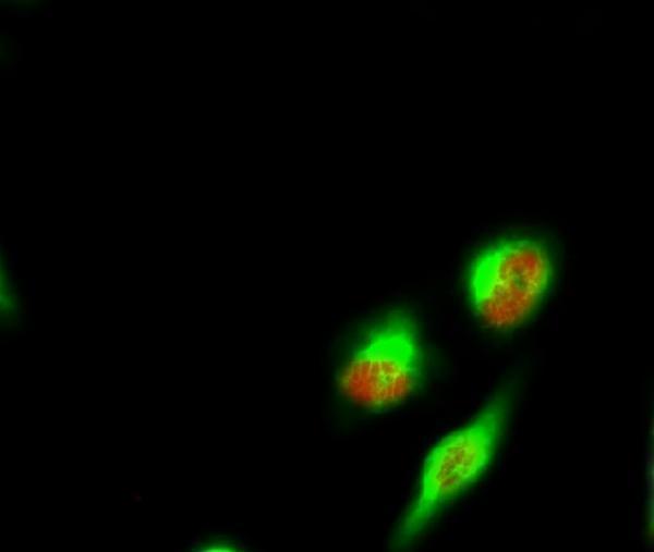
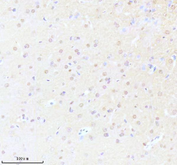
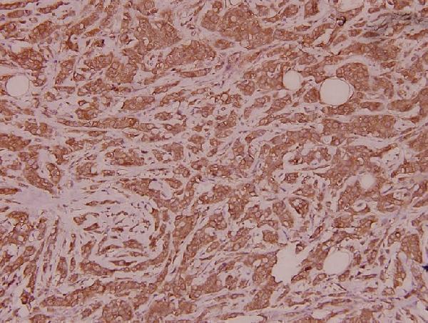
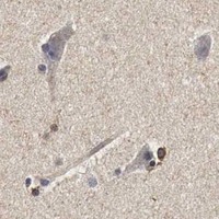
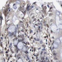
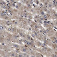
Anti-SPIN1 IHC images show paraffin sections of human breast cancer, human breast carcinoma and rat brain (catalog IHC captions); an IF image shows HeLa cells (A07281 IF caption).

Real IHC data Immunohistochemical analysis of paraffin-embedded Human breast cancer. Antibody was diluted at 1:100 (4° overnight). High-pressure and temperature Tris-EDTA, pH8.0 was used for antigen retrieval. Negetive contrl (right) obtaned from antibody was pre-absorbed by immunogen peptide.
Anti-Spindlin-1 SPIN1 Antibody
Cat # A07281
Real IHC data IHC analysis of SPIN1 using anti-SPIN1 antibody (A07281-2). SPIN1 was detected in a paraffin-embedded section of rat brain tissue. Heat mediated antigen retrieval was performed in EDTA buffer (pH 8.0, epitope retrieval solution). The tissue section was blocked with 10% goat serum. The tissue section was then incubated with 1:200 rabbit anti-SPIN1 Antibody (A07281-2) overnight at 4°C. Peroxidase Conjugated Goat Anti-rabbit IgG was used as secondary antibody and incubated for 30 minutes at 37°C. The tissue section was developed using HRP Conjugated Rabbit IgG Super Vision Assay Kit (Catalog # SV0002) with DAB as the chromogen.
Anti-SPIN1 antibody
Cat # A07281-2
Real IHC data Immunohistochemistry (IHC) analyzes of Spindlin-1 (H139) pAb in paraffin-embedded human breast carcinoma tissue at 1:100.
Anti-Spindlin-1 (H139) SPIN1 Antibody
Cat # A07281-1

A07281 is shown in human breast cancer IHC and HeLa cell IF (A07281 image captions), while A07281-1 is shown in human breast carcinoma IHC (A07281-1 IHC caption). A07281-2 is shown in rat brain IHC (A07281-2 IHC caption) and lists IF/ICC among its applications (A07281-2 catalog applications).

Which to pick: For human paraffin-section IHC, pick A07281 or A07281-1 based on their own tissue images; for rat brain IHC, A07281-2 has an image, although IHC is absent from its listed applications (catalog IHC captions and applications). For IF, A07281 has a HeLa cell image and lists IF; A07281-2 lists IF/ICC without a supplied IF image (catalog image captions and applications). All three list human, mouse and rat reactivity, but the supplied IHC images show only human or rat tissue, and their captions do not report the fixative (catalog reactivity and IHC captions).

Each figure is that product's own IHC / IF validation image from its datasheet.

References

  1. UniProt Consortium. UniProt entry Q9Y657 (SPIN1_HUMAN, Spindlin-1).
  2. Human Protein Atlas. SPIN1 tissue IHC expression (reliability: Uncertain).
  3. Human Protein Atlas. SPIN1 subcellular location (ICC-IF): Mainly localized to the nucleoplasm..
  4. Human Protein Atlas. SPIN1 antibody validation summary (2 antibodies).
  5. The histone code reader SPIN1 controls RET signaling in liposarcoma. Oncotarget 2015 — PMC4467114.
  6. The microRNA-381(miR-381)/Spindlin1(SPIN1) axis contributes to cell proliferation and invasion of colorectal cancer cells by regulating the Wnt/β-catenin pathway. Bioengineered 2021 — PMC8810024.
  7. miR-409 Inhibits Human Non-Small-Cell Lung Cancer Progression by Directly Targeting SPIN1. Molecular therapy. Nucleic acids 2018 — PMC6171160.
  8. SPIN1 accelerates tumorigenesis and confers radioresistance in non-small cell lung cancer by orchestrating the FOXO3a/FOXM1 axis. Cell death & disease 2024 — PMC11568276.
  9. PubMed PMID:16098913 — UniProt-cited evidence.
  10. PubMed PMID:11230166 — UniProt-cited evidence.
  11. PubMed PMID:14702039 — UniProt-cited evidence.