This website uses cookies to ensure you get the best experience on our website.


- Table of Contents
This guide plans chromogenic IHC-P for SPNS1 with the catalog antibody starting at 2.5 μg/mL (datasheet). Assess granular cytoplasmic staining in lung macrophages and placental Hofbauer cells (HPA tissue IHC), with lysosomal membrane localization as a molecular expectation (UniProt).

Expected localisation, validated protocols, controls and antibodies — the at-a-glance facts below, then the full design guide.
| Expected localisation | Granular cytoplasm (HPA tissue IHC); lysosomal membrane expected (UniProt) | |
| Staining pattern | Granular cytoplasm; high in lung macrophages and Hofbauer cells (HPA tissue IHC) | |
| Antigen retrieval | Citrate pH 6.0 HIER, 95–98 °C, 20 min (rule: cytoplasmic / membrane antigen) | |
| Positive control | Lung+4 more · see all | |
| Negative control | Adipose tissue+4 more · see all |
| Fixation | Keep fixation consistent across sections (standard IHC practice; not target-specific) | |
| Caveat | Macrophage peroxidase may raise chromogenic background (HPA tissue IHC; standard IHC practice) | |
| Regulation | Expression varies by cell type (HPA tissue IHC) | |
| Isoform / epitope | Five isoforms; epitope position is unspecified (UniProt) |
The catalog antibody has an IHC-P protocol (datasheet); the published skin melanoma protocol below provides a separate SPNS1 staining example (PMC8183723).
| Sample | Tissue sections; selected-image fixative not specified (standard IHC workflow) |
| Fixation | Image fixative and duration unreported (datasheet A09902); verify before use. |
| Sectioning | 4–5 µm sections on charged slides (standard) |
| Deparaffinisation | Xylene, graded ethanol series to water (standard) |
| Antigen retrieval | Heat-induced epitope retrieval in citrate buffer, pH 6.0, 20 min at 95–98 °C (standard rule: cytoplasmic / membrane antigen) |
| Peroxidase block | 3% H2O2, 10 min, room temperature (standard) |
| Blocking | 10% normal serum of the secondary host, 30 min, room temperature (standard) |
| Primary antibody | Rabbit anti-SPNS1, 2.5 μg/mL (datasheet A09902) |
| Primary incubation | Overnight at 4 °C (standard) |
| Detection | HRP-polymer secondary, DAB chromogen 5–10 min (standard) |
| Counterstain | Hematoxylin, blue, dehydrate and mount (standard) |
| Expected result | SPNS1-positive staining in macrophages of lung (HPA tissue IHC: High). HPA tissue profile: General cytoplasmic expression with a granular pattern, highly expressed in lung macrophages and placental trophoblasts. No signal in the no-primary control. |
SPNS1 is a 12-pass membrane protein assigned mainly to lysosomes, with occasional mitochondrial localization (UniProt Q9H2V7 localization and topology). In paraffin-section IHC, expect granular cytoplasmic staining, especially in lung macrophages and placental Hofbauer cells (HPA tissue IHC: High in both; Enhanced reliability). HPA describes medium consistency between antibody staining and RNA expression, so interpret intensity alongside cell identity and staining pattern (HPA tissue IHC: Enhanced reliability description).
| Granular cytoplasmic staining is strongest in lung macrophages or placental Hofbauer cells (HPA tissue IHC: High in both). | This matches the reported tissue pattern and is compatible with membrane-bound intracellular compartments (HPA tissue IHC profile; UniProt Q9H2V7 localization and topology). Compare the expected cells with adjacent tissue and controls before scoring intensity (general IHC practice). |
| A dominant nuclear rim, uniform nuclear fill, or surface outline replaces granular cytoplasmic staining. | These patterns do not match the reported tissue IHC profile (HPA tissue IHC profile). Review morphology, counterstain, and control sections for an artefact (general IHC practice). HPA lists nucleolar staining only as an uncertain additional ICC-IF location, so it does not establish a nuclear IHC pattern (HPA subcellular ICC-IF). |
| Strong staining appears in adipocytes, esophageal squamous cells, or skeletal myocytes while expected positive cells stain. | Those cell types were not detected in the HPA tissue IHC record (HPA tissue IHC: adipose tissue, esophagus, skeletal muscle). Consider antibody cross-reactivity or endogenous chromogen activity; compare primary-omission and detection controls before assigning SPNS1 expression (general IHC practice). |
| Color spreads evenly across stroma, lumina, or most cells without a granular cell-associated pattern. | Diffuse color is less consistent with HPA's granular cytoplasmic profile (HPA tissue IHC profile). Assess background with a primary-omission control, then inspect blocking, wash stringency, chromogen development, and tissue pigment (general IHC practice). A diffuse result alone cannot establish a new SPNS1 location. |
| No staining appears in lung macrophages or placental Hofbauer cells (HPA tissue IHC: High in both). | This is an uninformative negative result until section quality, antibody detection, and a known-positive control are checked (general IHC practice). HPA reports high staining in these cell types, but its Enhanced rating still carries only medium staining–RNA consistency (HPA tissue IHC and reliability description). |
| Compartment and topology (UniProt Q9H2V7 localization and topology) | SPNS1 has 12 transmembrane segments and is assigned to the lysosome membrane, with occasional mitochondrial localization (UniProt Q9H2V7 localization and topology). This supports an intracellular granular interpretation, while chromogenic IHC alone cannot identify which individual stained granules are lysosomes (general IHC practice). |
| Cell and tissue context (HPA tissue IHC) | HPA reports High staining in lung macrophages and placental Hofbauer cells, Medium staining in several other listed cell types, and Not detected staining in others (HPA tissue IHC). Score the identified cell population rather than treating every cell in a tissue as equally positive (general IHC practice). |
| Strength of tissue evidence (HPA tissue IHC and antibodies) | The tissue IHC reliability is Enhanced, and both listed antibodies have Enhanced IHC status (HPA tissue IHC; HPA antibodies: HPA041995, HPA042988). HPA also reports only medium consistency with RNA expression, so unexpected staining still needs control-based review (HPA tissue IHC reliability description; general IHC practice). |
| Isoform coverage (UniProt Q9H2V7 isoforms) | UniProt lists five SPNS1 isoforms (UniProt Q9H2V7 isoforms). The supplied record gives no antibody epitope or isoform-specific IHC evidence; the presence or absence of a stained cell therefore cannot be assigned to a particular isoform from this section alone (UniProt Q9H2V7 isoforms; supplied HPA IHC record). |
| IF/ICC Q: Should its image set guide this IHC call? (HPA subcellular ICC-IF) | A: HPA's approved main ICC-IF location is vesicles; its nucleolar fibrillar center, Golgi, and cytosol locations are uncertain (HPA subcellular ICC-IF). Use that record as localization context, while judging paraffin-section results against HPA's granular cytoplasmic tissue IHC pattern (HPA tissue IHC profile). |
| Situation | Likely cause | Next action |
|---|---|---|
| Known-positive macrophages or Hofbauer cells have no visible stain (HPA tissue IHC: High in both). | The staining run, retrieval conditions, antibody application, or detection may have failed; the absent signal does not identify which step failed (general IHC practice). | Check a known-positive section processed in the same run, section integrity, retrieval log, primary-antibody step, and detection reagents before interpreting a negative sample (general IHC practice). |
| A positive tissue stains, but its signal is faint and hard to score. | Weak development or suboptimal assay conditions are possible (general IHC practice); HPA's High category does not specify an intensity threshold for this assay (HPA tissue IHC: High). | Compare the positive control and cell morphology first; then review the documented antibody dilution and retrieval conditions for the assay without assuming an SPNS1-specific optimum (general IHC practice). |
| Most of the section has diffuse brown color without discrete granules. | Background from nonspecific binding, insufficient washing, or overdevelopment is possible (general IHC practice); the pattern differs from HPA's granular cytoplasmic profile (HPA tissue IHC profile). | Inspect a primary-omission control and review blocking, wash steps, and chromogen time; rescore only cell-associated staining with preserved morphology (general IHC practice). |
| Color appears in HPA Not detected cells while positive cells also stain (HPA tissue IHC negative entries). | Cross-reactivity, endogenous enzyme activity, or pigment may mimic a positive result (general IHC practice). HPA's Not detected category is an observation, not proof that every specimen lacks SPNS1 (HPA tissue IHC). | Compare the suspect cells with primary-omission and detection controls, check pigment and morphology, and document the unexpected pattern separately from expected positive cells (general IHC practice). |
| The most conspicuous signal is nuclear or outlines cell surfaces. | That distribution conflicts with the reported granular cytoplasmic IHC profile (HPA tissue IHC profile). HPA's additional nucleolar ICC-IF location is uncertain and does not validate nuclear IHC staining (HPA subcellular ICC-IF). | Recheck compartment assignment using the counterstain and adjacent cells, then inspect assay controls; withhold a positive call based only on the atypical compartment (general IHC practice). |
| Granular cytoplasmic staining varies between fields or specimens. | Different fields may contain different proportions of the cell types that HPA scores at High, Medium, or Not detected levels (HPA tissue IHC). Assay variation is also possible (general IHC practice). | Identify and score the same cell type across fields, compare control sections from the run, and record intensity and distribution separately (general IHC practice; HPA tissue IHC cell-specific levels). |
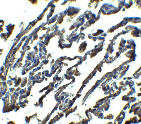
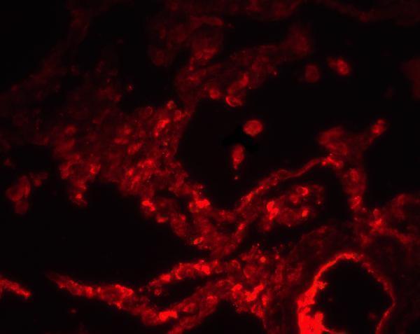


Comprehensive Human Protein Atlas IHC scoring per tissue (reliability: Enhanced — Medium consistency between antibody staining and RNA expression data.). Rows are taken directly from the HPA tissue chart — click any row's HPA link to view the source.
| Tissue | Cell type | Level | Evidence | Source |
|---|---|---|---|---|
| Lung | Macrophages | High | Protein (IHC) | HPA → |
| Placenta | Hofbauer cells | High | Protein (IHC) | HPA → |
| Adrenal gland | Glandular cells | Medium | Protein (IHC) | HPA → |
| Appendix | Glandular cells | Medium | Protein (IHC) | HPA → |
| Breast | Myoepithelial cells | Medium | Protein (IHC) | HPA → |
| Tissue | Cell type | Level | Evidence | Source |
|---|---|---|---|---|
| Adipose tissue | Adipocytes | Not detected | Protein (IHC) | HPA → |
| Esophagus | Squamous epithelial cells | Not detected | Protein (IHC) | HPA → |
| Oral mucosa | Squamous epithelial cells | Not detected | Protein (IHC) | HPA → |
| Ovary | Follicle cells | Not detected | Protein (IHC) | HPA → |
| Salivary gland | Glandular cells | Not detected | Protein (IHC) | HPA → |
SPNS1 staining is best assessed as granular cytoplasmic signal in the expected cells, with compartment and control checks guiding interpretation (HPA tissue IHC; UniProt Q9H2V7 localisation).
The catalog antibody has IHC and IF images from human lung tissue (catalog image captions) and lists human reactivity (catalog: A09902).
A09902 lists IHC-P and IF applications with human reactivity (catalog: A09902). Its IHC image shows human lung tissue at 2.5 μg/mL, and its IF image shows human lung tissue at 20 μg/mL (catalog image captions: A09902).
Which to pick: Choose A09902 for human paraffin-section IHC because IHC-P is listed and its own IHC image shows human lung tissue; the caption does not report the fixative (catalog: A09902 applications; A09902 IHC image caption). Choose A09902 for tissue IF based on its human lung IF image; ICC validation and clonality are unreported (catalog: A09902 applications and IF image caption; catalog: clone unspecified). No cross-species choice is supported because A09902 lists human reactivity only (catalog: A09902 reactivity).