This website uses cookies to ensure you get the best experience on our website.


- Table of Contents
Plan paraffin-section SREBF2 IHC around the nuclear staining reported in most tissues (HPA tissue IHC). Colon glandular cells show high staining (HPA tissue IHC), while sterol-dependent processing can affect nuclear signal (UniProt).

Expected localisation, validated protocols, controls and antibodies — the at-a-glance facts below, then the full design guide.
| Expected localisation | Nuclear staining in most tissues (HPA tissue IHC); ER/Golgi precursor (UniProt) | |
| Staining pattern | Nuclear staining across most tissue cell types (HPA tissue IHC) | |
| Antigen retrieval | Tris-EDTA pH 9.0 HIER, 95–98 °C, 20 min (rule: nuclear antigen) | |
| Positive control | Appendix+4 more · see all | |
| Negative control | Cerebellum+4 more · see all |
| Fixation | Matched tissue-IHC evidence does not establish the fixation claim. Validate the specimen-specific method before use. (selected-SKU IHC image A01678) | |
| Caveat | Sterol status can change processing and nuclear signal (UniProt) | |
| Regulation | Low sterols promote nuclear SREBF2 (UniProt) | |
| Isoform / epitope | 2 isoforms; processed 1–484 lacks C-terminal epitopes (UniProt) |
The catalog antibody’s IHC-P protocol is accompanied by published paraffin-section methods for colon adenocarcinoma and cervical tumors (PMC10723182; PMC12992596).
| Sample | Tissue sections; selected-image fixative not specified (standard IHC workflow) |
| Fixation | Image fixative and duration unreported (datasheet A01678); verify before use. |
| Sectioning | 4–5 µm sections on charged slides (standard) |
| Deparaffinisation | Xylene, graded ethanol series to water (standard) |
| Antigen retrieval | Heat-induced epitope retrieval in Tris-EDTA buffer, pH 9.0, 20 min at 95–98 °C (standard rule: nuclear antigen) |
| Peroxidase block | 3% H2O2, 10 min, room temperature (standard) |
| Blocking | 10% normal serum of the secondary host, 30 min, room temperature (standard) |
| Primary antibody | Rabbit anti-SREBF2, 5 μg/mL (datasheet A01678) |
| Primary incubation | Overnight at 4 °C (standard) |
| Detection | HRP-polymer secondary, DAB chromogen 5–10 min (standard) |
| Counterstain | Hematoxylin, blue, dehydrate and mount (standard) |
| Expected result | SREBF2-positive staining in lymphoid tissue of appendix (HPA tissue IHC: High). HPA tissue profile: Nuclear expression in most tissues. No signal in the no-primary control. |
SREBF2 occupies ER and Golgi membranes and can enter nuclei after processing (UniProt Q12772: localization, processing). HPA reports nuclear staining in most tissues, including high staining in colon glandular cells (HPA: tissue IHC). Its IHC profile is Approved, with low consistency between antibody staining and RNA expression (HPA: reliability).
| High nuclear stain in colon glands (HPA: tissue IHC). | Expected pattern; HPA reports nuclear expression in most tissues (HPA: tissue IHC). |
| Stain confined to extracellular spaces (general IHC practice). | Suspect artefact; SREBF2 localizes inside cells (UniProt Q12772: localization). |
| Strong stain in pancreatic exocrine cells (HPA: not detected). | Check cross-reactivity or endogenous detection activity (general IHC practice). |
| Diffuse stain across tissue and blank areas (general IHC practice). | Background is likely; compare a no-primary control (general IHC practice). |
| No stain in colon glands (HPA: High). | Review the assay before scoring these cells negative (general IHC practice). |
| Sterol-dependent processing (UniProt Q12772). | Low sterol releases a nuclear form; high sterol retains the precursor in ER (UniProt: processing). |
| Membrane topology (UniProt Q12772). | The precursor has 2 transmembrane spans; the processed chain is residues 1–484 (UniProt: topology). |
| IHC evidence (HPA: antibodies). | HPA031962 is IHC Approved; tissue staining has low RNA consistency (HPA: antibodies, reliability). |
| IF/ICC: where is signal? | Nucleoplasm and vesicles; ER also seen (HPA: supported ICC-IF localization). |
| Situation | Likely cause | Next action |
|---|---|---|
| No nuclear DAB in colon glands (HPA: High). | Assay failure is possible (general IHC practice). | Check positive control, retrieval and detection steps (general IHC practice). |
| Signal in a no-primary section (general IHC practice). | Endogenous activity or reagent background (general IHC practice). | Block endogenous activity and repeat the control (general IHC practice). |
| Diffuse tissue haze (general IHC practice). | Blocking or washing may be inadequate (general IHC practice). | Adjust blocking, antibody dilution and washes (general IHC practice). |
| Strong prostate gland stain (HPA: not detected). | Cross-reactivity or detection background is possible (general IHC practice). | Compare no-primary control and verify antibody specificity (general IHC practice). |
| Cytoplasmic stain without nuclear stain (HPA: nuclear in most tissues). | ER precursor may contribute (UniProt Q12772: localization, processing). | Check counterstain and score compartments separately (general IHC practice). |


Comprehensive Human Protein Atlas IHC scoring per tissue (reliability: Approved — Low consistency between antibody staining and RNA expression data.). Rows are taken directly from the HPA tissue chart — click any row's HPA link to view the source.
| Tissue | Cell type | Level | Evidence | Source |
|---|---|---|---|---|
| Appendix | Lymphoid tissue | High | Protein (IHC) | HPA → |
| Bone marrow | Hematopoietic cells | High | Protein (IHC) | HPA → |
| Colon | Glandular cells | High | Protein (IHC) | HPA → |
| Duodenum | Glandular cells | High | Protein (IHC) | HPA → |
| Esophagus | Squamous epithelial cells | High | Protein (IHC) | HPA → |
| Tissue | Cell type | Level | Evidence | Source |
|---|---|---|---|---|
| Cerebellum | Cells in granular layer | Not detected | Protein (IHC) | HPA → |
| Heart muscle | Cardiomyocytes | Not detected | Protein (IHC) | HPA → |
| Liver | Cholangiocytes | Not detected | Protein (IHC) | HPA → |
| Pancreas | Exocrine glandular cells | Not detected | Protein (IHC) | HPA → |
| Parathyroid gland | Glandular cells | Not detected | Protein (IHC) | HPA → |
Troubleshoot chromogenic SREBF2 IHC by checking retrieval, compartment-specific staining and cell-level controls before interpreting signal as sterol-dependent processing.
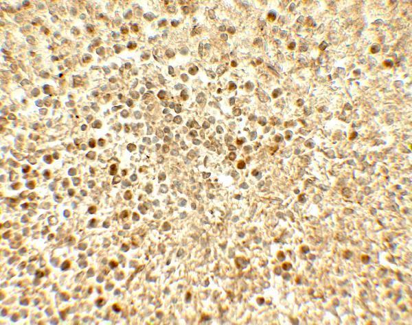
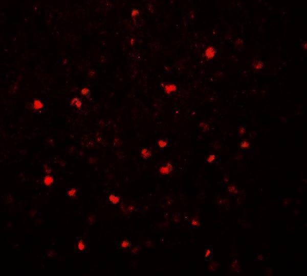
A01678 has IHC and IF images from human spleen (catalog: IHC and IF image captions); Human and Mouse reactivity is listed (catalog: reactivity).
A01678 is listed for IHC-P and IF, with Human and Mouse reactivity (catalog: applications and reactivity). Its IHC and IF images show human spleen at 5 μg/mL and 20 μg/mL, respectively (catalog: IHC and IF image captions).
Which to pick: Choose A01678 for paraffin-section IHC (catalog: IHC-P); its human spleen IHC caption reports 5 μg/mL but does not report the fixative (catalog: IHC image caption). A01678 is also the IF choice (catalog: IF application and human spleen IF image caption); ICC validation is unreported (catalog: applications). For Mouse samples, A01678 has listed reactivity but no Mouse IHC or IF image in the payload; its host is Rabbit and clone type is unreported (catalog: reactivity, image captions, host, clone).