This website uses cookies to ensure you get the best experience on our website.


- Table of Contents
Plan SRPX2 chromogenic IHC in paraffin sections using the mainly cytoplasmic tissue pattern as a reference (HPA tissue IHC). The catalog antibody is listed for IHC-P at 5 μg/mL (datasheet); assess staining with secreted SRPX2 in mind (UniProt).

Expected localisation, validated protocols, controls and antibodies — the at-a-glance facts below, then the full design guide.
| Expected localisation | Mainly cytoplasmic staining across tissues (HPA tissue IHC) | |
| Staining pattern | Mainly cytoplasmic staining across tissue cells (HPA tissue IHC) | |
| Antigen retrieval | Citrate pH 6.0 HIER, 95–98 °C, 20 min (rule: cytoplasmic / membrane antigen) | |
| Positive control | Adipose tissue+4 more · see all | |
| Negative control | None in HPA (detected in all 45 tissues); use no-primary + isotype controls |
| Fixation | Matched tissue-IHC evidence does not establish the fixation claim. Validate the specimen-specific method before use. (selected-SKU IHC image A06268) | |
| Caveat | Secreted SRPX2 may extend beyond producing cells (UniProt) | |
| Regulation | Higher in colorectal cancer than normal mucosa (UniProt) | |
| Isoform / epitope | No isoforms listed; map epitopes to the mature chain (24–465) when assessing secreted versus cytoplasmic signal (UniProt) |
The catalog antibody’s IHC-P protocol is accompanied by published SRPX2 staining methods for prostate cancer, oral squamous cell carcinoma, and gastric carcinoma specimens (PMC5983007; PMC7279144; PMC6413340).
| Sample | Tissue sections; selected-image fixative not specified (standard IHC workflow) |
| Fixation | Image fixative and duration unreported (datasheet A06268); verify before use. |
| Sectioning | 4–5 µm sections on charged slides (standard) |
| Deparaffinisation | Xylene, graded ethanol series to water (standard) |
| Antigen retrieval | Heat-induced epitope retrieval in citrate buffer, pH 6.0, 20 min at 95–98 °C (standard rule: cytoplasmic / membrane antigen) |
| Peroxidase block | 3% H2O2, 10 min, room temperature (standard) |
| Blocking | 10% normal serum of the secondary host, 30 min, room temperature (standard) |
| Primary antibody | Rabbit anti-SRPX2, 5 μg/mL (datasheet A06268) |
| Primary incubation | Overnight at 4 °C (standard) |
| Detection | HRP-polymer secondary, DAB chromogen 5–10 min (standard) |
| Counterstain | Hematoxylin, blue, dehydrate and mount (standard) |
| Expected result | SRPX2-positive staining in adipocytes of adipose tissue (HPA tissue IHC: High). HPA tissue profile: Mainly cytoplasmic expression in all tissues including plasma. No signal in the no-primary control. |
SRPX2 is secreted and also annotated at the cytoplasm, cell surface and synapse, with no transmembrane segment (UniProt O60687 topology). In tissue IHC, expect mainly cytoplasmic staining, including in adipocytes, respiratory epithelial cells, neurons and endothelial cells (HPA tissue IHC). HPA rates its tissue evidence Approved while noting that secreted variants complicate RNA–protein comparisons and that external verification is pending (HPA tissue IHC).
| Distinct cytoplasmic stain in adipocytes, bronchial respiratory epithelium or colon endothelium. | These cell types have High reported staining (HPA tissue IHC). Judge the pattern by cell identity and compartment together; secretion means extracellular or plasma-associated signal may coexist (UniProt O60687 subcellular; HPA tissue IHC). |
| Strong, exclusively nuclear stain with little cytoplasmic signal. | This conflicts with the mainly cytoplasmic tissue profile and the reported secreted, cytoplasmic and cell-surface locations (HPA tissue IHC; UniProt O60687 subcellular). Treat it as suspect; compare the negative reagent control and a known-positive tissue section (general IHC practice). |
| Stain appears chiefly in an unexpected cell population while expected cells are faint. | The distribution warrants a specificity check against the reported High cell populations; it alone does not prove cross-reactivity because HPA describes expression across tissues (HPA tissue IHC). Check antibody omission and detection controls for endogenous activity (general IHC practice). |
| Uniform haze covers tissue and surrounding space, obscuring cell outlines. | Some extracellular or plasma-associated staining is plausible for a secreted protein (UniProt O60687 subcellular; HPA tissue IHC). Haze that also appears in reagent controls points to background; review blocking, washes and detection chemistry (general IHC practice). |
| No convincing stain in an adipose section containing adipocytes. | Adipocytes are reported High in adipose tissue (HPA tissue IHC). First verify tissue preservation and run controls, then review the validated IHC procedure and detection reagents (general IHC practice). A blank section alone cannot establish absent SRPX2 expression. |
| Secreted protein and compartment | SRPX2 has a signal peptide at residues 1–23, a mature chain at 24–465, and no transmembrane segment (UniProt O60687 processing/topology). Interpret cell-associated stain alongside possible extracellular or plasma signal; do not require a continuous membrane rim (UniProt O60687 subcellular; HPA tissue IHC). |
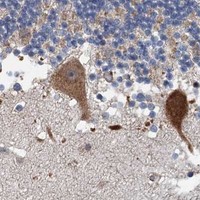
| Choice of tissue and cell population | High staining is reported in adipocytes, adrenal glandular cells, marrow hematopoietic cells, bronchial epithelium, caudate neurons, Purkinje cells and colon endothelium (HPA tissue IHC). Ovarian stromal and vaginal squamous epithelial cells are reported Low, so compare like cell types when judging intensity (HPA tissue IHC). |
| Evidence strength | The tissue profile is Approved, with external verification pending; one listed rabbit polyclonal antibody, HPA038786, has IHC Approved status (HPA tissue IHC; HPA antibodies). Approval supports use of the reported pattern as a reference, but does not make every unexpected stain a confirmed biological finding. |
| Retrieval and detection conditions | General IHC practice: use the IHC-validated antibody's documented paraffin-section procedure as the starting point; check retrieval, blocking, reagent concentration, washes and chromogen development when optimizing. The supplied UniProt and HPA records give no SRPX2-specific fixation or retrieval response. |
| IF/ICC evidence | Q: Does the tissue IHC pattern establish an IF/ICC protocol? A: No. HPA lists SRPX2 as secreted but provides no main ICC/IF location or cell-line images, and the listed antibody has no ICC status (HPA subcellular; HPA antibodies). Interpret IF/ICC on its own guide page. |
| Situation | Likely cause | Next action |
|---|---|---|
| A reported High population stains weakly or not at all. | Section or detection failure is possible; the reported High category is a reference, not a guarantee for every specimen (HPA tissue IHC; general IHC practice). | Check morphology and run a positive tissue plus reagent controls; then review the IHC-validated antibody's documented retrieval and detection steps (general IHC practice). |
| Nuclei dominate while cytoplasm stays pale. | An exclusively nuclear result conflicts with the mainly cytoplasmic tissue pattern (HPA tissue IHC). | Compare a known-positive section and omission control; reassess antibody concentration, blocking and chromogen development before assigning a nuclear location (general IHC practice). |
| Unexpected cells stain more than the expected population. | Cross-reactivity or endogenous detection activity is possible, though broad tissue expression prevents a conclusion from location alone (HPA tissue IHC; general IHC practice). | Confirm cell identity and compare adjacent controls; test the detection system without primary antibody (general IHC practice). |
| Diffuse staining masks cellular boundaries. | Secreted SRPX2 can contribute extracellular or plasma-associated signal; broad control staining instead suggests background (UniProt O60687 subcellular; HPA tissue IHC; general IHC practice). | Compare primary-omission controls, then review blocking, washes and detection development (general IHC practice). |
| Ovarian stroma or vaginal squamous epithelium looks faint. | Both cell populations are reported Low (HPA tissue IHC). | Score them against their own expected level and examine a reported High population in parallel before changing the assay (HPA tissue IHC; general IHC practice). |
| IF/ICC shows a pattern that differs from paraffin IHC. | HPA provides no ICC/IF images or main intracellular location, while its tissue IHC profile is mainly cytoplasmic (HPA subcellular; HPA tissue IHC). | Interpret IF/ICC with application-specific controls and its separate guide; do not infer an IF/ICC protocol from tissue IHC approval (HPA antibodies; general IF practice). |



Comprehensive Human Protein Atlas IHC scoring per tissue (reliability: Approved — At least one protein variant secreted, tissue location of RNA and protein might differ and correlation is complex. Pending external verification.). Rows are taken directly from the HPA tissue chart — click any row's HPA link to view the source.
| Tissue | Cell type | Level | Evidence | Source |
|---|---|---|---|---|
| Adipose tissue | Adipocytes | High | Protein (IHC) | HPA → |
| Adrenal gland | Glandular cells | High | Protein (IHC) | HPA → |
| Bone marrow | Hematopoietic cells | High | Protein (IHC) | HPA → |
| Breast | Adipocytes | High | Protein (IHC) | HPA → |
| Bronchus | Respiratory epithelial cells | High | Protein (IHC) | HPA → |
| Tissue | Cell type | Level | Evidence | Source |
|---|---|---|---|---|
| None in HPA: SRPX2 is detected in all 45 scored tissues. Use a no-primary (secondary-only) and an isotype control instead. | ||||
Troubleshoot SRPX2 staining in paraffin sections by checking retrieval, cell identity, and compartment before comparing chromogenic signal across samples.
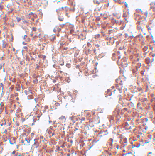
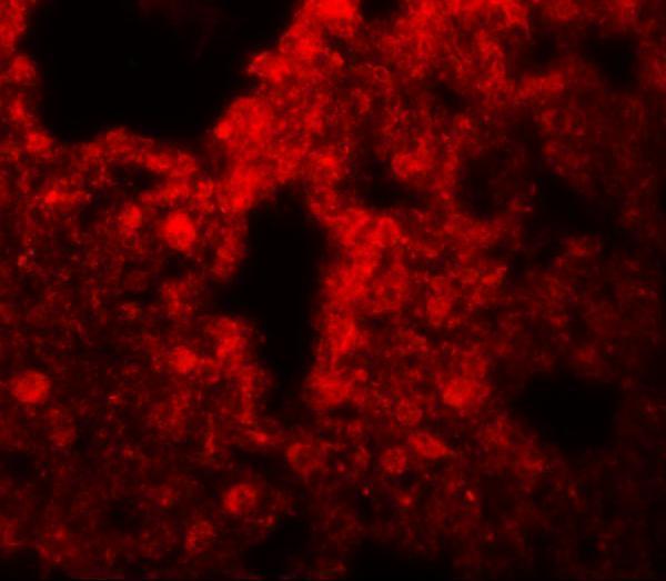
A06268 has IHC data in rat lung tissue and IF data in human lung tissue (catalog image captions); its listed reactivity covers human, mouse, and rat (catalog: reactivity).
A06268 is listed for IHC-P and IF (catalog: applications). Its images show IHC in rat lung tissue at 5 μg/mL and IF in human lung tissue at 20 μg/mL (catalog image captions).
Which to pick: Choose A06268 for tissue IHC-P: its IHC image shows rat lung tissue at 5 μg/mL (catalog: IHC-P application; IHC image caption); the fixative is unreported (catalog: IHC image caption). For IF, A06268 has a human lung tissue image at 20 μg/mL (catalog: IF image caption); ICC is not shown (catalog: applications and image captions). For cross-species work, A06268 lists human, mouse, and rat reactivity, though the supplied images cover rat IHC and human IF only (catalog: reactivity; image captions); its host is rabbit and clonality is unreported (catalog: host; clone).