This website uses cookies to ensure you get the best experience on our website.


- Table of Contents
Use this guide to plan SSR3 staining in paraffin sections and interpret its general cytoplasmic tissue pattern (HPA tissue IHC). Its ER membrane location provides molecular context (UniProt), while medium agreement between staining and RNA warrants careful interpretation (HPA tissue IHC).

Expected localisation, validated protocols, controls and antibodies — the at-a-glance facts below, then the full design guide.
| Expected localisation | Cytoplasmic tissue staining; ER membrane location (HPA tissue IHC; UniProt) | |
| Staining pattern | Cytoplasmic staining in glandular and hematopoietic cells (HPA tissue IHC) | |
| Antigen retrieval | EDTA pH 8.0 HIER, heat-mediated (datasheet PA1796) | |
| Positive control | Adipose tissue+4 more · see all | |
| Negative control | None in HPA (detected in all 43 tissues); use no-primary + isotype controls |
| Fixation | Matched tissue-IHC evidence does not establish the fixation claim. Validate the specimen-specific method before use. (selected-SKU IHC image PA1796) | |
| Caveat | Staining and RNA show only medium consistency (HPA tissue IHC) | |
| Regulation | No specific expression regulator reported (UniProt) | |
| Isoform / epitope | 2 isoforms; lumenal versus cytoplasmic epitope unknown (UniProt) |
The catalog antibody’s IHC protocol uses EDTA pH 8.0 heat retrieval (datasheet PA1796). One published mouse tumor IHC protocol provides additional staining details (PMC10191221).
| Sample | Paraffin-embedded human breast cancer tissue; fixative not specified (datasheet PA1796) |
| Fixation | Image fixative and duration unreported (datasheet PA1796); verify before use. |
| Sectioning | 4–5 µm sections on charged slides (standard) |
| Deparaffinisation | Xylene, graded ethanol series to water (standard) |
| Antigen retrieval | Heat retrieval: EDTA pH 8.0 (datasheet PA1796); 20 min, 95–100 °C (standard) |
| Peroxidase block | 3% H2O2, 10 min, room temperature (standard) |
| Blocking | 10% goat serum (datasheet PA1796) |
| Primary antibody | Rabbit anti-SSR3, 2-5μg/ml (datasheet PA1796) |
| Primary incubation | Overnight at 4 °C (datasheet PA1796) |
| Detection | HRP-conjugated secondary, DAB chromogen (datasheet PA1796) |
| Counterstain | Hematoxylin, blue, dehydrate and mount (standard) |
| Expected result | SSR3-positive staining in adipocytes of adipose tissue (HPA tissue IHC: High). HPA tissue profile: General cytoplasmic expression. No signal in the no-primary control. |
SSR3 is a four-pass endoplasmic reticulum (ER) membrane protein (UniProt Q9UNL2 topology). In paraffin-section IHC, expect predominantly cytoplasmic staining, consistent with the tissue survey (HPA: general cytoplasmic expression). Glandular cells in colon, duodenum, cervix and epididymis are useful high-staining examples (HPA: High in each). Interpret intensity cautiously: HPA rates tissue IHC Approved, with medium consistency between staining and RNA expression (HPA: reliability description).
| Cytoplasmic staining in colon or duodenal glandular cells. | This fits the reported high-staining cell types (HPA: High in colon and duodenal glandular cells). ER membrane localisation makes intracellular staining plausible (UniProt Q9UNL2 topology); chromogenic IHC alone does not prove that each stained structure is ER. |
| Predominantly nuclear or cell-surface staining, with little cytoplasmic signal. | Treat a compartment shift as suspect: the tissue pattern is generally cytoplasmic (HPA: tissue IHC profile), while SSR3 is assigned to the ER membrane (UniProt Q9UNL2 subcellular location). Review morphology, controls and detection conditions before assigning the signal to SSR3. |
| Strong signal in vaginal squamous cells or smooth muscle cells. | Those cells are reported as low staining (HPA: Vagina, squamous epithelial cells; smooth muscle, smooth muscle cells). Unexpected strong signal may reflect nonspecific antibody binding or endogenous detection activity (standard IHC practice); compare with a matched detection control. |
| Broad haze or precipitate across cells and empty areas. | A field-wide signal is difficult to reconcile with the reported cell-associated cytoplasmic pattern (HPA: tissue IHC profile). Check background in a control processed without primary antibody, then review blocking, washes and chromogen development (standard IHC practice). |
| No staining in glandular cells of a well-preserved colon section. | Colon glandular cells are reported as high staining (HPA: Colon, glandular cells). A negative result warrants a check of the antibody, retrieval and detection run (standard IHC practice). The HPA rating is Approved with medium RNA–staining consistency, so a single section is not an absolute biological standard (HPA: reliability description). |
| ER location and membrane topology | SSR3 has four transmembrane segments, with short cytoplasmic regions and lumenal regions (UniProt Q9UNL2 topology). An ER-associated target can appear cytoplasmic by chromogenic IHC (standard IHC interpretation); the topology does not establish which epitope the antibody recognises. |
| Reference tissue and cell type | High staining is reported in adipocytes, bone-marrow hematopoietic cells and several glandular epithelia (HPA: tissue IHC). Vaginal squamous cells, muscle cells and chondrocytes are listed as low (HPA: tissue IHC). These are useful comparisons, not guaranteed positive or negative controls for every specimen. |
| Strength of IHC evidence | The listed rabbit polyclonal antibody HPA014906 is IHC Approved (HPA: antibody validation). The tissue survey describes medium consistency with RNA expression (HPA: reliability description). Neither statement establishes antibody-specific ER colocalisation or identical staining in every sample. |
| Isoforms and epitope coverage | Two isoforms are listed (UniProt Q9UNL2 isoforms 1 and 2). The supplied evidence does not map the IHC antibody epitope to either isoform, so isoform-specific staining or a predicted change in intensity cannot be assigned. |
| ICC-IF evidence | The subcellular entry summarizes location as Membrane but supplies no main location or cell-line ICC-IF images (HPA: subcellular record). Use the ER assignment as a localisation hypothesis for IF (UniProt Q9UNL2); this record does not validate an IF staining pattern. |
| Fixation and retrieval evidence | Target-specific fixation sensitivity and an SSR3 retrieval condition are unreported in the supplied UniProt and HPA records. Retrieval may be checked against tissue controls as general paraffin-section IHC practice; do not infer a specific fixation effect from topology or tissue staining. |
| Situation | Likely cause | Next action |
|---|---|---|
| A high-staining reference tissue gives no signal. | A failed detection step, unsuitable antibody conditions or tissue processing are possible (standard IHC practice); colon glandular cells are a reported high-staining reference (HPA: Colon). | Confirm tissue morphology and a working detection control, then check the antibody instructions and compare retrieval conditions on adjacent sections (standard IHC practice). Do not assign an SSR3-specific fixation cause from these sources. |
| The entire section is uniformly brown. | Background from the detection system, insufficient blocking or excessive chromogen development is possible (standard IHC practice). Uniform color lacks the reported cytoplasmic cell pattern (HPA: tissue IHC profile). | Inspect a section run without primary antibody; adjust the relevant blocking, washing or chromogen step according to that control (standard IHC practice). |
| Signal is strongest in nuclei or on the outer cell edge. | The dominant compartment conflicts with general cytoplasmic tissue staining (HPA: tissue IHC profile) and ER membrane assignment (UniProt Q9UNL2). Artefact or nonspecific binding is possible (standard IHC practice). | Compare a high-staining reference section with a matched detection control; score SSR3 only where cell-associated cytoplasmic staining is credible (HPA: tissue IHC profile; standard IHC practice). |
| Vaginal squamous cells stain as strongly as the positive reference. | Vaginal squamous cells are reported as low staining (HPA: Vagina), whereas colon glandular cells are high (HPA: Colon). Strong equal signal may indicate background or antibody cross-reactivity (standard IHC practice). | Compare sections processed in the same run, including a control without primary antibody; assess cell-specific staining after background is accounted for (standard IHC practice). |
| Intensity varies among otherwise comparable sections. | Section handling, retrieval or development can vary between IHC runs (standard IHC practice). HPA reports medium consistency between staining and RNA expression, so intensity alone is a limited biological readout (HPA: reliability description). | Process reference and study sections together, keep development and scoring consistent, and interpret differences alongside tissue morphology (standard IHC practice). |
| An IF image is requested to confirm the IHC pattern. | The supplied HPA subcellular record has no cell-line ICC-IF images or specified main location (HPA: subcellular record). It cannot establish an SSR3 IF reference pattern. | Use the separate IF/ICC guide for assay design. For localisation interpretation, treat ER membrane residence as the UniProt assignment (UniProt Q9UNL2), without claiming IF validation from this record. |


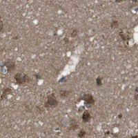


Comprehensive Human Protein Atlas IHC scoring per tissue (reliability: Approved — Medium consistency between antibody staining and RNA expression data.). Rows are taken directly from the HPA tissue chart — click any row's HPA link to view the source.
| Tissue | Cell type | Level | Evidence | Source |
|---|---|---|---|---|
| Adipose tissue | Adipocytes | High | Protein (IHC) | HPA → |
| Bone marrow | Hematopoietic cells | High | Protein (IHC) | HPA → |
| Cerebellum | GLUC cells - cytoplasm/membrane | High | Protein (IHC) | HPA → |
| Cervix | Glandular cells | High | Protein (IHC) | HPA → |
| Colon | Glandular cells | High | Protein (IHC) | HPA → |
| Tissue | Cell type | Level | Evidence | Source |
|---|---|---|---|---|
| None in HPA: SSR3 is detected in all 43 scored tissues. Use a no-primary (secondary-only) and an isotype control instead. | ||||
Troubleshoot SSR3 staining in paraffin sections by checking retrieval, cellular compartment, controls and scoring before interpreting signal intensity.
PA1796 has paraffin-section IHC images from human breast cancer, placenta, lung cancer and mouse cerebral cortex, plus IF images from human placenta and mouse and rat brain (catalog image captions).
PA1796 is the sole card; its IHC images show paraffin sections of human breast cancer, placenta and lung cancer, and mouse cerebral cortex (PA1796 IHC captions). Its IF images show human placenta and mouse and rat brain; an ICC image shows HELA cells with DAB detection (PA1796 IF/ICC captions).
Which to pick: Choose PA1796 for tissue IHC in paraffin sections: its IHC captions document EDTA pH 8.0 retrieval, 2 μg/ml primary antibody and DAB detection (PA1796 IHC captions). PA1796 also supports IF and ICC, with images for each application (PA1796 application list and IF/ICC captions). Human, Monkey, Mouse and Rat are listed as reactive, while the pictured tissue IHC covers Human and Mouse; the fixative and clonality are unreported (PA1796 reactivity list, IHC captions and clone field).