This website uses cookies to ensure you get the best experience on our website.


- Table of Contents
Plan STAT3 staining and scoring in paraffin sections using the variable cytoplasmic and nuclear tissue pattern (HPA tissue IHC). Placental cytotrophoblasts provide a high-staining reference (HPA tissue IHC); score nuclear and cytoplasmic staining separately because LIF can shift STAT3 into nuclei (UniProt).

Expected localisation, validated protocols, controls and antibodies — the at-a-glance facts below, then the full design guide.
| Expected localisation | Variable cytoplasmic and nuclear staining (HPA tissue IHC) | |
| Staining pattern | Cytotrophoblasts stain strongly; compartments vary (HPA tissue IHC) | |
| Antigen retrieval | Tris-EDTA pH 9.0 HIER, 95–98 °C, 20 min (rule: nuclear antigen) | |
| Positive control | Placenta+4 more · see all | |
| Negative control | Heart muscle+3 more · see all |
| Fixation | Keep fixation consistent across paraffin sections. (standard IHC practice; not target-specific) | |
| Caveat | Nuclear staining alone does not prove Tyr705 activation (UniProt) | |
| Regulation | LIF promotes nuclear accumulation (UniProt) | |
| Isoform / epitope | 3 isoforms; check epitope coverage across variants (UniProt) |
The catalog antibody protocol is followed by four published STAT3 IHC protocols, including phospho-STAT3 staining in kidney, lung, glioblastoma and salivary gland samples (PMC7114280; PMC5374098; PMC6829927; PMC4287356).
| Sample | Paraffin-embedded tissue sections; fixative not specified (datasheet M00007-1; sample unspecified) |
| Fixation | Image fixative and duration unreported (datasheet M00007-1); verify before use. |
| Sectioning | 4–5 µm sections on charged slides (standard) |
| Deparaffinisation | Xylene, graded ethanol series to water (standard) |
| Antigen retrieval | Heat-induced epitope retrieval in Tris-EDTA buffer, pH 9.0, 20 min at 95–98 °C (standard rule: nuclear antigen) |
| Peroxidase block | 3% H2O2, 10 min, room temperature (standard) |
| Blocking | 10% normal serum of the secondary host, 30 min, room temperature (standard) |
| Primary antibody | Rabbit monoclonal (clone CAB-19) anti-STAT3, 1:50-1:200 (datasheet M00007-1) |
| Primary incubation | Overnight at 4 °C (standard) |
| Detection | HRP-polymer secondary, DAB chromogen 5–10 min (standard) |
| Counterstain | Hematoxylin, blue, dehydrate and mount (standard) |
| Expected result | STAT3-positive staining in cytotrophoblasts of placenta (HPA tissue IHC: High). HPA tissue profile: General cytoplasmic and nuclear expression at variable levels. No signal in the no-primary control. |
STAT3 stains cytoplasm/nuclei in cytotrophoblasts (HPA: High, Supported); lacks a TM segment (UniProt P40763).
| Cytoplasmic and nuclear stain in cytotrophoblasts (HPA: High) | Expected distribution (HPA: tissue IHC); nuclear enrichment can vary with signaling (UniProt P40763). |
| Membrane-only staining | Suspect artefact; STAT3 has no TM segment (UniProt P40763). |
| Strong stain in prostate glandular cells | Unexpected; check cross-reactivity or endogenous detection activity (HPA: Not detected; general IHC practice). |
| Diffuse stain across cells and stroma | Background may reflect incomplete blocking or excess primary antibody (general IHC practice). |
| No stain in placental cytotrophoblasts | Unexpected for a positive control; assess retrieval and detection (HPA: High; general IHC practice). |
| Signaling state (UniProt P40763) | Usually cytoplasmic without stimuli; nuclear STAT3 can increase after activation (UniProt P40763). |
| Tissue context (HPA: tissue IHC) | Cytotrophoblasts are High; adipocytes are Medium; prostate glandular cells are Not detected (HPA). |
| Antibody choice (HPA: antibody data) | Five listed antibodies have IHC Supported status (HPA: antibody data). |
| IF/ICC: expected location? | Nucleoplasm and cytosol (HPA: ICC-IF); follow the separate IF/ICC guide for method. |
| Situation | Likely cause | Next action |
|---|---|---|
| No cytotrophoblast signal (HPA: High) | Retrieval or detection failure is possible (general IHC practice). | Check the positive control, retrieval and detection steps (general IHC practice). |
| Diffuse section background | Incomplete blocking or excess primary antibody is possible (general IHC practice). | Use a no-primary control; adjust blocking or dilution (general IHC practice). |
| Membrane-only signal | Localization conflicts with STAT3 topology (UniProt P40763: no TM). | Check staining controls and antibody specificity (general IHC practice). |
| Prostate glandular cells stain strongly | Unexpected signal (HPA: Not detected in prostate glandular cells). | Check controls; compare another IHC Supported antibody (HPA: antibody data). |
| No nuclear enrichment after stimulation | A signaling-dependent shift may be absent (UniProt P40763). | Verify the stimulus and score nuclei with a counterstain (general IHC practice). |
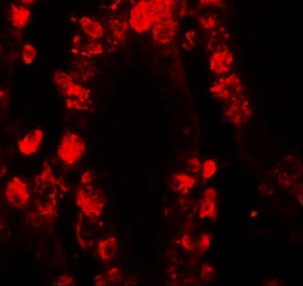


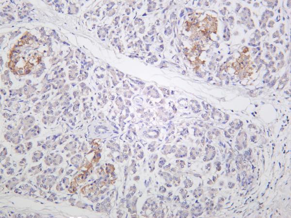
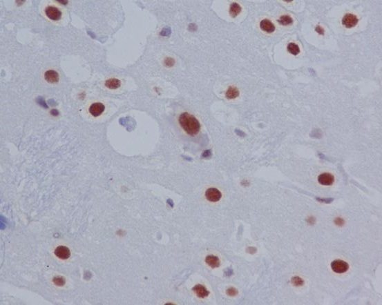

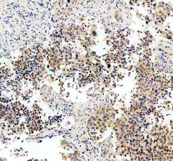


Comprehensive Human Protein Atlas IHC scoring per tissue (reliability: Supported — Medium consistency between antibody staining and RNA expression data.). Rows are taken directly from the HPA tissue chart — click any row's HPA link to view the source.
| Tissue | Cell type | Level | Evidence | Source |
|---|---|---|---|---|
| Placenta | Cytotrophoblasts | High | Protein (IHC) | HPA → |
| Adipose tissue | Adipocytes | Medium | Protein (IHC) | HPA → |
| Adrenal gland | Glandular cells | Medium | Protein (IHC) | HPA → |
| Appendix | Glandular cells | Medium | Protein (IHC) | HPA → |
| Breast | Adipocytes | Medium | Protein (IHC) | HPA → |
| Tissue | Cell type | Level | Evidence | Source |
|---|---|---|---|---|
| Heart muscle | Cardiomyocytes | Not detected | Protein (IHC) | HPA → |
| Hippocampus | Glial cells | Not detected | Protein (IHC) | HPA → |
| Parathyroid gland | Glandular cells | Not detected | Protein (IHC) | HPA → |
| Prostate | Glandular cells | Not detected | Protein (IHC) | HPA → |
Troubleshoot STAT3 staining by checking retrieval, cell identity, and nuclear versus cytoplasmic signal before comparing chromogenic scores across sections.
The catalog lists IHC and IF applications for STAT3 and phospho-STAT3 antibodies (catalog applications), with IHC images from human tissues, mouse liver, and rat brain (catalog IHC captions).
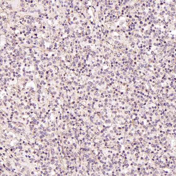
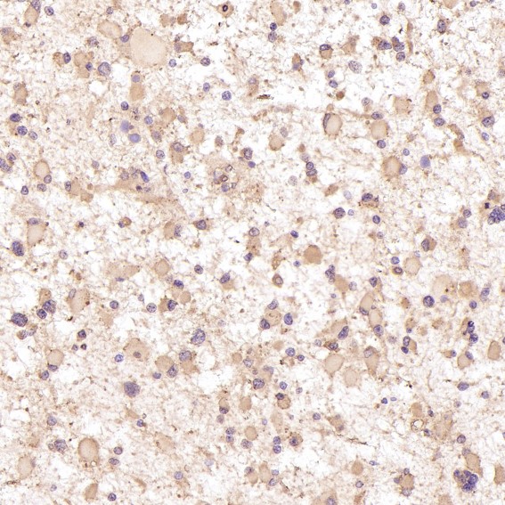
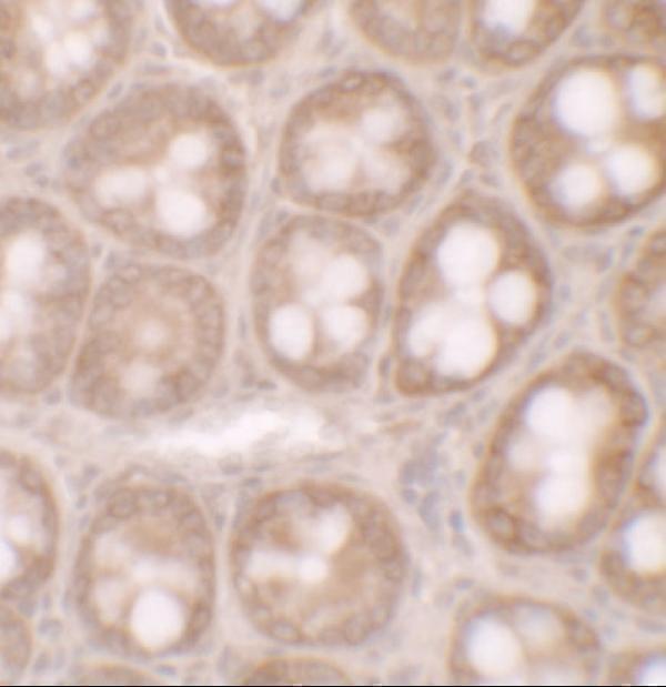
M00007-1 shows IHC in human Hodgkin’s lymphoma (catalog M00007-1 IHC caption), A00007-1 in human small intestine (catalog A00007-1 IHC caption), and M00007 in human thyroid carcinoma (catalog M00007 IHC caption). M00007-2 shows mouse liver (catalog M00007-2 IHC caption), M00007-4 shows human pancreas (catalog M00007-4 IHC caption), and phospho-STAT3 (S727) P00007-1 shows rat brain (catalog P00007-1 IHC caption).
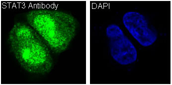
Which to pick: For paraffin-section IHC, M00007-1 is a rabbit monoclonal with an IHC image of paraffin-embedded human Hodgkin’s lymphoma at 1:50 (catalog M00007-1 title and IHC caption). For IF/ICC, M00007 lists both applications and has a HeLa IF image (catalog M00007 applications and IF caption). For cross-species IHC, M00007-1 lists human, mouse, and rat reactivity and has human and rat IHC images; the listed mouse reactivity lacks a mouse IHC image for this SKU, and its paraffin-section caption does not report the fixative (catalog M00007-1 reactivity and IHC captions).