This website uses cookies to ensure you get the best experience on our website.
- Table of Contents
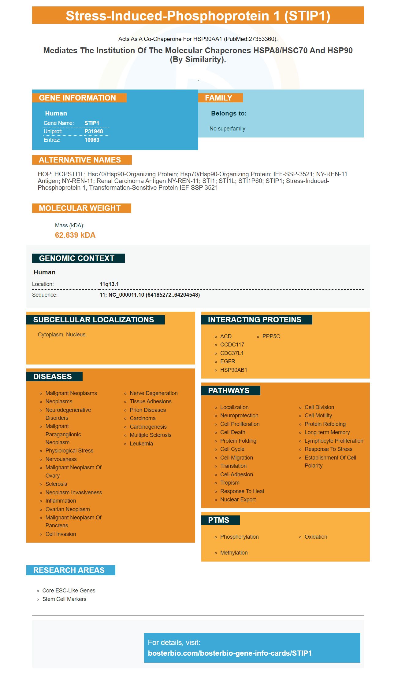
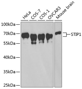
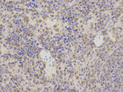
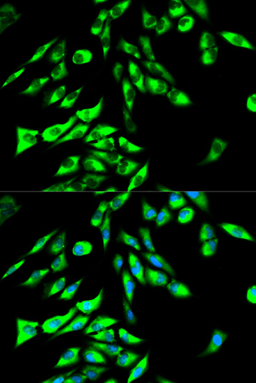
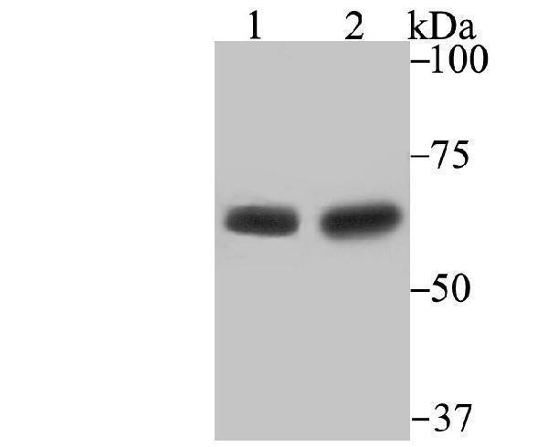
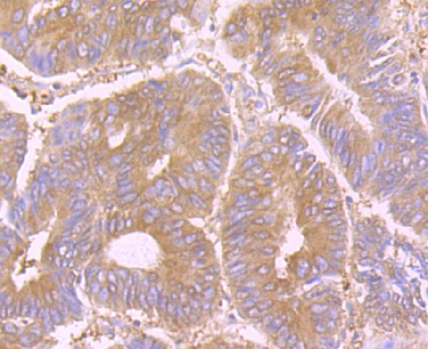
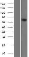
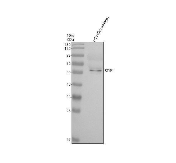
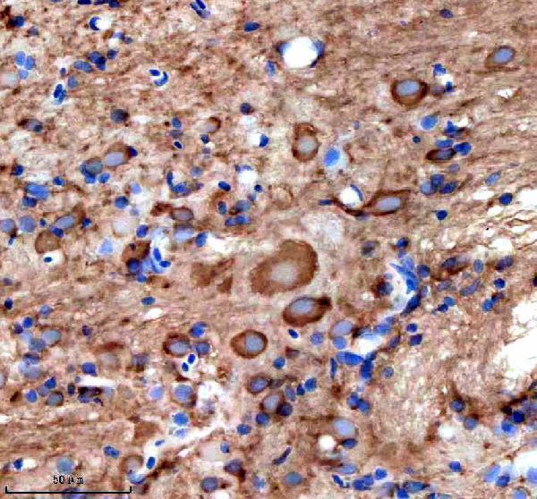
Facts about Stress-induced-phosphoprotein 1.
Acts as a co-chaperone for HSP90AA1 (PubMed:27353360).
Mediates the institution of the molecular chaperones HSPA8/HSC70 and HSP90 (By similarity)..
Human | |
---|---|
Gene Name: | STIP1 |
Uniprot: | P31948 |
Entrez: | 10963 |
Belongs to: |
---|
No superfamily |
HOP; HOPSTI1L; Hsc70/Hsp90-organizing protein; Hsp70/Hsp90-organizing protein; IEF-SSP-3521; NY-REN-11 antigen; NY-REN-11; Renal carcinoma antigen NY-REN-11; STI1; STI1L; STI1P60; STIP1; stress-induced-phosphoprotein 1; Transformation-sensitive protein IEF SSP 3521
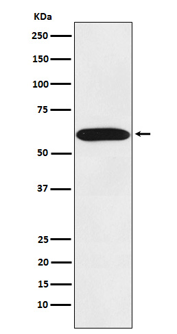
Mass (kDA):
62.639 kDA
Human | |
---|---|
Location: | 11q13.1 |
Sequence: | 11; NC_000011.10 (64185272..64204548) |
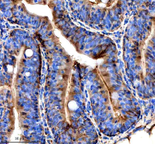
Cytoplasm. Nucleus.
This article will discuss the expression of STIP1 in osteosarcoma CSCs, how STIP1 knockdown inhibits the PI3K/Akt and ERK1/2 pathways, and possible therapeutic targets of STIP1. Read on for more information. Also, read our previous article, "Stip1 in Osteosarcoma CSCs: A New Target for Treatment" to learn more about this protein.
The knockdown of STIP1 in osteosarcoma CSCs inhibits invasion of these cells through the PI3K/Akt and ERK1/2 pathways. STIP1 expression in osteosarcoma cells can also decrease the expression of MMPs, which are associated with cell migration and invasion. Inhibition of STIP1 expression in osteosarcoma cells may provide new targets for targeted therapies.
We tested the effects of knockdown of STIP1 in CD133+ osteosarcoma CSCs in culture. We first isolated osteosarcoma MG63 cells from the China Center for Type Culture Collection at Wuhan University. Cells were cultured in DMEM/F12 supplemented with 10% FBS and grown in humidified incubators at 37degC. We then digested cells with 2.5 g/l trypsin and passaged them every five to six days.
We further confirmed that knockdown of STIP1 inhibited the phosphorylation of ERK1/2 and Akt. We also tested the inhibition of JAK2 by modifying STIP1 with the JAK2 mutant Y134/152A. This double mutant inhibited the phosphorylation of STIP1 by the JAK2 protein. This suggests that STIP1 knockdown in osteosarcoma CSCs inhibits JAK2 and may be a useful therapeutic strategy.
Our results suggest that STIP1 is required for the JAK2/STAT3 signaling pathway. We found that knocking down STIP1 with JAK2 may be a viable way to inhibit this pathway and increase the sensitivity of osteosarcoma CSCs to chemotherapy drugs. If you're thinking about pursuing this therapeutic option, STIP1 knockdown will be your best bet.
Knockdown of STIP1 by siRNA in osteosarcoma CSCs has potential for drug delivery. The knockdown of osteosarcoma CSCs might provide new opportunities for treating and preventing the tumor. The authors have used two different methods to isolate osteosarcoma CSCs: the serum-free medium suspension cell sphere culture method and the self-renewal behavior test. The combined methods improved the efficiency and purity of the process.
Although standard chemotherapy has shown significant improvements in long-term survival, metastasis remains a major challenge for cancer treatment. The emergence of a CSC model is supported by a growing body of evidence. While some believe that a single cancer-initiating cell is responsible for the development of all tumors, others argue that metastasis and resistance to chemotherapy are the result of a different subpopulation of cancer cells.
Interestingly, STIP1 expression in osteosarcomats is independent of the activity of BMPs. BMP-2 has an anti-metastasis role in osteosarcoma, which was further studied by Geng et al. This mechanism explains why a small amount of BMP-2 can inhibit osteosarcoma metastasis. However, it must be noted that the inhibitory effect of STIP1 is not universal, and the effects of different BMPs on osteosarcoma cells will vary.
To investigate the role of STIP1 in osteosarcoma CSCs, researchers first examined the levels of CD133+ cells in the osteosarcoma tumor microenvironment. When CD133+ cells were cultured in serum-free DMEM/F12, osteosarcoma CSCs were isolated as spheres. These spheres were similar to MG63 cell spheres.
One recent study suggests that STIP1 is an important transcription factor that regulates MMP-9 expression. It inhibits the migration and invasion of pancreatic cancer cells via its effect on MMP-2. STIP1 knockdown did not alter the expression of MMP-9 or the client proteins HSP70 and HSP90. It also reduced the expression of phosphorylated Akt and ERK1/2, suggesting a possible role for STIP1 in tumor cells.
Akt signaling plays an important role in several biological processes and is involved in regulating various cellular processes, including proliferation and metabolism. This pathway also controls angiogenesis. It regulates different cellular processes, including angiogenesis, cell survival, and adhesion. This is why targeting STIP1 can help block cancer cell proliferation. Moreover, it inhibits the phosphorylation of Akt, a component of the ERK and PI3K signaling.
In cancer cells, STIP1 expression correlates with poor prognosis and advanced tumor progression. It also regulates tumor invasion and metastasis. By inhibiting STIP1, cancer cells cannot sustain a high metabolic rate. Though the exact role of STIP1 is unknown, it is implicated in many aspects of cancer cell survival, including tumor growth and metastasis.
The effects of STIP1 knockdown on the PI3K/Akt and ERk1/2 pathways were determined by transfecting CSCs with siRNA against STIP1. The most significant inhibition was seen in the group expressing STIP1 siRNA-643 while the others exhibited a moderate inhibition of STIP1. Additionally, the various interference levels differed from one another.
The authors of this study used an antiserum to detect follitropin. They used a rabbit antiserum, which was affinity-purified for high purity. The antibody can be stored at -20!C or at 4!C for up to a month. The antibodies are available from Tebu-bio. They are available for research use.
For this study, CSCs were seeded into six-well culture plates at a density of 4x10⁴ cells/well. After overnight culture, these cells were transfected with siRNA. The negative control was also used. Cells were incubated in the CO2 incubator. After 48 h, the samples were evaluated using a Cell Counting Kit-8 manufactured by Beyotime Institute of Biotechnology. Using a 450 nm filter, the absorbance was measured on a plate reader. The data presented were obtained from six independent experiments.
The results of these studies suggest that STIP1 knockdown increases autophagy by increasing the expression of CHOP and AP-1. These results indicate that this protein may be used as a potential chemopreventive agent for hepatocellular carcinoma. However, the authors caution that these studies may not prove to be conclusive.
Most cancers progress through metastasis. Invasive subpopulations of cancer cells may initiate invasion of surrounding tissues. Rare tumor cells may escape the heterogeneous primary tumor. In this study, invasive subpopulations of human non-small cell lung cancer (NSCLC) H460 and h2299 cell lines were isolated. Invasive cells exhibited DNA damage repair, cell survival signaling, and epigenetic signatures of an epithelial-mesenchymal transition.
The development of novel therapeutic agents based on STIP1 has been slow but steady. A new therapeutic approach is targeting STIP1 intracellularly through the delivery of an anti-STIP1 antibody. The purpose of STIP1-targeting therapies is to inhibit the proliferation and survival of cancer cells by targeting the protein. The mechanism of STIP1 action has been a source of much speculation for years. Several recent studies have demonstrated its potential as a therapeutic target.
In a recent study, we analyzed the immunoreactivity of STIP1 in 19 protein specimens, including primary RCC tumors and bone metastatic samples. We electrophoresed the proteins in two 10% SDS-PAGE gels and transferred them to PVDF membranes. We quantified the intensity of STIP1 in both samples and normalized it against GAPDH. We then analyzed the results of the western blots by cropping the images to show the protein of interest.
The results of our study suggest that STIP1 inhibition promotes the catabolism of the targeted protein, LSD1. Further, TRIM21 mediates the degradation of the targeted proteins. Intracellular delivery of anti-STIP1 antibodies inhibited the growth of ovarian cancer cells. The effects of anti-STIP1 antibodies on cancer cell lines were similar in mouse models, and they significantly reduced the expression of apoptosis-related genes and caspase-3.
While extracellular molecules are still considered the most popular cancer druggable targets, intracellular approaches are gaining momentum. STIP1 is a transcription factor that coordinates the functions of chaperones and facilitates protein folding within the cell. Several solid malignancies exhibit overexpression of STIP1, suggesting that it has an oncogenic role. Furthermore, STIP1 is expressed on the cell surface and in the cytosol, indicating its oncogenic potential.
Anti-STIP1 antibodies reduced the mortality of mice bearing experimental tumors. To determine the efficacy of anti-STIP1 antibodies, anti-STIP1-antibodies were injected three times into C57BL/6 mice. Bioluminescent images were obtained after each treatment. In both groups, anti-STIP1-treated mice displayed significantly reduced mortality compared to the Ctrl-IgG group.
The role of STIP1 in cancer cell proliferation is unclear. The enzyme promotes tumor cell proliferation and migration. However, despite its role in tumor growth, it remains unknown whether STIP1 inhibition could be a worthwhile antitumor strategy. This study has a promising future for cancer research and the development of new anti-STIP1 drugs. While STIP1 has already been identified as a potential therapeutic target, there are currently no effective anticancer drugs that use it.
In addition to regulating the growth of tumor cells, STIP1 also promotes cell migration and invasion. However, this effect was only seen in cells that were pretreated with STIP1 siRNA or plasmids expressing the gene. In the presence of STIP1 siRNA, tumor cells were more likely to migrate toward the lower chamber. After treatment, tumor cells migrated towards the lower chamber and did not invade the upper chamber.
PMID: 1569099 by Honore B., et al. Molecular cloning and expression of a transformation-sensitive human protein containing the TPR motif and sharing identity to the stress- inducible yeast protein STI1.
PMID: 9195923 by Silverstein A.M., et al. Protein phosphatase 5 is a major component of glucocorticoid receptor.hsp90 complexes with properties of an FK506-binding immunophilin.