This website uses cookies to ensure you get the best experience on our website.
- Table of Contents
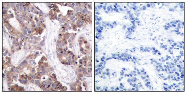
2 Citations 1 Q&As


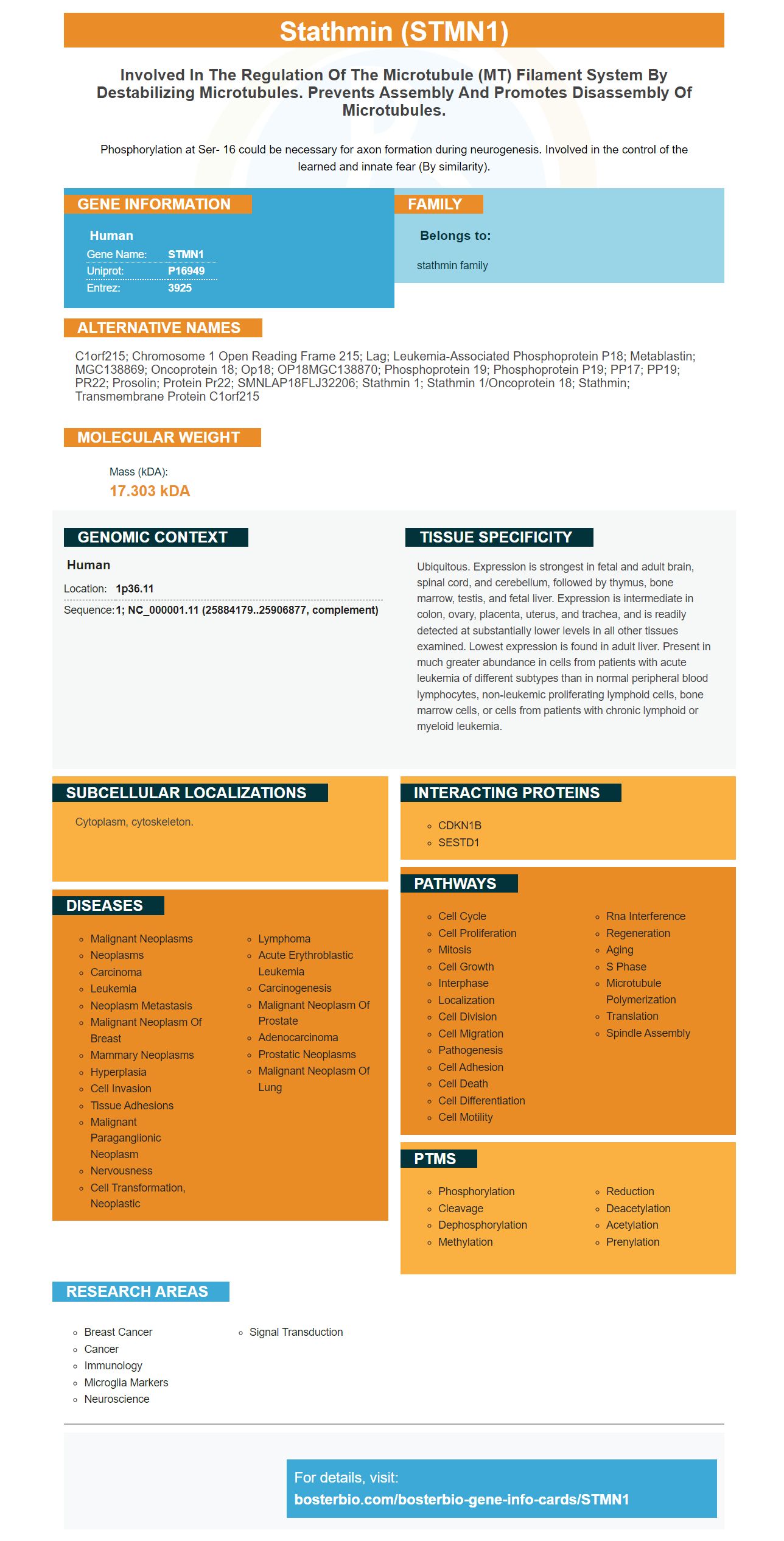
Facts about Stathmin.

Phosphorylation at Ser- 16 could be necessary for axon formation during neurogenesis. Involved in the control of the learned and innate fear (By similarity).
| Human | |
|---|---|
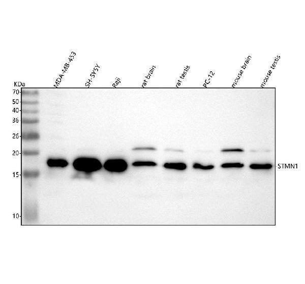
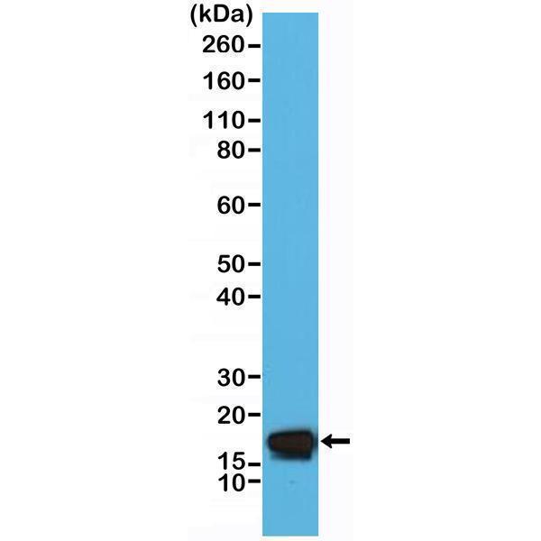
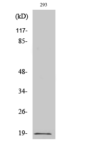
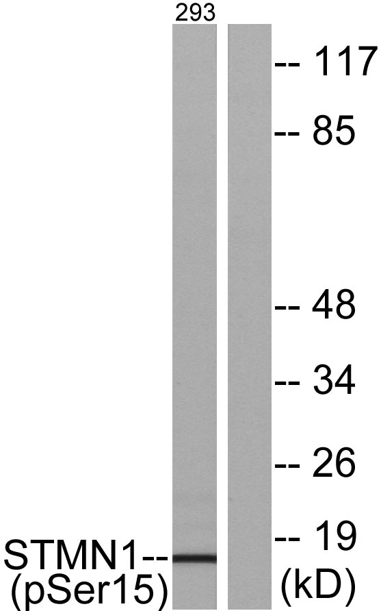
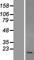
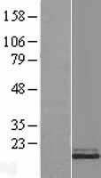
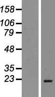
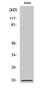
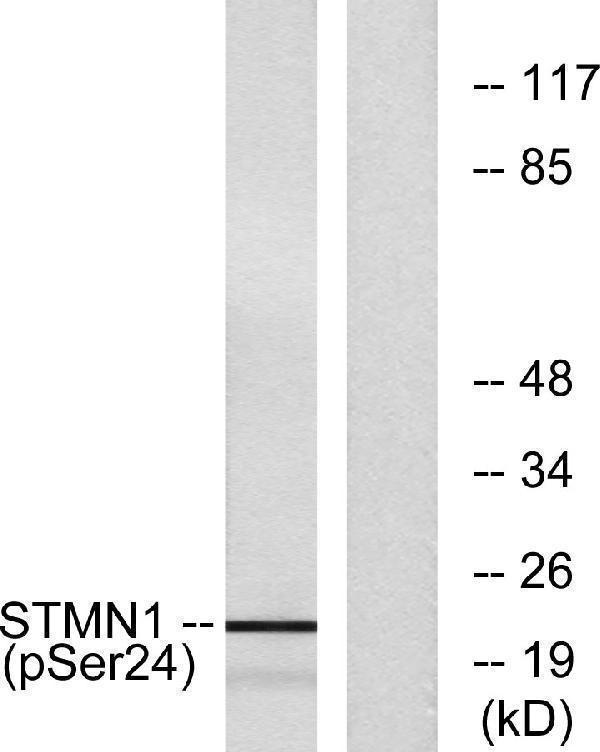
| Gene Name: | STMN1 |
| Uniprot: | P16949 |
| Entrez: | 3925 |

| Belongs to: |
|---|
| stathmin family |

C1orf215; chromosome 1 open reading frame 215; Lag; Leukemia-associated phosphoprotein p18; metablastin; MGC138869; Oncoprotein 18; Op18; OP18MGC138870; phosphoprotein 19; Phosphoprotein p19; PP17; PP19; PR22; prosolin; Protein Pr22; SMNLAP18FLJ32206; stathmin 1; stathmin 1/oncoprotein 18; stathmin; transmembrane protein C1orf215
Mass (kDA):
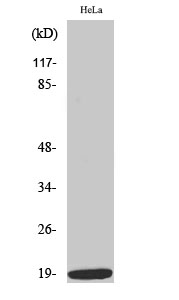
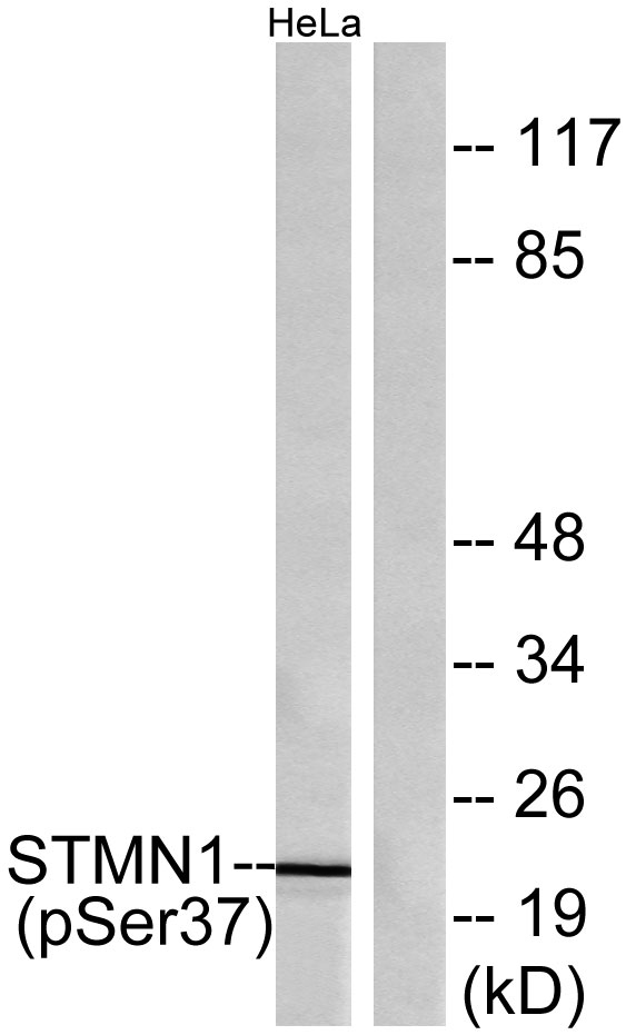
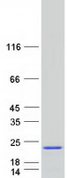
17.303 kDA

| Human | |
|---|---|
| Location: | 1p36.11 |
| Sequence: | 1; NC_000001.11 (25884179..25906877, complement) |
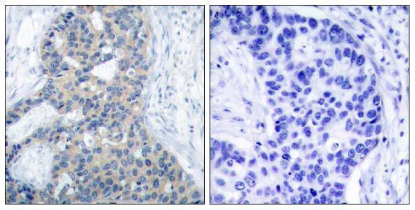
Ubiquitous. Expression is strongest in fetal and adult brain, spinal cord, and cerebellum, followed by thymus, bone marrow, testis, and fetal liver. Expression is intermediate in colon, ovary, placenta, uterus, and trachea, and is readily detected at substantially lower levels in all other tissues examined. Lowest expression is found in adult liver. Present in much greater abundance in cells from patients with acute leukemia of different subtypes than in normal peripheral blood lymphocytes, non-leukemic proliferating lymphoid cells, bone marrow cells, or cells from patients with chronic lymphoid or myeloid leukemia.
Cytoplasm, cytoskeleton.


PMID: 2760073 by Zhu X.-X., et al. Molecular cloning of a novel human leukemia-associated gene. Evidence of conservation in animal species.
PMID: 2358074 by Maucuer A., et al. A single amino acid difference distinguishes the human and the rat sequences of stathmin, a ubiquitous intracellular phosphoprotein associated with cell regulations.