This website uses cookies to ensure you get the best experience on our website.


- Table of Contents
Plan STRIP2 chromogenic IHC in paraffin sections using cytoplasmic staining in neuronal and stromal cells as a reference (HPA tissue IHC). Compare cell types within each section and titrate the catalog antibody within 2–5 μg/ml (datasheet A12139).

Expected localisation, validated protocols, controls and antibodies — the at-a-glance facts below, then the full design guide.
| Expected localisation | Cytoplasmic in neuronal and stromal cells (HPA tissue IHC) | |
| Staining pattern | Neuronal and stromal cells show cytoplasmic staining (HPA tissue IHC) | |
| Antigen retrieval | EDTA pH 8.0 HIER, heat-mediated (datasheet A12139) | |
| Positive control | Cerebellum+4 more · see all | |
| Negative control | Adipose tissue+4 more · see all |
| Fixation | Keep fixation consistent across sections (standard IHC practice; not target-specific) | |
| Caveat | Adipocytes may be unstained (HPA tissue IHC) | |
| Regulation | Expression regulation not annotated (UniProt) | |
| Isoform / epitope | 2 isoforms; epitope differences unspecified (UniProt) |
The catalog antibody’s IHC-P protocol is accompanied by published STRIP2 IHC methods for NSCLC and HCC specimens (PMC9837939; PMC12598350).
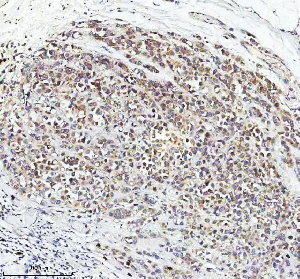
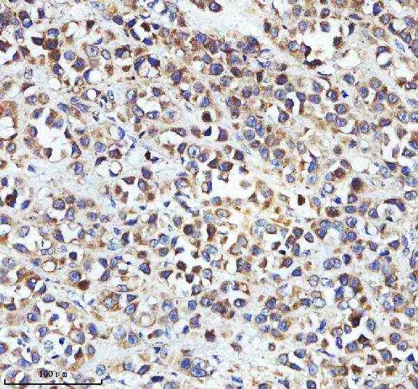
| Sample | Paraffin-embedded human breast cancer tissue; fixative not specified (datasheet A12139) |
| Fixation | Image fixative and duration unreported (datasheet A12139); verify before use. |
| Sectioning | 4–5 µm sections on charged slides (standard) |
| Deparaffinisation | Xylene, graded ethanol series to water (standard) |
| Antigen retrieval | Heat retrieval: EDTA pH 8.0 (datasheet A12139); 20 min, 95–100 °C (standard) |
| Peroxidase block | 3% H2O2, 10 min, room temperature (standard) |
| Blocking | 10% goat serum (datasheet A12139) |
| Primary antibody | Rabbit anti-STRIP2, 2-5 μg/ml (datasheet A12139) |
| Primary incubation | Overnight at 4 °C (datasheet A12139) |
| Detection | HRP-conjugated secondary, DAB chromogen (datasheet A12139) |
| Counterstain | Hematoxylin, blue, dehydrate and mount (standard) |
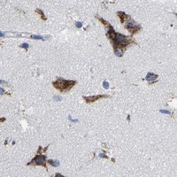
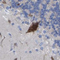
| Expected result | STRIP2-positive staining in purkinje cells of cerebellum (HPA tissue IHC: High). HPA tissue profile: Cytoplasmic expression in neuronal cells and stromal compartments. No signal in the no-primary control. |
STRIP2 is cytoplasmic and has no transmembrane segment (UniProt Q9ULQ0). In tissue IHC, expect cytoplasmic staining in neuronal cells and stromal compartments, including Purkinje cells and testicular peritubular cells with High reported staining (HPA tissue IHC). HPA rates the tissue profile Approved, pending external verification; use it as a reference pattern rather than a universal result for every antibody (HPA tissue IHC).
| Cytoplasmic staining is strong in cerebellar Purkinje cells, cerebral cortical or hippocampal neurons, or testicular peritubular cells (HPA tissue IHC). | This matches HPA's High staining in those specific cell populations and its cytoplasmic tissue profile (HPA tissue IHC). Score the named cells and their compartment, rather than treating all cells in a positive tissue as equally positive (general IHC practice). |
| The dominant signal is nuclear or sharply confined to a cell membrane instead of cytoplasmic (UniProt Q9ULQ0; HPA tissue IHC). | That pattern conflicts with UniProt's cytoplasmic location and lack of a transmembrane segment, and with HPA's cytoplasmic IHC profile (UniProt Q9ULQ0; HPA tissue IHC). Check counterstain, morphology and detection controls before assigning the signal to STRIP2 (general IHC practice). |
| Strong staining appears in adipocytes or in cervical glandular cells (HPA tissue IHC). | HPA reports STRIP2 as Not detected in those particular cell types, so this is a discordant result, not proof of target expression (HPA tissue IHC). Examine antibody-independent detection activity and possible cross-reactivity using appropriate controls (general IHC practice). |
| Colour spreads across extracellular space, tissue edges or many unrelated cell types, obscuring cell boundaries (general IHC practice). | This is diffuse background, which cannot establish STRIP2 localisation. Review detection and blocking controls, reagent concentration, washing and section morphology before scoring cells; those checks are general IHC practice, not an HPA finding. |
| A cerebellar Purkinje-cell section has no convincing cytoplasmic signal (HPA tissue IHC). | The result conflicts with HPA's High Purkinje-cell staining, but does not by itself show that STRIP2 is absent (HPA tissue IHC). Confirm the expected cells are present, then examine the assay controls and retrieval or detection conditions (general IHC practice). |
| Cell-specific reference pattern | HPA reports High staining in Purkinje cells, cortical and hippocampal neurons, and testicular peritubular cells; caudate neurons are Medium (HPA tissue IHC). Compare identified cells, since intensity across whole tissues is not an interchangeable measure (general IHC practice). |
| Reference limits and antibody validation | The tissue profile is Approved but pending external verification; antibody HPA019657 has IHC Approved status, not IHC Enhanced status (HPA tissue IHC; HPA antibodies). This supports a reference pattern, not independent confirmation for a different catalog antibody. |
| Cellular location and topology | UniProt places STRIP2 in cytoplasm, notes enrichment in lamellipodia, and lists no transmembrane segment (UniProt Q9ULQ0). The HPA tissue profile is cytoplasmic; it does not require visibly resolved lamellipodia in paraffin sections (HPA tissue IHC). |
| Isoforms and protein processing | UniProt lists two isoforms, no signal peptide or propeptide, and one annotated chain spanning residues 1–834 (UniProt Q9ULQ0). Without an epitope map for the IHC antibody, these facts cannot establish which isoforms it detects or predict a tissue-specific loss of staining. |
| IF/ICC Q&A: where should signal appear? | HPA reports approved cytosolic localisation in ICC-IF images from A-431, U-251MG and U2OS; UniProt also notes lamellipodial enrichment (HPA subcellular; UniProt Q9ULQ0). These IF observations can guide compartment interpretation, but do not specify an IHC-P retrieval or staining protocol. |
| Situation | Likely cause | Next action |
|---|---|---|
| No staining in morphologically identifiable cerebellar Purkinje cells (HPA tissue IHC). | Target-specific fixation effects are not established by the supplied assay evidence. Verify with a matched IHC source before attributing a result to fixation. | Verify section identity and morphology, positive-control performance, primary-antibody application and detection; review retrieval according to the antibody's IHC-P instructions (general IHC practice). |
| Predominantly nuclear staining in cells expected to show cytoplasmic signal (UniProt Q9ULQ0; HPA tissue IHC). | The compartment disagrees with both reference sources; counterstain or nonspecific detection may be mistaken for target staining (general IHC practice). | Check the counterstain and a control omitting primary antibody, then assess whether cytoplasmic signal remains in identifiable positive cells (general IHC practice). |
| Strong colour in cervical glandular cells or adipocytes (HPA tissue IHC). | Those specified cell types are Not detected in HPA; cross-reactivity or antibody-independent detection activity is possible (HPA tissue IHC; general IHC practice). | Confirm cell identity, compare a primary-omission control, and reassess the signal alongside a known-positive cell population (general IHC practice). |
| Diffuse colour obscures individual cells across the section (general IHC practice). | Background from detection reagents, inadequate blocking or washing, or an overly concentrated primary antibody may obscure localisation (general IHC practice). | Inspect reagent-only controls; adjust blocking, washing or antibody concentration according to the assay controls and IHC-P instructions (general IHC practice). |
| A low-signal cell population is treated as a negative control (HPA tissue IHC). | HPA lists low staining in lung macrophages and several epithelial or glandular cell populations, which differs from its Not detected category (HPA tissue IHC). | Use the HPA cell-level category when choosing comparators, and record cell identity before judging a slide negative (HPA tissue IHC; general IHC practice). |
| An IF/ICC image looks cytosolic, but a tissue IHC section seems to show a different compartment (HPA subcellular; HPA tissue IHC). | HPA's ICC-IF location is approved cytosol, while its tissue profile is cytoplasmic; image type and cell context differ (HPA subcellular; HPA tissue IHC). | Judge each assay against its own controls and identifiable cells; use the shared cytoplasmic expectation without importing an IF protocol into IHC-P (HPA subcellular; HPA tissue IHC; general IHC practice). |
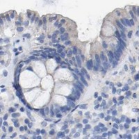
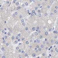


Comprehensive Human Protein Atlas IHC scoring per tissue (reliability: Approved — Pending external verification.). Rows are taken directly from the HPA tissue chart — click any row's HPA link to view the source.
| Tissue | Cell type | Level | Evidence | Source |
|---|---|---|---|---|
| Cerebellum | Purkinje cells | High | Protein (IHC) | HPA → |
| Cerebral cortex | Neuronal cells | High | Protein (IHC) | HPA → |
| Hippocampus | Neuronal cells | High | Protein (IHC) | HPA → |
| Testis | Peritubular cells | High | Protein (IHC) | HPA → |
| Caudate | Neuronal cells | Medium | Protein (IHC) | HPA → |
| Tissue | Cell type | Level | Evidence | Source |
|---|---|---|---|---|
| Adipose tissue | Adipocytes | Not detected | Protein (IHC) | HPA → |
| Breast | Adipocytes | Not detected | Protein (IHC) | HPA → |
| Cervix | Glandular cells | Not detected | Protein (IHC) | HPA → |
| Colon | Endothelial cells | Not detected | Protein (IHC) | HPA → |
| Fallopian tube | Glandular cells | Not detected | Protein (IHC) | HPA → |
Troubleshoot STRIP2 staining in paraffin sections by checking retrieval, cell type, cytoplasmic localisation, and controls before scoring chromogenic signal.
A12139 has paraffin-section IHC images from human breast cancer and mouse cerebellum (catalog image captions). No IF/ICC image or application is listed (catalog applications and images).
A12139 is listed for IHC in human, mouse and rat samples (catalog applications and reactivity). Its IHC images show human breast cancer and mouse cerebellum paraffin sections (catalog image captions).
Which to pick: For tissue IHC, choose A12139: its paraffin-section images use 2 μg/ml antibody after EDTA retrieval at pH 8.0 (A12139 image captions). A12139 lists human, mouse and rat reactivity, but the pictured IHC examples cover human and mouse only (catalog reactivity; A12139 image captions). No listed SKU supports an IF/ICC recommendation; A12139 is rabbit hosted, its clonality is unreported, and the fixative used for its paraffin sections is unreported (catalog host and applications; A12139 image captions).