Click image to see more details
-

-
-
-
-
+4
Product Info Summary
| SKU: | A12139 |
|---|---|
| Size: | 100 μg/vial |
| Reactive Species: | Human, Mouse, Rat |
| Host: | Rabbit |
| Application: | ELISA, Flow Cytometry, IHC, WB |
Customers Who Bought This Also Bought

Product info
Product Name
Anti-FAM40B/STRIP2 Antibody Picoband®
SKU/Catalog Number
A12139
Size
100 μg/vial
Form
Lyophilized
Description
Boster Bio Anti-FAM40B/STRIP2 Antibody Picoband® catalog # A12139. Tested in WB, IHC, FCM, ELISA applications. This antibody reacts with Human, Mouse, Rat. The brand Picoband indicates this is a premium antibody that guarantees superior quality, high affinity, and strong signals with minimal background in Western blot applications. Only our best-performing antibodies are designated as Picoband, ensuring unmatched performance.
Storage & Handling
At -20°C for one year from date of receipt. After reconstitution, at 4°C for one month. It can also be aliquotted and stored frozen at -20°C for six months. Avoid repeated freezing and thawing.
Cite This Product
Anti-FAM40B/STRIP2 Antibody Picoband® (Boster Biological Technology, Pleasanton CA, USA, Catalog # A12139)
Host
Rabbit
Contents
Each vial contains 4 mg Trehalose, 0.9 mg NaCl, 0.2 mg Na2HPO4.
Clonality
Polyclonal
Immunogen
E.coli-derived human FAM40B/STRIP2 recombinant protein (Position: H173-Q507). Human STRIP2 shares 96.4% amino acid (aa) sequence identity with mouse STRIP2.
Reactive Species
A12139 is reactive to STRIP2 in Human, Mouse, Rat
Observed Molecular Weight
100 kDa
Calculated molecular weight
95.4 kDa
Background of STRIP2
The STRIP2 gene encodes a protein known as striatin-interacting protein 2, which interacts with striatin, a scaffolding protein involved in diverse cellular processes such as signaling, cell adhesion, and transcriptional regulation. STRIP2 is implicated in cardiac development and function, where it modulates cardiac hypertrophy, contractility, and arrhythmogenesis. Additionally, it plays roles in neuronal development and synaptic plasticity in the brain. Dysregulation of STRIP2 expression or function has been associated with various cardiovascular disorders, including hypertrophic cardiomyopathy and arrhythmias, as well as neurodevelopmental disorders such as autism spectrum disorders. Elucidating the molecular mechanisms underlying STRIP2 function is essential for understanding its physiological roles in cardiac and neural tissues and its potential implications for disease pathology and therapeutic targeting.
Antibody Validation
Boster validates all antibodies on WB, IHC, ICC, Immunofluorescence, and ELISA with known positive control and negative samples to ensure specificity and high affinity, including thorough antibody incubations.
Application & Images
Applications
A12139 is guaranteed for ELISA, Flow Cytometry, IHC, WB Boster Guarantee
Assay Dilutions Recommendation
The recommendations below provide a starting point for assay optimization. The actual working concentration varies and should be decided by the user.
Western blot, 0.25-0.5 μg/ml, Human, Rat
Immunohistochemistry(Paraffin-embedded Section), 2-5 μg/ml, Human, Mouse, Rat
Flow Cytometry (Fixed), 1-3 μg/1x106 cells, Human
ELISA, 0.1-0.5 μg/ml, -
Positive Control
WB: human K562 whole cell, human Hacat whole cell, human A431 whole cell, human SiHa whole cell, rat testis tissue
IHC: human breast cancer tissue, human breast cancer tissue, mouse cerebellum tissue, mouse cerebellum tissue, rat cerebellum tissue, rat cerebellum tissue
FCM: U87 cell
Validation Images & Assay Conditions
Click image to see more details
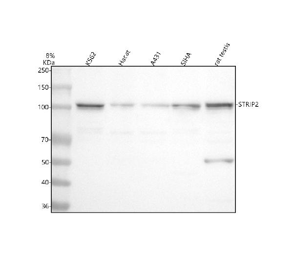
Western blot analysis of FAM40B/STRIP2 using anti-FAM40B/STRIP2 antibody (A12139).
Electrophoresis was performed on a 5-20% SDS-PAGE gel at 70V (Stacking gel) / 90V (Resolving gel) for 2-3 hours. The sample well of each lane was loaded with 30 ug of sample under reducing conditions.
Lane 1: human K562 whole cell lysates,
Lane 2: human Hacat whole cell lysates,
Lane 3: human A431 whole cell lysates,
Lane 4: human SiHa whole cell lysates,
Lane 5: rat testis tissue lysates.
After electrophoresis, proteins were transferred to a nitrocellulose membrane at 150 mA for 50-90 minutes. Blocked the membrane with 5% non-fat milk/TBS for 1.5 hour at RT. The membrane was incubated with rabbit anti-FAM40B/STRIP2 antigen affinity purified polyclonal antibody (Catalog # A12139) at 0.5 μg/mL overnight at 4°C, then washed with TBS-0.1%Tween 3 times with 5 minutes each and probed with a goat anti-rabbit IgG-HRP secondary antibody at a dilution of 1:5000 for 1.5 hour at RT. The signal is developed using an Enhanced Chemiluminescent detection (ECL) kit (Catalog # EK1002) with Tanon 5200 system. A specific band was detected for FAM40B/STRIP2 at approximately 100 kDa. The expected band size for FAM40B/STRIP2 is at 95 kDa.

Click image to see more details
IHC analysis of FAM40B/STRIP2 using anti-FAM40B/STRIP2 antibody (A12139).
FAM40B/STRIP2 was detected in a paraffin-embedded section of human breast cancer tissue. Heat mediated antigen retrieval was performed in EDTA buffer (pH 8.0, epitope retrieval solution). The tissue section was blocked with 10% goat serum. The tissue section was then incubated with 2 μg/ml rabbit anti-FAM40B/STRIP2 Antibody (A12139) overnight at 4°C. Peroxidase Conjugated Goat Anti-rabbit IgG was used as secondary antibody and incubated for 30 minutes at 37°C. The tissue section was developed using HRP Conjugated Rabbit IgG Super Vision Assay Kit (Catalog # SV0002) with DAB as the chromogen.

Click image to see more details
IHC analysis of FAM40B/STRIP2 using anti-FAM40B/STRIP2 antibody (A12139).
FAM40B/STRIP2 was detected in a paraffin-embedded section of mouse cerebellum tissue. Heat mediated antigen retrieval was performed in EDTA buffer (pH 8.0, epitope retrieval solution). The tissue section was blocked with 10% goat serum. The tissue section was then incubated with 2 μg/ml rabbit anti-FAM40B/STRIP2 Antibody (A12139) overnight at 4°C. Peroxidase Conjugated Goat Anti-rabbit IgG was used as secondary antibody and incubated for 30 minutes at 37°C. The tissue section was developed using HRP Conjugated Rabbit IgG Super Vision Assay Kit (Catalog # SV0002) with DAB as the chromogen.

Click image to see more details
IHC analysis of FAM40B/STRIP2 using anti-FAM40B/STRIP2 antibody (A12139).
FAM40B/STRIP2 was detected in a paraffin-embedded section of rat cerebellum tissue. Heat mediated antigen retrieval was performed in EDTA buffer (pH 8.0, epitope retrieval solution). The tissue section was blocked with 10% goat serum. The tissue section was then incubated with 2 μg/ml rabbit anti-FAM40B/STRIP2 Antibody (A12139) overnight at 4°C. Peroxidase Conjugated Goat Anti-rabbit IgG was used as secondary antibody and incubated for 30 minutes at 37°C. The tissue section was developed using HRP Conjugated Rabbit IgG Super Vision Assay Kit (Catalog # SV0002) with DAB as the chromogen.

Click image to see more details
Flow Cytometry analysis of U87 cells using anti-FAM40B/STRIP2 antibody (A12139).
Overlay histogram showing U87 cells stained with A12139 (Blue line). To facilitate intracellular staining, cells were fixed with 4% paraformaldehyde and permeabilized with permeabilization buffer. The cells were blocked with 10% normal goat serum. And then incubated with rabbit anti-FAM40B/STRIP2 Antibody (A12139, 1 μg/1x106 cells) for 30 min at 20°C. DyLight®488 conjugated goat anti-rabbit IgG (BA1127, 5-10 μg/1x106 cells) was used as secondary antibody for 30 minutes at 20°C. Isotype control antibody (Green line) was rabbit IgG (1 μg/1x106) used under the same conditions. Unlabelled sample without incubation with primary antibody and secondary antibody (Red line) was used as a blank control.
Specific Publications For Anti-FAM40B/STRIP2 Antibody Picoband® (A12139)
Loading publications
Recommended Resources
Here are featured tools and databases that you might find useful.
- Boster's Pathways Library
- Protein Databases
- Bioscience Research Protocol Resources
- Data Processing & Analysis Software
- Photo Editing Software
- Scientific Literature Resources
- Research Paper Management Tools
- Molecular Biology Software
- Primer Design Tools
- Bioinformatics Tools
- Phylogenetic Tree Analysis
Customer Reviews
Have you used Anti-FAM40B/STRIP2 Antibody Picoband®?
Share your experimental results or join a short interview to earn up to $1,000 in product credits or other rewards.
0 Reviews For Anti-FAM40B/STRIP2 Antibody Picoband®
Customer Q&As
Have a question?
Find answers in Q&As, reviews.
Can't find your answer?
Submit your question
