This website uses cookies to ensure you get the best experience on our website.


- Table of Contents
Plan STX4 staining in paraffin sections using the catalog antibody at 2–5 μg/ml (datasheet A05345-1). Compare cytoplasmic and membranous staining with the reported tissue pattern (HPA tissue IHC).

Expected localisation, validated protocols, controls and antibodies — the at-a-glance facts below, then the full design guide.
| Expected localisation | Cytoplasmic and membranous tissue staining (HPA tissue IHC) | |
| Staining pattern | Glandular cells show cytoplasmic and membranous staining (HPA tissue IHC) | |
| Antigen retrieval | EDTA pH 8.0 HIER, heat-mediated (datasheet A05345-1) | |
| Positive control | Colon+4 more · see all | |
| Negative control | Adipose tissue+4 more · see all |
| Fixation | Keep fixation conditions consistent across sections. (standard IHC practice; not target-specific) | |
| Caveat | Staining has low consistency with tissue RNA levels (HPA tissue IHC) | |
| Regulation | Rises with neutrophil differentiation (UniProt) | |
| Isoform / epitope | 2 isoforms; map the epitope to cytoplasmic or external sequence (UniProt) |
The catalog antibody’s IHC-P protocol is accompanied by three published STX4 staining protocols for ovarian cancer and clinical specimens (PMC8798739; PMC12144715; PMC10788513).
| Sample | Paraffin-embedded human colon cancer tissue; fixative not specified (datasheet A05345-1) |
| Fixation | Image fixative and duration unreported (datasheet A05345-1); verify before use. |
| Sectioning | 4–5 µm sections on charged slides (standard) |
| Deparaffinisation | Xylene, graded ethanol series to water (standard) |
| Antigen retrieval | Heat retrieval: EDTA pH 8.0 (datasheet A05345-1); 20 min, 95–100 °C (standard) |
| Peroxidase block | 3% H2O2, 10 min, room temperature (standard) |
| Blocking | 10% goat serum (datasheet A05345-1) |
| Primary antibody | Rabbit anti-STX4, 2-5 μg/ml (datasheet A05345-1) |
| Primary incubation | Overnight at 4 °C (datasheet A05345-1) |
| Detection | HRP-conjugated secondary, DAB chromogen (datasheet A05345-1) |
| Counterstain | Hematoxylin, blue, dehydrate and mount (standard) |
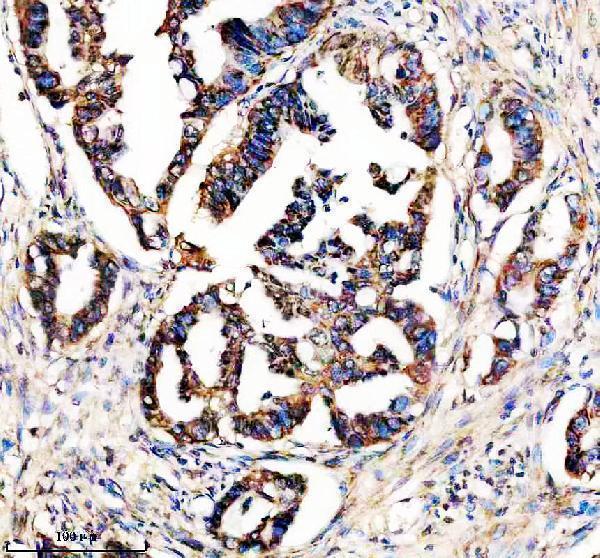
| Expected result | STX4-positive staining in glandular cells of colon (HPA tissue IHC: High). HPA tissue profile: Cytoplasmic and membranous expression in several tissues. No signal in the no-primary control. |
STX4 should appear chiefly at the cell membrane, with cytoplasmic staining also reported in tissue sections (UniProt Q12846 localization; HPA tissue IHC). Strong examples include gastrointestinal glandular cells, kidney tubular cells, and lymph-node germinal-center cells (HPA tissue IHC: High). HPA rates the tissue pattern Approved while noting low consistency between antibody staining and RNA expression; interpret intensity alongside morphology and controls (HPA tissue IHC).
| Membranous staining, with some cytoplasmic signal, in glandular epithelium or renal tubules. | This fits the reported tissue pattern and the membrane location of STX4 (HPA tissue IHC; UniProt Q12846 localization). Score the identified cell population and compartment together; a brown field alone does not establish a positive result (general IHC practice). |
| Strong staining is confined to nuclei, with little membrane or cytoplasmic signal. | An exclusively nuclear pattern conflicts with the reported membrane and cytoplasmic locations (HPA tissue IHC; UniProt Q12846 localization). Treat it as suspect and check morphology, detection background, and a known-positive section before interpreting it as STX4 (general IHC practice). |
| Adipocytes or cardiomyocytes stain strongly while expected positive cells do not. | HPA reports STX4 as Not detected in adipocytes and cardiomyocytes under its IHC conditions (HPA tissue IHC). Consider cross-reactivity or endogenous detection activity, then compare a reagent control and a known-positive tissue; one unexpected stain does not establish expression (general IHC practice). |
| Pale, widespread color obscures cell borders across positive and comparison tissue. | This is difficult to reconcile with a cell-associated membranous and cytoplasmic pattern (HPA tissue IHC). Check blocking, washing, chromogen development, and detection controls before scoring; diffuse background can make weak cells appear positive (general IHC practice). |
| No convincing signal appears in glandular cells of colon or in kidney tubules. | These are High-staining populations in HPA tissue IHC, so a blank run warrants a technical check (HPA tissue IHC: High). Review section quality, retrieval, primary-antibody conditions, and detection with controls; HPA's low staining–RNA consistency cautions against treating one negative slide as proof of absence (HPA tissue IHC; general IHC practice). |
| Membrane topology and compartment | STX4 has a transmembrane segment at residues 276–296 and a cytoplasmic region at 1–275 (UniProt Q12846 topology). A membrane-associated pattern is therefore biologically plausible. The antibody epitope is not given, so topology cannot identify which antigen-retrieval condition will work. |
| Tissue and cell selection | Colon, duodenum, gallbladder, rectum, small intestine, and stomach glandular cells; kidney tubules; and lymph-node germinal-center cells are reported High (HPA tissue IHC). Select and score the named cells, since neighboring cells within a section need not share that reported level (HPA tissue IHC). |
| Strength of the tissue evidence | HPA calls tissue IHC Approved but reports low consistency between staining and RNA expression; antibody HPA001330 is IHC Approved (HPA tissue IHC; HPA antibody validation). Use the observed pattern as a guide, and resolve unexpected staining with controls rather than treating an HPA level as a universal cutoff (general IHC practice). |
| Processing and isoforms | UniProt lists a full-length 1–297 chain, no signal peptide or propeptide, and 2 isoforms (UniProt Q12846 processing; isoforms). No antibody epitope or isoform-specific staining evidence is supplied, so these annotations cannot predict separate IHC patterns or justify a shedding explanation. |
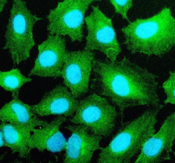
| IF/ICC Q: What localization should a separate IF/ICC assay show? | A: Predominant plasma-membrane signal; HPA approves that subcellular location and rates antibody HPA001330 ICC Enhanced (HPA subcellular ICC-IF; HPA antibody validation). UniProt also lists perinuclear localization and neurite tips in neuronal cells (UniProt Q12846 localization). These observations do not specify an IF/ICC protocol. |
| Situation | Likely cause | Next action |
|---|---|---|
| Positive tissue is blank. | A technical failure is possible when HPA High glandular or tubular cells lack signal (HPA tissue IHC; general IHC practice). | Confirm the named cells are present, then check the run's retrieval, primary-antibody conditions, and detection controls (general IHC practice). No STX4-specific retrieval setting or dilution is supplied. |
| Only nuclei are colored. | This disagrees with reported membrane and cytoplasmic localization (HPA tissue IHC; UniProt Q12846 localization). | Compare with a known-positive section and a detection control; reassess nonspecific color and cell identification before assigning STX4 positivity (general IHC practice). |
| The whole section has diffuse brown haze. | Background can obscure the cell-associated pattern expected for STX4 (HPA tissue IHC; general IHC practice). | Review blocking, washes, and chromogen exposure; use an appropriate negative reagent control to locate background in the detection workflow (general IHC practice). |
| Comparison cells stain as strongly as expected positive cells. | HPA reports adipocytes and cardiomyocytes as Not detected, while several glandular and tubular populations are High (HPA tissue IHC). | Verify cell identity and compare controlled sections. Investigate nonspecific antibody binding or endogenous detection activity; do not infer a new tissue distribution from one section (general IHC practice). |
| A positive cell population shows cytoplasmic color without a crisp surface rim. | HPA describes tissue staining as cytoplasmic and membranous, so cytoplasmic color alone is not automatically incompatible (HPA tissue IHC). | Judge whether staining remains cell-associated and occurs in the expected cells; compare a controlled positive section before rejecting or accepting the result (HPA tissue IHC; general IHC practice). |
| An HPA Not detected tissue has faint staining. | Not detected describes the HPA observation under its conditions, not a proven universal absence of STX4 (HPA tissue IHC). | Score intensity and cell type against the same run's positive and reagent controls; report the observation cautiously if it persists (general IHC practice). |
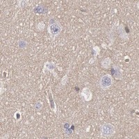

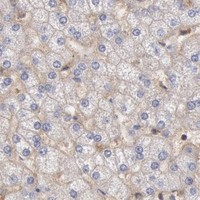


Comprehensive Human Protein Atlas IHC scoring per tissue (reliability: Approved — Low consistency between antibody staining and RNA expression data.). Rows are taken directly from the HPA tissue chart — click any row's HPA link to view the source.
| Tissue | Cell type | Level | Evidence | Source |
|---|---|---|---|---|
| Colon | Glandular cells | High | Protein (IHC) | HPA → |
| Duodenum | Glandular cells | High | Protein (IHC) | HPA → |
| Gallbladder | Glandular cells | High | Protein (IHC) | HPA → |
| Kidney | Cells in tubules | High | Protein (IHC) | HPA → |
| Lymph node | Germinal center cells | High | Protein (IHC) | HPA → |
| Tissue | Cell type | Level | Evidence | Source |
|---|---|---|---|---|
| Adipose tissue | Adipocytes | Not detected | Protein (IHC) | HPA → |
| Caudate | Glial cells | Not detected | Protein (IHC) | HPA → |
| Heart muscle | Cardiomyocytes | Not detected | Protein (IHC) | HPA → |
| Hippocampus | Glial cells | Not detected | Protein (IHC) | HPA → |
| Soft tissue | Fibroblasts | Not detected | Protein (IHC) | HPA → |
Troubleshoot STX4 staining in paraffin sections by checking retrieval, signal location, background and scoring against the documented IHC conditions.
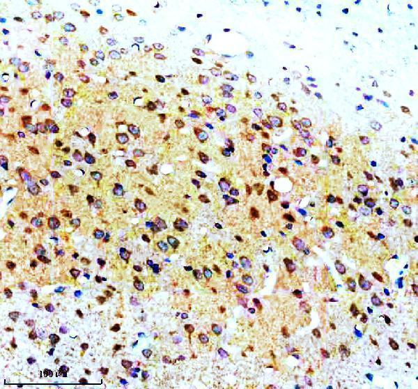
Anti-STX4 antibody A05345-1 has IHC images from human colon cancer and rat brain paraffin sections, plus IF data from A549 cells (catalog image captions). Human, mouse and rat reactivity is listed (catalog).
A05345-1 will render with its IHC figure from a human colon cancer paraffin section; the catalog also shows IHC in a rat brain paraffin section (catalog IHC captions). The same SKU is listed for IHC and IF/ICC and has an IF image from A549 cells (catalog applications and IF caption).
Which to pick: Choose A05345-1 for paraffin-section IHC: its human colon cancer and rat brain captions document EDTA retrieval at pH 8.0 and 2 μg/ml primary antibody (catalog IHC captions); the fixative is unreported. Choose the same SKU for IF/ICC in A549 cells, supported by its 5 μg/ml IF caption and IF/ICC application listing (catalog). For cross-species planning, human, mouse and rat reactivity is listed, while the supplied IHC images document human and rat samples; the antibody is rabbit-hosted, and clonality is unreported (catalog reactivity, host and IHC captions).