This website uses cookies to ensure you get the best experience on our website.


- Table of Contents
Plan SUMO3 staining in paraffin sections around the predominantly nuclear tissue pattern (HPA tissue IHC). The guide covers fixation, staining controls and interpretation, including possible SUMO2/3 cross-reactivity (HPA tissue IHC; datasheet: IHC image caption).

Expected localisation, validated protocols, controls and antibodies — the at-a-glance facts below, then the full design guide.
| Expected localisation | Predominantly nuclear in tissue IHC (HPA tissue IHC) | |
| Staining pattern | Nuclear staining in most tissues (HPA tissue IHC) | |
| Antigen retrieval | Tris-EDTA pH 9.0 HIER, 95–98 °C, 20 min (rule: nuclear antigen) | |
| Positive control | Adipose tissue+4 more · see all | |
| Negative control | None in HPA (detected in all 45 tissues); use no-primary + isotype controls |
| Fixation | Keep formalin fixation consistent across sections (standard IHC practice; not target-specific) | |
| Caveat | SUMO2/3 cross-reactivity may confound staining (HPA tissue IHC; datasheet: IHC image caption) | |
| Regulation | Conjugation requires SAE1–SAE2 and UBE2I (UniProt) | |
| Isoform / epitope | 2 isoforms; processing removes residues 93–103; check epitope (UniProt) |
The catalog antibody’s IHC-P protocol is accompanied by 2 published paraffin-section IHC examples (PMC11345958; PMC11496881).
| Sample | Tissue sections; selected-image fixative not specified (standard IHC workflow) |
| Fixation | Image fixative and duration unreported (datasheet A05193); verify before use. |
| Sectioning | 4–5 µm sections on charged slides (standard) |
| Deparaffinisation | Xylene, graded ethanol series to water (standard) |
| Antigen retrieval | Heat-induced epitope retrieval in Tris-EDTA buffer, pH 9.0, 20 min at 95–98 °C (standard rule: nuclear antigen) |
| Peroxidase block | 3% H2O2, 10 min, room temperature (standard) |
| Blocking | 10% normal serum of the secondary host, 30 min, room temperature (standard) |
| Primary antibody | Rabbit anti-SUMO3, 5 μg/mL (datasheet A05193) |
| Primary incubation | Overnight at 4 °C (standard) |
| Detection | HRP-polymer secondary, DAB chromogen 5–10 min (standard) |
| Counterstain | Hematoxylin, blue, dehydrate and mount (standard) |
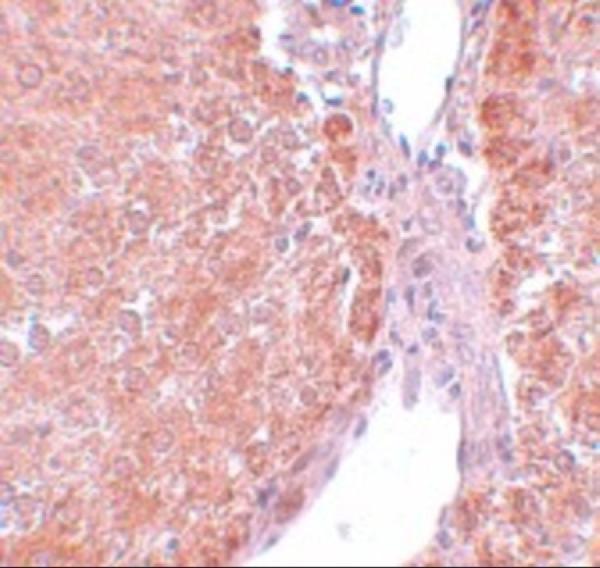
| Expected result | SUMO3-positive staining in adipocytes of adipose tissue (HPA tissue IHC: High). HPA tissue profile: Nuclear expression in most tissues. No signal in the no-primary control. |
SUMO3 is reported in the nucleus, cytoplasm and PML bodies, and has no transmembrane segment (UniProt P55854). In tissue IHC, expect mainly nuclear staining across many cell types, including adipocytes and respiratory epithelial cells (HPA: nuclear expression in most tissues; High in adipocytes and respiratory epithelial cells). HPA rates the tissue pattern Supported, with medium agreement between staining and RNA data, and cautions that the antibodies may detect protein from more than one gene (HPA: reliability description).
| Distinct nuclear staining in adipocytes or bronchial respiratory epithelial cells. | Consistent with the reported IHC pattern and High staining in those cells (HPA: nuclear expression; High in adipocytes and respiratory epithelial cells). Compare nuclei with the surrounding tissue before scoring intensity; HPA's cross-gene caution limits SUMO3-specific attribution (HPA: reliability description). |
| Strong staining confined to cell membranes, with little nuclear staining. | Treat this as a compartment mismatch requiring investigation: SUMO3 has no transmembrane segment (UniProt P55854 topology), while HPA reports predominantly nuclear tissue staining (HPA: tissue IHC profile). Cytoplasmic signal alone is less decisive because UniProt also lists cytoplasmic localisation (UniProt P55854 subcellular location). |
| The strongest signal is in a cell population outside the expected pattern for the selected reference tissue. | Check cell identity and compare with the relevant HPA image; the supplied HPA list is not an exhaustive map of negative cell types (HPA: tissue IHC profile; no negative entries). Unexpected staining can suggest cross-reactivity or endogenous detection activity, but its appearance alone does not establish either cause. |
| Brown colour spreads across nuclei, cytoplasm and extracellular areas without clear cell boundaries. | Treat diffuse signal as background until controls show a defined pattern. HPA reports nuclear expression in most tissues (HPA: tissue IHC profile), but this does not validate indiscriminate colour. Review blocking, washing, chromogen development and the no-primary control as general IHC checks. |
| No convincing signal in a reference area expected to stain strongly. | A blank adipocyte or bronchial epithelial reference area conflicts with HPA's High staining entries (HPA: High in adipocytes and respiratory epithelial cells). First confirm that the expected cells are present and that the detection control worked; a blank section alone cannot distinguish a technical failure from antibody-specific behaviour. |
| Compartment used for scoring | Prioritise nuclear localisation in paraffin tissue because that is the observed IHC profile (HPA: nuclear expression in most tissues). UniProt also lists cytoplasm and PML bodies (UniProt P55854 subcellular location); the supplied tissue profile does not assign a separate expected IHC intensity to either. |
| Choice of positive reference | Adipocytes, hematopoietic cells and bronchial respiratory epithelial cells are reported High, while cardiomyocytes and smooth muscle cells are reported Low (HPA: tissue IHC positive and low lists). Compare the same identified cell type within a section; Low is not a validated negative control. |
| Antibody specificity | HPA describes the tissue evidence as Supported with medium staining–RNA agreement and warns that the antibodies target protein from more than one gene (HPA: reliability description). A matching nuclear pattern therefore supports interpretation of the stain, but cannot by itself establish that every stained molecule is SUMO3. |
| SUMO3 processing and variants | The record lists two isoforms and processing from a 103-residue precursor to a 1–92 chain (UniProt P55854 isoforms and processing). Epitope placement and variant coverage are not supplied, so do not infer that a chosen antibody detects both isoforms or the removed segment. |
| Conjugated SUMO3 | SUMO3 can attach covalently to target proteins as a monomer or polymer (UniProt P55854 function). That biology is compatible with staining at multiple intracellular sites, but tissue IHC localisation alone cannot identify a conjugated substrate or separate conjugated from unconjugated SUMO3. |
| Situation | Likely cause | Next action |
|---|---|---|
| Nuclear staining is weak in a High reference cell type. | The reference expectation may be unmet, or detection may be weak; HPA reports High staining in adipocytes and bronchial respiratory epithelial cells (HPA: tissue IHC positive list). | Confirm the cells and section quality, then review the catalog antibody's IHC-P instructions, retrieval settings and detection control. Treat retrieval adjustments as general IHC optimisation; no SUMO3-specific fixation sensitivity is established here. |
| Colour persists in the no-primary control. | Endogenous detection activity or nonspecific reagent deposition can produce chromogen without primary antibody; this is a general chromogenic IHC failure mode. | Check the detection system's endogenous activity block, blocking and wash steps. Compare the no-primary and primary-stained sections before assigning the colour to SUMO3. |
| Staining is diffuse across the section. | Excess background can obscure the expected mainly nuclear pattern (HPA: tissue IHC profile); the appearance does not identify a single cause. | Inspect background controls and tissue edges, then optimise the catalog antibody's IHC-P dilution, washing and chromogen development within its instructions. Score only cells whose boundaries and nuclei remain interpretable. |
| Membrane-only staining dominates. | That compartment conflicts with the reported mainly nuclear tissue pattern (HPA: tissue IHC profile) and lack of a transmembrane segment (UniProt P55854 topology). | Check the no-primary control, cell boundaries and antibody identification. Repeat against an HPA High reference cell type before treating membrane colour as target staining (HPA: tissue IHC positive list). |
| A Low reference cell type shows visible nuclear colour. | Low does not mean absent: HPA lists cardiomyocytes and smooth muscle cells as Low, without defining them as negative controls (HPA: tissue IHC low list). | Compare intensity and compartment with a High reference on a comparable run (HPA: tissue IHC positive and low lists). Do not call low-level nuclear colour a false positive solely because the reference is labelled Low. |
| What should an IF/ICC image show? | HPA reports supported nucleoplasm and nuclear-body localisation in ICC-IF, with additional approved localisation in sperm structures (HPA: subcellular summary and locations). | Look for nucleoplasmic and nuclear-body signal when interpreting IF/ICC (HPA: supported subcellular locations). Use the separate IF/ICC guide for that application; the supplied IHC evidence does not establish an IF/ICC protocol. |


Comprehensive Human Protein Atlas IHC scoring per tissue (reliability: Supported — Medium consistency between antibody staining and RNA expression data. Caution, targets protein from more than one gene.). Rows are taken directly from the HPA tissue chart — click any row's HPA link to view the source.
| Tissue | Cell type | Level | Evidence | Source |
|---|---|---|---|---|
| Adipose tissue | Adipocytes | High | Protein (IHC) | HPA → |
| Bone marrow | Hematopoietic cells | High | Protein (IHC) | HPA → |
| Bronchus | Respiratory epithelial cells | High | Protein (IHC) | HPA → |
| Caudate | Neuronal cells | High | Protein (IHC) | HPA → |
| Cervix | Glandular cells | High | Protein (IHC) | HPA → |
| Tissue | Cell type | Level | Evidence | Source |
|---|---|---|---|---|
| None in HPA: SUMO3 is detected in all 45 scored tissues. Use a no-primary (secondary-only) and an isotype control instead. | ||||
Use nuclear localisation and controlled chromogenic staining to assess SUMO3, while accounting for antibody cross-reactivity with SUMO2 (UniProt P55854; HPA tissue IHC).
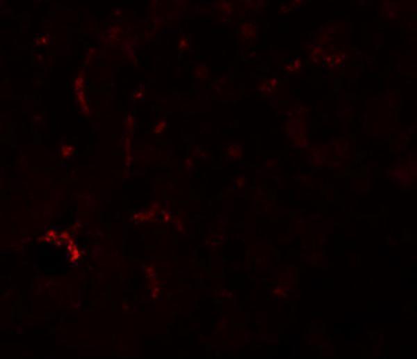
A05193 has real IHC data from rat liver and IF data from mouse liver (IHC and IF captions); its listed reactivity covers human, mouse and rat (catalog reactivity).
A05193 is listed for IHC-P and IF (catalog applications). Its images show rat liver IHC at 5 μg/mL and mouse liver IF at 20 μg/mL (IHC and IF captions).
Which to pick: For paraffin-section IHC, choose A05193 (catalog: IHC-P; IHC caption: rat liver at 5 μg/mL); the fixative is unreported in the caption. For IF/ICC planning, A05193 has mouse liver IF evidence at 20 μg/mL (IF caption), while ICC validation and clonality are unreported (catalog applications and clone field). For work across species, A05193 lists human, mouse and rat reactivity (catalog reactivity); the pictured IHC and IF examples are rat and mouse, respectively (IHC and IF captions).