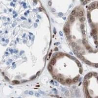
SYK / Tyrosine-protein kinase SYK · IHC design guide

Design Immunohistochemistry for SYK

Plan chromogenic IHC for SYK in paraffin sections using the catalog antibody’s validated conditions. Compare cytoplasmic staining across annotated cell types, and score immune cells separately from surrounding tissue (HPA tissue IHC).

Evidence assembled Oct 2026 · For research use; verify linked source records and product datasheet before use
Immunohistochemistry protocol sheet for SYK (IHC for SYK): expected localisation Cytoplasmic staining, with some membrane signal (HPA tissue IHC), antibody A00490-3, validated IHC image, and IHC protocol steps
Printable SYK IHC protocol sheet — expected localisation Cytoplasmic staining, with some membrane signal (HPA tissue IHC), antibody A00490-3, controls and protocol steps. Open the full SYK IHC guide →

SYK Immunohistochemistry Experimental Design Guide

Expected localisation, validated protocols, controls and antibodies — the at-a-glance facts below, then the full design guide.

Must know before staining
Expected localisation Cytoplasmic staining, with some membrane signal (HPA tissue IHC)
Staining pattern Cytoplasmic in glial, glandular and immune cells (HPA tissue IHC)
Antigen retrieval EDTA pH 8.0 HIER, heat-mediated (datasheet A00490-3)
Positive control ⓘ Appendix+4 more · see all
Negative control ⓘ Adrenal gland+4 more · see all
Important caveats
Reasons your staining may differ from the expected pattern.
Fixation Keep fixation consistent across sections (standard IHC practice; not target-specific); Matched tissue-IHC evidence does not establish the fixation claim. Validate the specimen-specific method before use. (selected-SKU IHC image A00490-3)
Caveat Immune-cell admixture can confound tissue scoring (HPA tissue IHC)
Regulation Expressed from pro-B to plasma cells (UniProt)
Isoform / epitope Two isoforms, Long and Short; verify epitope coverage (UniProt)
Section 1

Recommended SYK IHC & IF Protocols

The catalog antibody’s IHC-P protocol (datasheet A00490-3) is paired with four published SYK IHC workflows (PMC11775117; PMC5257279; PMC8576038; PMC7072502).

Recommended immunohistochemistry (IHC-P) protocol parameters
SampleParaffin-embedded human lymphoma tissue; fixative not specified (datasheet A00490-3)
FixationImage fixative and duration unreported (datasheet A00490-3); verify before use.
Sectioning4–5 µm sections on charged slides (standard)
DeparaffinisationXylene, graded ethanol series to water (standard)
Antigen retrievalHeat retrieval: EDTA pH 8.0 (datasheet A00490-3); 20 min, 95–100 °C (standard)
Peroxidase block3% H2O2, 10 min, room temperature (standard)
Blocking10% goat serum (datasheet A00490-3)
Primary antibodyRabbit anti-SYK, 2-5μg/ml (datasheet A00490-3)
Primary incubationOvernight at 4 °C (datasheet A00490-3)
DetectionHRP-conjugated secondary, DAB chromogen (datasheet A00490-3)
CounterstainHematoxylin, blue, dehydrate and mount (standard)
Expected resultSYK-positive staining in glandular cells of appendix (HPA tissue IHC: High). HPA tissue profile: Distinct cytoplasmic expression in most tissues, highly abundant in glial cells of CNS, glandular and immune cells in several tissues. No signal in the no-primary control.
💡Decision noteStart with heat-mediated EDTA at pH 8.0 for the catalog antibody (datasheet A00490-3). Use the published citrate conditions when adapting those articles’ antibody workflows (PMC5257279; PMC7072502).
Section 2

What Is the Expected SYK Staining Pattern?

SYK is a non-receptor kinase found in the cytosol and at the cell membrane; it has no transmembrane segment (UniProt P43405 topology and subcellular location). In paraffin-section IHC, expect predominantly cytoplasmic staining in appropriate cells, including bone-marrow hematopoietic cells, lung macrophages and lymph-node germinal-center cells (HPA: High in each). HPA rates its tissue staining profile Enhanced, reflecting consistency between antibody staining and RNA expression (HPA: tissue IHC reliability).

What am I looking at on my slide?
Distinct cytoplasmic stain in lung macrophages or bone-marrow hematopoietic cells (HPA: High in each).This fits the reported tissue pattern; membrane-associated signal can also fit SYK localization (HPA: tissue IHC; UniProt P43405 subcellular location). Judge the named cells, rather than treating every cell in the section as equally positive.
Nuclear staining dominates while the expected cytoplasmic pattern is absent (HPA: tissue IHC profile).Treat the compartment mismatch as a possible artifact and review morphology, background and controls before scoring it as SYK. The supplied localization evidence supports cytosol and membrane, with no nuclear location listed (UniProt P43405; HPA: subcellular).
Strong stain appears in cardiomyocytes or adrenal glandular cells (HPA: Not detected in each).These cell-specific results conflict with the HPA tissue profile. Consider antibody cross-reactivity or endogenous chromogen-generating activity, and check a reagent control; staining elsewhere in the same tissue needs separate interpretation (HPA: tissue IHC; general IHC practice).
Diffuse color covers cells and surrounding tissue without clear cellular boundaries.This is background until controls show a reproducible cellular pattern. Check reagent-only controls and washing, then compare the suspected positive cells with the cytoplasmic HPA pattern (general IHC practice; HPA: tissue IHC profile).
No stain appears in bone-marrow hematopoietic cells or lung macrophages (HPA: High in each).A blank known-positive cell population makes the run inconclusive. First check section quality, antibody and detection steps, and a positive control; HPA staining levels alone do not identify a fixation effect (general IHC practice; HPA: tissue IHC).
💡Expected SYK appearanceCall a positive result when identifiable hematopoietic, macrophage or germinal-center cells show distinct, often strong cytoplasmic stain, with possible membrane-associated signal; dominant nuclear color or indiscriminate diffuse color warrants review (HPA: High in named cells and cytoplasmic tissue profile; UniProt P43405 subcellular location).
How each factor affects the staining
Compartment (UniProt P43405; HPA: subcellular)SYK is cytosolic and membrane-associated without a transmembrane segment (UniProt P43405 topology and subcellular location). Interpret membrane accentuation alongside cytoplasmic signal, rather than requiring an exclusively membrane-bound outline.
Cell-specific tissue distribution (HPA: tissue IHC)High staining is reported in lymph-node germinal-center cells and several glandular cell populations, while prostate and salivary glandular cells are Not detected (HPA: tissue IHC). Select and score the documented cell population, not the organ as a whole.
Antibody evidence (HPA: tissue IHC and antibodies)The HPA tissue profile is rated Enhanced; HPA001384 and CAB007773 each have Enhanced IHC status (HPA: reliability and antibody validation). This supports the reference pattern, but a catalog antibody used here still needs its own controls.
Isoforms and modification (UniProt P43405)UniProt lists Long and Short isoforms and multiple modified residues, including phosphotyrosines (UniProt P43405 isoforms and modified residues). The supplied record does not establish which forms a particular IHC antibody recognizes; avoid assigning stain intensity to activation state.
Why is my staining missing, weak or wrong?
SituationLikely causeNext action
Expected positive cells are blank (HPA: High in bone-marrow hematopoietic cells).The run may have failed at staining or detection; HPA does not report target-specific fixation sensitivity (general IHC practice; HPA: tissue IHC).Confirm the positive-control section, antibody identity, retrieval and detection sequence; adjust conditions using the antibody's IHC instructions (general IHC practice).
Nuclear color dominates (HPA: cytoplasmic tissue profile).This conflicts with the supplied cytosol and membrane localization (UniProt P43405; HPA: subcellular).Check the reagent control and cell boundaries before interpretation; repeat with controlled staining conditions if the mismatch persists (general IHC practice).
Macrophages stain, but the whole lung section also looks positive (HPA: High in macrophages).HPA's High designation applies to macrophages, not every lung cell (HPA: tissue IHC).Score macrophages separately and inspect the other cells against morphology and controls (general IHC practice).
Negative-reference cells show strong chromogen (HPA: cardiomyocytes Not detected).Cross-reactivity or endogenous detection activity is possible (general IHC practice); the result conflicts with HPA's cell-specific observation (HPA: tissue IHC).Review no-primary and detection controls; address endogenous enzyme activity as appropriate for the chromogenic system (general IHC practice).
Diffuse stain obscures cellular localization.Nonspecific reagent binding or inadequate washing can produce background (general IHC practice).Compare reagent controls, then review blocking, antibody concentration and washes while retaining a documented positive-cell control (general IHC practice; HPA: tissue IHC).
How should an IF/ICC image be judged?HPA reports plasma membrane and cytosol as main locations, with vesicles additionally observed (HPA: subcellular ICC-IF).Judge fluorescence against those compartments and appropriate controls; use the separate IF/ICC guide for method choices (HPA: subcellular; general IF practice).

Sample controls for SYK IHC & IF

🧪Run bone marrow first: hematopoietic cells should stain (HPA: High in bone marrow hematopoietic cells). Use heart muscle cardiomyocytes as the negative tissue (HPA: Not detected in cardiomyocytes); on the bone marrow slide, cells outside the stained population should show only background signal, assessed against the no-primary control.
Positive control tissue: Appendix (Glandular cells, HPA High)
Negative control tissue: Adrenal gland (HPA Not detected)
ICC-IF cell lines (HPA subcellular resource): HPA ICC-IF images show SYK in A-431, HEK293, U2OS, with annotated localisation: Plasma membrane (approved), Cytosol (approved) (HPA subcellular).
Technical controls: Include a no-primary, secondary-only control and a rabbit IgG isotype control matched to the primary antibody’s class and clonality where known (selected-SKU caption: rabbit primary). Confirm target specificity with SYK knockout material or a validated peptide-block control; suppress endogenous peroxidase activity in bone marrow before HRP/DAB detection (selected-SKU caption: HRP/DAB; HPA: bone marrow hematopoietic cells).
⚠️Feasibility: The selected-SKU caption demonstrates paraffin-section IHC after heat retrieval in EDTA at pH 8.0, but reports no fixative; a target-specific fixation window and the effect of fixation on SYK staining are unreported (selected-SKU tissue-IHC caption). Retrieval is part of that demonstrated workflow, but its necessity relative to other retrieval conditions is unreported (selected-SKU tissue-IHC caption). There is no supplied comparison establishing that frozen sections or IF/ICC are easier; for bone marrow, endogenous peroxidase can produce chromogenic background and warrants a no-primary control (HPA: bone marrow hematopoietic cells; selected-SKU caption: HRP/DAB).

HPA tissue IHC evidence for SYK

Comprehensive Human Protein Atlas IHC scoring per tissue (reliability: Enhanced — High consistency between antibody staining and RNA expression data. Antibody staining in cells/structures not annotated, view images.). Rows are taken directly from the HPA tissue chart — click any row's HPA link to view the source.

Positive expression · recommended positive controls

TissueCell typeLevelEvidenceSource
Appendix Glandular cells High Protein (IHC) HPA →
Bone marrow Hematopoietic cells High Protein (IHC) HPA →
Cerebellum Granular cells - cytoplasm/membrane High Protein (IHC) HPA →
Duodenum Glandular cells High Protein (IHC) HPA →
Fallopian tube Glandular cells High Protein (IHC) HPA →

Undetected expression · recommended negative controls

TissueCell typeLevelEvidenceSource
Adrenal gland Glandular cells Not detected Protein (IHC) HPA →
Heart muscle Cardiomyocytes Not detected Protein (IHC) HPA →
Liver Cholangiocytes Not detected Protein (IHC) HPA →
Ovary Follicle cells Not detected Protein (IHC) HPA →
Placenta Decidual cells Not detected Protein (IHC) HPA →
Section 3

Advanced SYK IHC Tips

Troubleshoot SYK staining in paraffin sections by checking retrieval, cell identity and subcellular pattern before interpreting chromogenic signal.

How should I retrieve SYK antigen when staining paraffin sections?
Use heat-mediated EDTA pH 8.0 retrieval for SYK in paraffin sections (datasheet A00490-3). The selected tissue image used this retrieval before overnight incubation with 2 μg/ml primary antibody at 4°C (datasheet A00490-3). If staining is weak, compare a modest range of heating times while holding section thickness, antibody concentration and detection conditions constant (standard IHC practice). Include a positive control section in every retrieval run; hematopoietic cells in bone marrow are a documented high-staining population (HPA: High in bone marrow hematopoietic cells). Evaluate improved cellular signal alongside tissue damage and background, rather than choosing the hottest condition automatically (standard IHC practice).
Could fixation explain weak or uneven SYK staining?
The selected SYK tissue caption describes a paraffin section but does not report its fixative, so target-specific fixation sensitivity is unknown (datasheet A00490-3). Record the specimen fixative, fixation duration and time to fixation before comparing sections; those variables can change antigen accessibility and tissue morphology (standard IHC practice). For a controlled comparison, stain sections with differing documented processing histories in the same run using EDTA pH 8.0 retrieval (datasheet A00490-3). Include a high-staining reference population, such as bone marrow hematopoietic cells, when available (HPA: High in bone marrow hematopoietic cells). Interpret a processing-associated difference as a specimen effect until repeat sections and matched controls support it (standard IHC practice).
Where should convincing SYK staining appear in a paraffin section?
Assess SYK chiefly as cytoplasmic staining, with possible membrane-associated accentuation, rather than requiring a nuclear pattern (UniProt P43405: cytosol and cell membrane; HPA: distinct cytoplasmic tissue expression). SYK has no transmembrane segment, so membrane-adjacent signal should be interpreted as localization of a signaling protein, not an integral membrane outline (UniProt P43405 topology). The selected chromogenic example detects SYK in human lymphoma tissue, but its caption does not assign staining to a particular cell compartment (datasheet A00490-3). Compare individual cells with adjacent unstained cells and section morphology to separate cellular signal from diffuse chromogen deposition (standard IHC practice). Confirm unexpected compartment patterns with a second validated reagent or orthogonal evidence before scoring them as SYK (standard IHC practice).
Can the antibody distinguish SYK isoforms or modified epitopes?
SYK has Long and Short isoforms and contains 2 SH2 domains plus a kinase domain spanning residues 371–631 (UniProt P43405). Its record also lists numerous modified residues, including phosphotyrosines at positions 28, 47 and 131 (UniProt P43405). The supplied tissue caption gives antibody concentration and retrieval but no mapped epitope or isoform specificity, so a positive section cannot identify either isoform or phosphorylation state (datasheet A00490-3). Check the antibody's epitope documentation before comparing specimens with different processing histories (standard IHC practice). For isoform-specific or activation claims, pair chromogenic staining with an independently validated assay that actually resolves the feature of interest (standard IHC practice).
How would I examine SYK by multiplex immunofluorescence?
For IF, pair SYK with a marker identifying the expected cell population, such as a macrophage marker in lung, where macrophages show high SYK staining (HPA: High in lung macrophages). Select spectrally separated fluorophores after imaging an unstained section in each channel, because tissue autofluorescence can resemble weak signal (standard IF practice). Use a permeabilisation condition that allows access to cytosolic epitopes while preserving membrane detail; SYK localizes to cytosol and plasma membrane and lacks a transmembrane segment (UniProt P43405 topology; HPA: approved subcellular locations). Check single-label controls for bleed-through and secondary-antibody cross-reactivity before interpreting overlap (standard IF practice). The supplied chromogenic tissue caption supplies no IF fixation or antibody validation conditions (datasheet A00490-3).
How can I reduce diffuse or misleading DAB background?
First compare the stained section with a no-primary control to identify signal from the detection system or tissue itself (standard IHC practice). The selected method used 10% goat serum blocking and a peroxidase-linked secondary antibody with DAB detection (datasheet A00490-3). Apply an endogenous peroxidase block as a general chromogenic IHC step, and inspect controls before attributing brown deposits to SYK (standard IHC practice). If background remains broad, titrate the primary around the documented 2 μg/ml condition and optimize wash stringency without changing multiple variables at once (datasheet A00490-3; standard IHC practice). Preserve recognizable cytoplasmic staining in expected cells while reducing diffuse or edge-associated signal (HPA: cytoplasmic tissue expression; standard IHC practice).
How should I score SYK staining across tissue sections? ⚠ ANSWER MARKED FOR VERIFICATION
Define the cell population and compartment before scoring, because SYK appears in both immune and glandular populations across tissues (HPA: tissue IHC profile). For identifiable cells, report the percentage positive and an intensity-weighted H-score using the same thresholds and exposure-independent DAB assessment across slides (standard IHC practice). For dispersed immune cells, report positive-cell density per mm² of viable analyzed tissue and state how cells were classified (standard IHC practice). Normalize comparisons to the area or number of eligible cells, excluding folds, necrosis and damaged edges by a preset rule (standard IHC practice). Include a reference section across runs; lymph node germinal center cells show high staining in the supplied tissue profile (HPA: High in lymph node germinal center cells).
What distinguishes true SYK positivity from staining artefact?
A convincing result follows cell boundaries and shows predominantly cytoplasmic signal, sometimes with membrane-associated signal, in an appropriate cell population (HPA: cytoplasmic tissue profile; UniProt P43405 localization). High staining in lung macrophages or bone marrow hematopoietic cells supports a positive-control comparison, while cardiomyocytes are listed as not detected (HPA: tissue IHC). Treat isolated nuclear-only signal, uniform edge staining and deposits over necrotic areas as findings that need control review (UniProt P43405 localization; standard IHC practice). Check a no-primary section and endogenous peroxidase blocking before assigning DAB precipitate to SYK (standard IHC practice). Interpret staining as protein detection, not proof of kinase activation or a particular isoform (UniProt P43405: phosphorylation sites and Long/Short isoforms).
Boster reagents

Best SYK / Tyrosine-protein kinase SYK IHC Antibodies

Two anti-SYK antibodies have IHC images from human tissue (catalog image captions); their stated reactivity covers human, mouse, and rat (catalog reactivity). One also lists IF validation (catalog applications).

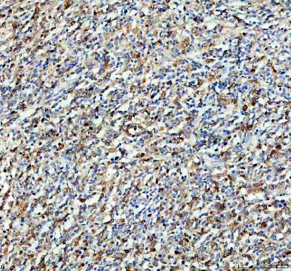
Real IHC data IHC analysis of SYK using anti-SYK antibody (A00490-3). SYK was detected in a paraffin-embedded section of human lymphoma tissue. Heat mediated antigen retrieval was performed in EDTA buffer (pH 8.0, epitope retrieval solution). The tissue section was blocked with 10% goat serum. The tissue section was then incubated with 2 μg/ml rabbit anti-SYK Antibody (A00490-3) overnight at 4°C. Peroxidase Conjugated Goat Anti-rabbit IgG was used as secondary antibody and incubated for 30 minutes at 37°C. The tissue section was developed using HRP Conjugated Rabbit IgG Super Vision Assay Kit (Catalog # SV0002) with DAB as the chromogen.
Anti-SYK Antibody ®
Cat # A00490-3
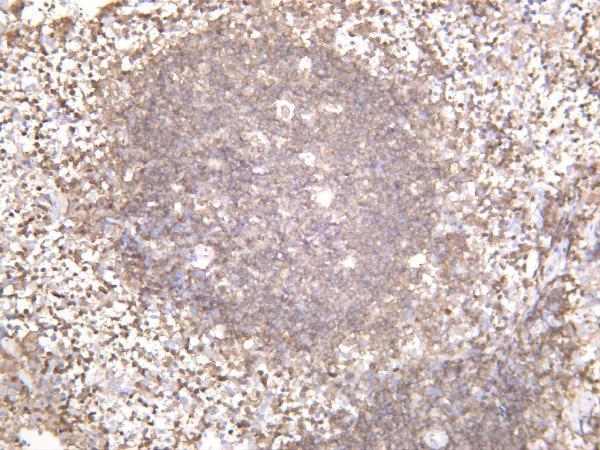
Real IHC data Human spleen was stained with anti-Syk rabbit antibody
Anti-Syk Rabbit Monoclonal Antibody
Cat # M00490-1

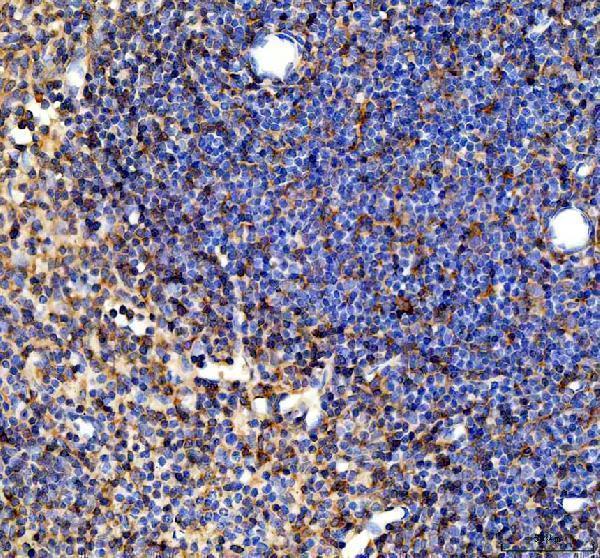
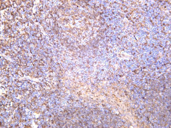
A00490-3 will render with a human lymphoma paraffin-section IHC image; its additional captions show mouse and rat spleen paraffin sections (A00490-3 image captions). M00490-1 will render with a human spleen IHC image; its additional captions show mouse and rat spleen, and its application list includes IF (M00490-1 image captions; catalog applications).

Which to pick: For paraffin-section IHC, choose A00490-3: its own caption specifies EDTA retrieval at pH 8.0 and 2 μg/ml primary antibody; the fixative is unreported, so the caption does not establish fixation or tissue validation (A00490-3 image caption). For IF, choose M00490-1 at the listed 1:200–1:1000 dilution; its application list names IF but does not separately name ICC, and its IHC spleen captions do not report processing or fixative (M00490-1 catalog dilution and applications; M00490-1 image captions). For cross-species tissue IHC, both list human, mouse, and rat reactivity and have IHC captions for those species; M00490-1 is monoclonal, while A00490-3 has no clonality stated (catalog reactivity and descriptions; image captions). The selected A00490-3 tissue-IHC caption documents paraffin sections, but does not specify the fixative (selected-SKU IHC image A00490-3).

Each figure is that product's own IHC / IF validation image from its datasheet.

References

  1. UniProt Consortium. UniProt entry P43405 (KSYK_HUMAN, Tyrosine-protein kinase SYK).
  2. Human Protein Atlas. SYK tissue IHC expression (reliability: Enhanced).
  3. Human Protein Atlas. SYK subcellular location (ICC-IF): Mainly localized to the plasma membrane and cytosol. In addition localized to vesicles..
  4. Human Protein Atlas. SYK antibody validation summary (2 antibodies).
  5. Primary intestinal T-cell and natural killer-cell lymphomas: Clinicopathologic and prognostic features of 79 cases in South China. American journal of clinical pathology 2025 — PMC11775117.
  6. Inhibition of Spleen Tyrosine Kinase Potentiates Paclitaxel-Induced Cytotoxicity in Ovarian Cancer Cells by Stabilizing Microtubules. Cancer cell 2015 — PMC5257279.
  7. The FcεRI signaling pathway is involved in the pathogenesis of lacrimal gland benign lymphoepithelial lesions as shown by transcriptomic analysis. Scientific reports 2021 — PMC8576038.
  8. Synergistic Anti-Tumour Effect of Syk Inhibitor and Olaparib in Squamous Cell Carcinoma: Roles of Syk in EGFR Signalling and PARP1 Activation. Cancers 2020 — PMC7072502.
  9. PubMed PMID:7513161 — UniProt-cited evidence.
  10. PubMed PMID:8163536 — UniProt-cited evidence.
  11. PubMed PMID:15164053 — UniProt-cited evidence.