Click image to see more details
-

-
-
-
-
+2
Product Info Summary
| SKU: | A00490-3 |
|---|---|
| Size: | 100 μg/vial |
| Reactive Species: | Human, Mouse, Rat |
| Host: | Rabbit |
| Application: | ELISA, Flow Cytometry, IHC, WB |
Customers Who Bought This Also Bought

Product info
Product Name
Anti-SYK Antibody Picoband®
SKU/Catalog Number
A00490-3
Size
100 μg/vial
Form
Lyophilized
Description
Boster Bio Anti-SYK Antibody Picoband® catalog # A00490-3. Tested in ELISA, Flow Cytometry, IHC, WB applications. This antibody reacts with Human, Mouse, Rat. The brand Picoband indicates this is a premium antibody that guarantees superior quality, high affinity, and strong signals with minimal background in Western blot applications. Only our best-performing antibodies are designated as Picoband, ensuring unmatched performance.
Storage & Handling
Store at -20˚C for one year from date of receipt. After reconstitution, at 4˚C for one month. It can also be aliquotted and stored frozen at -20˚C for six months. Avoid repeated freeze-thaw cycles.
Cite This Product
Anti-SYK Antibody Picoband® (Boster Biological Technology, Pleasanton CA, USA, Catalog # A00490-3)
Host
Rabbit
Contents
Each vial contains 4 mg Trehalose, 0.9 mg NaCl and 0.2 mg Na2HPO4.
Clonality
Polyclonal
Isotype
Rabbit IgG
Immunogen
E.coli-derived human SYK recombinant protein (Position: A10-N635).
Cross-reactivity
No cross-reactivity with other proteins.
Reactive Species
A00490-3 is reactive to SYK in Human, Mouse, Rat
Observed Molecular Weight
72 kDa
Calculated molecular weight
72.1 kDa
Background of SYK
Spleen tyrosine kinase, also known as Syk, is an enzyme which in humans is encoded by the SYK gene. It is mapped to 9q22.2. This gene encodes a member of the family of non-receptor type Tyr protein kinases. This protein is widely expressed in hematopoietic cells and is involved in coupling activated immunoreceptors to downstream signaling events that mediate diverse cellular responses, including proliferation, differentiation, and phagocytosis. It is thought to be a modulator of epithelial cell growth and a potential tumour suppressor in human breast carcinomas. Alternatively spliced transcript variants encoding different isoforms have been found for this gene.
Antibody Validation
Boster validates all antibodies on WB, IHC, ICC, Immunofluorescence, and ELISA with known positive control and negative samples to ensure specificity and high affinity, including thorough antibody incubations.
Application & Images
Applications
A00490-3 is guaranteed for ELISA, Flow Cytometry, IHC, WB Boster Guarantee
Recommend Dilution
| Application | Dilution | Species |
|---|---|---|
| Western blot | 0.25-0.5μg/ml | Human, Mouse, Rat |
| Immunohistochemistry(Paraffin-embedded Section) | 2-5μg/ml | Human, Mouse, Rat |
| Flow Cytometry (Fixed) | 1-3μg/1x106 cells | Human |
| ELISA | 0.1-0.5μg/ml | - |
Tested application
Suggested blocking solution with 5% non-fat milk or BSA; (*)Recommended protein loading: 20-40 µg per lane
Use TE buffer pH 9.0 for antigen retrieval; (*) citrate buffer pH 6.0 is an alternative.
Validation Images & Assay Conditions
Click image to see more details
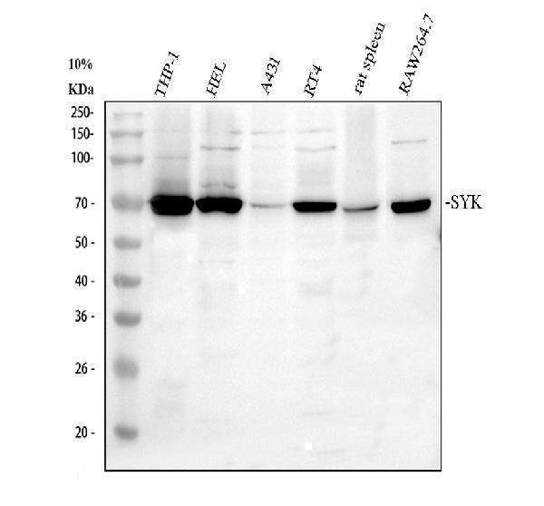
Western blot analysis of SYK using anti-SYK antibody (A00490-3).
Electrophoresis was performed on a 10% SDS-PAGE gel at 80V (Stacking gel) / 120V (Resolving gel) for 2 hours. The sample well of each lane was loaded with 30 ug of sample under reducing conditions.
Lane 1: human THP-1 whole cell lysates,
Lane 2: human HEL whole cell lysates,
Lane 3: human A431 whole cell lysates,
Lane 4: human RT4 whole cell lysates,
Lane 5: rat spleen tissue lysates,
Lane 6: mouse RAW264.7 whole cell lysates.
After electrophoresis, proteins were transferred to a nitrocellulose membrane at 150 mA for 50-90 minutes. Blocked the membrane with 5% non-fat milk/TBS for 1.5 hour at RT. The membrane was incubated with rabbit anti-SYK antigen affinity purified polyclonal antibody (A00490-3) at 0.5 μg/mL overnight at 4°C, then washed with TBS-0.1%Tween 3 times with 5 minutes each and probed with a goat anti-rabbit IgG-HRP secondary antibody (Catalog # BA1054) at a dilution of 1:5000 for 1.5 hour at RT. The signal is developed using an ECL Plus Western Blotting Substrate (Catalog # AR1196-200) with Tanon 5200 system. A specific band was detected for SYK at approximately 72 kDa. The expected band size for SYK is at 72 kDa.

Click image to see more details
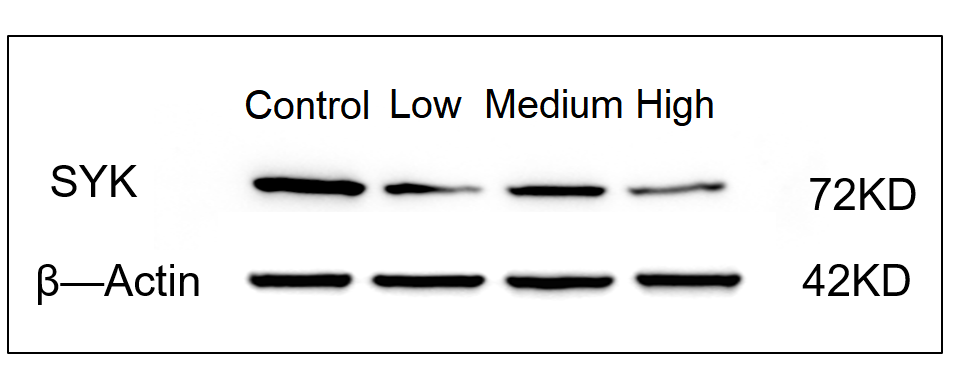
Western blot analysis of SYK using anti-SYK antibody (A00490-3).
Electrophoresis was performed on a 10% SDS-PAGE gel at 80V (Stacking gel) / 120V (Resolving gel) for 2 hours. The sample well of each lane was loaded with 30 ug of sample under reducing conditions.
Lane 1: mouse HT22 whole cell lysates,
Lane 2: low concentration drug treamented-mouse HT22 whole cell lysates,
Lane 3: medium concentration drug treamented-mouse HT22 whole cell lysates,
Lane 4: High concentration drug treamented-mouse HT22 whole cell lysates.
After electrophoresis, proteins were transferred to a nitrocellulose membrane at 150 mA for 50-90 minutes. Blocked the membrane with 5% non-fat milk/TBS for 1.5 hour at RT. The membrane was incubated with rabbit anti-SYK antigen affinity purified polyclonal antibody (A00490-3) at 1:1000 overnight at 4°C, then washed with TBS-0.1%Tween 3 times with 5 minutes each and probed with a goat anti-rabbit IgG-HRP secondary antibody (Catalog # BA1054) at a dilution of 1:2000 for 1 hour at RT. The signal is developed using an ECL Plus Western Blotting Substrate (Catalog # AR1196-200) with Tanon 5200 system. A specific band was detected for SYK at approximately 72 kDa. The expected band size for SYK is at 72 kDa.

Click image to see more details
IHC analysis of SYK using anti-SYK antibody (A00490-3).
SYK was detected in a paraffin-embedded section of human lymphoma tissue. Heat mediated antigen retrieval was performed in EDTA buffer (pH 8.0, epitope retrieval solution). The tissue section was blocked with 10% goat serum. The tissue section was then incubated with 2 μg/ml rabbit anti-SYK Antibody (A00490-3) overnight at 4°C. Peroxidase Conjugated Goat Anti-rabbit IgG was used as secondary antibody and incubated for 30 minutes at 37°C. The tissue section was developed using HRP Conjugated Rabbit IgG Super Vision Assay Kit (Catalog # SV0002) with DAB as the chromogen.

Click image to see more details
IHC analysis of SYK using anti-SYK antibody (A00490-3).
SYK was detected in a paraffin-embedded section of mouse spleen tissue. Heat mediated antigen retrieval was performed in EDTA buffer (pH 8.0, epitope retrieval solution). The tissue section was blocked with 10% goat serum. The tissue section was then incubated with 2 μg/ml rabbit anti-SYK Antibody (A00490-3) overnight at 4°C. Peroxidase Conjugated Goat Anti-rabbit IgG was used as secondary antibody and incubated for 30 minutes at 37°C. The tissue section was developed using HRP Conjugated Rabbit IgG Super Vision Assay Kit (Catalog # SV0002) with DAB as the chromogen.

Click image to see more details
IHC analysis of SYK using anti-SYK antibody (A00490-3).
SYK was detected in a paraffin-embedded section of rat spleen tissue. Heat mediated antigen retrieval was performed in EDTA buffer (pH 8.0, epitope retrieval solution). The tissue section was blocked with 10% goat serum. The tissue section was then incubated with 2 μg/ml rabbit anti-SYK Antibody (A00490-3) overnight at 4°C. Peroxidase Conjugated Goat Anti-rabbit IgG was used as secondary antibody and incubated for 30 minutes at 37°C. The tissue section was developed using HRP Conjugated Rabbit IgG Super Vision Assay Kit (Catalog # SV0002) with DAB as the chromogen.

Click image to see more details
Flow Cytometry analysis of RT4 cells using anti-SYK antibody (A00490-3).
Overlay histogram showing RT4 cells stained with A00490-3 (Blue line). To facilitate intracellular staining, cells were fixed with 4% paraformaldehyde and permeabilized with permeabilization buffer. The cells were blocked with 10% normal goat serum. And then incubated with rabbit anti-SYK Antibody (A00490-3, 1 μg/1x106 cells) for 30 min at 20°C. DyLight®488 conjugated goat anti-rabbit IgG (BA1127, 5-10 μg/1x106 cells) was used as secondary antibody for 30 minutes at 20°C. Isotype control antibody (Green line) was rabbit IgG (1 μg/1x106) used under the same conditions. Unlabelled sample without incubation with primary antibody and secondary antibody (Red line) was used as a blank control.
Specific Publications For Anti-SYK Antibody Picoband® (A00490-3)
Loading publications
Recommended Resources
Here are featured tools and databases that you might find useful.
- Boster's Pathways Library
- Protein Databases
- Bioscience Research Protocol Resources
- Data Processing & Analysis Software
- Photo Editing Software
- Scientific Literature Resources
- Research Paper Management Tools
- Molecular Biology Software
- Primer Design Tools
- Bioinformatics Tools
- Phylogenetic Tree Analysis
Customer Reviews
Have you used Anti-SYK Antibody Picoband®?
Share your experimental results or join a short interview to earn up to $1,000 in product credits or other rewards.
1 Reviews For Anti-SYK Antibody Picoband®
This antibody is highly specific and efficient, suitable for Western blot detection of Anti-SYK Antibody protein in HT22 cells, with only minor nonspecific bands observed.
Excellent
| SKU | A00318-1 |
|---|---|
| Application | Western Blot |
| Sample | Mouse hippocampal tissue |
| Sample Processing Description | Mouse brain tissues were lysed in RIPA buffer containing protease inhibitors at 4°C for 2 hours, followed by centrifugation to collect the supernatant. Protein concentration was then determined, and after adjusting to the desired concentration, samples were mixed with 5× protein loading buffer and denatured by heating for 10 minutes. Fifteen microliters of each sample were loaded per lane for electrophoresis. |
| Other Reagents | Blocking buffer |
| Primary Antibody | PI 3 Kinase p85 alpha/PIK3R1 Antibody Picoband® |
| Primary Incubation | 1:2000, overnight at 4 ℃ |
| Secondary Antibody | HRP Goat Anti-Rabbit IgG |
| Secondary Incubation | 1:10000, 1 hour in room temperature |
| Detection | Substrate: ECL, Imaging system:ChemiDoc MP |
| Results Summary | The figure shows a schematic of the WB results for PIK3R1 and the loading control β-actin in brain tissues from normal mice, model mouse, and mouse treated with high and low doses of AB drug. Although the expression differences between experimental groups are not obvious, the WB results with this antibody are still clear and well-defined. |
Yu Tian, Institute of Materia Medica, Beijing
Verified customer
Submitted 2025-12-25
Customer Q&As
Have a question?
Find answers in Q&As, reviews.
Can't find your answer?
Submit your question
5 Customer Q&As for Anti-SYK Antibody Picoband®
Question
We have seen staining in mouse leukocyte. Do you have any suggestions? Is anti-SYK antibody supposed to stain leukocyte positively?
Verified Customer
Verified customer
Asked: 2020-04-30
Answer
From literature leukocyte does express SYK. From Uniprot.org, SYK is expressed in leukocyte, eye lymph, tonsil, platelet, leukemic t-cell, b-cell, among other tissues. Regarding which tissues have SYK expression, here are a few articles citing expression in various tissues:
B-cell, Pubmed ID: 21469132
Eye, and Lymph, Pubmed ID: 15489334
Leukemic T-cell, Pubmed ID: 19690332
Platelet, Pubmed ID: 18088087
Tonsil, Pubmed ID: 8168854
Boster Scientific Support
Answered: 2020-04-30
Question
We bought anti-SYK antibody for WB on tonsil in the past. I am using rat, and We intend to use the antibody for Flow Cytometry next. We want examining tonsil as well as platelet in our next experiment. Do you have any suggestion on which antibody would work the best for Flow Cytometry?
Verified Customer
Verified customer
Asked: 2020-04-03
Answer
I looked at the website and datasheets of our anti-SYK antibody and it seems that A00490-3 has been validated on rat in both WB and Flow Cytometry. Thus A00490-3 should work for your application. Our Boster satisfaction guarantee will cover this product for Flow Cytometry in rat even if the specific tissue type has not been validated. We do have a comprehensive range of products for Flow Cytometry detection and you can check out our website bosterbio.com to find out more information about them.
Boster Scientific Support
Answered: 2020-04-03
Question
We were satisfied with the WB result of your anti-SYK antibody. However we have been able to see positive staining in leukocyte cell membrane. cytoplasm, using this antibody. Is that expected? Could you tell me where is SYK supposed to be expressed?
Verified Customer
Verified customer
Asked: 2020-03-12
Answer
From literature, leukocyte does express SYK. Generally SYK expresses in cell membrane. cytoplasm,. Regarding which tissues have SYK expression, here are a few articles citing expression in various tissues:
B-cell, Pubmed ID: 21469132
Eye, and Lymph, Pubmed ID: 15489334
Leukemic T-cell, Pubmed ID: 19690332
Platelet, Pubmed ID: 18088087
Tonsil, Pubmed ID: 8168854
Boster Scientific Support
Answered: 2020-03-12
Question
We are currently using anti-SYK antibody A00490-3 for human tissue, and we are happy with the WB results. The species of reactivity given in the datasheet says human, mouse, rat. Is it true that the antibody can work on goat tissues as well?
Verified Customer
Verified customer
Asked: 2020-02-03
Answer
The anti-SYK antibody (A00490-3) has not been validated for cross reactivity specifically with goat tissues, but there is a good chance of cross reactivity. We have an innovator award program that if you test this antibody and show it works in goat you can get your next antibody for free. Please contact me if I can help you with anything.
Boster Scientific Support
Answered: 2020-02-03
Question
I am interested in using your anti-SYK antibody for role of phospholipids in phagocytosis studies. Has this antibody been tested with western blotting on a431 cells? We would like to see some validation images before ordering.
Verified Customer
Verified customer
Asked: 2019-11-20
Answer
Thank you for your inquiry. This A00490-3 anti-SYK antibody is validated on a431 cells, mouse lung. It is guaranteed to work for ELISA, Flow Cytometry, WB in human, mouse, rat. Our Boster guarantee will cover your intended experiment even if the sample type has not been be directly tested.
Boster Scientific Support
Answered: 2019-11-20
