TCF7L2 / Transcription factor 7-like 2 · IHC design guide

Design Immunohistochemistry for TCF7L2

Plan TCF7L2 paraffin IHC around nuclear staining across tissues (HPA tissue IHC) and the catalog antibody’s documented 1:200 tissue protocol (datasheet A00431-2). This guide covers fixation, antigen retrieval, chromogenic detection and interpretation.

Evidence assembled Oct 2026 · For research use; verify linked source records and product datasheet before use
Immunohistochemistry protocol sheet for TCF7L2 (IHC for TCF7L2): expected localisation Nuclear staining across tissues (HPA tissue IHC), antibody A00431-2, validated IHC image, and IHC protocol steps
Printable TCF7L2 IHC protocol sheet — expected localisation Nuclear staining across tissues (HPA tissue IHC), antibody A00431-2, controls and protocol steps. Open the full TCF7L2 IHC guide →

TCF7L2 Immunohistochemistry Experimental Design Guide

Expected localisation, validated protocols, controls and antibodies — the at-a-glance facts below, then the full design guide.

Must know before staining
Expected localisation Nuclear staining across tissues (HPA tissue IHC)
Staining pattern Broad nuclear staining, including glandular and glial cells (HPA tissue IHC)
Antigen retrieval EDTA pH 8.0 HIER, heat-mediated (datasheet A00431-2)
Positive control ⓘ Adrenal gland+4 more · see all
Negative control ⓘ None in HPA (detected in all 45 tissues); use no-primary + isotype controls
Important caveats
Reasons your staining may differ from the expected pattern.
Fixation Keep fixation consistent across paraffin sections. (standard IHC practice; not target-specific)
Caveat Staining may not closely track tissue RNA levels (HPA tissue IHC)
Regulation Crypt-to-villus expression gradient (UniProt)
Isoform / epitope 17 isoforms; epitope coverage needs checking (UniProt)
Section 1

Recommended TCF7L2 IHC & IF Protocols

The catalog antibody’s IHC-P protocol (datasheet: A00431-2) accompanies four published TCF7L2 paraffin-section methods (PMC3748032; PMC4300120; PMC5946422; PMC5423960).

Recommended immunohistochemistry (IHC-P) protocol parameters
SampleParaffin-embedded human glioma tissue; fixative not specified (datasheet A00431-2)
FixationImage fixative and duration unreported (datasheet A00431-2); verify before use.
Sectioning4–5 µm sections on charged slides (standard)
DeparaffinisationXylene, graded ethanol series to water (standard)
Antigen retrievalHeat retrieval: EDTA pH 8.0 (datasheet A00431-2); 20 min, 95–100 °C (standard)
Peroxidase block3% H2O2, 10 min, room temperature (standard)
Blocking10% goat serum (datasheet A00431-2)
Primary antibodyRabbit anti-TCF7L2, 1:50 recommended; image 1:200 (datasheet A00431-2)
Primary incubationOvernight at 4 °C (datasheet A00431-2)
DetectionHRP-conjugated secondary, DAB chromogen (datasheet A00431-2)
CounterstainHematoxylin, blue, dehydrate and mount (standard)
Expected resultTCF7L2-positive staining in glandular cells of adrenal gland (HPA tissue IHC: High). HPA tissue profile: Ubiquitous nuclear expression. No signal in the no-primary control.
💡Decision noteStart with heat-mediated EDTA pH 8.0 retrieval (datasheet: A00431-2); one published method specifies 1 mM EDTA at 95°C for 20 min (PMC4300120).
Section 2

What Is the Expected TCF7L2 Staining Pattern?

TCF7L2 should appear predominantly in nuclei: UniProt reports diffuse nuclear localization with PML nuclear bodies, and HPA describes ubiquitous nuclear tissue staining (UniProt Q9NQB0 subcellular location; HPA tissue IHC: Supported). Look for signal in the cells present in the chosen section, including glandular cells in breast or hematopoietic cells in bone marrow, both reported as high staining (HPA tissue IHC). TCF7L2 has no transmembrane segment (UniProt Q9NQB0 topology).

What am I looking at on my slide?
Clear nuclear staining in breast glandular cells or bone marrow hematopoietic cells.This matches the reported high staining in those cell populations and the broadly nuclear tissue profile (HPA tissue IHC). Judge the named cell population and its nuclei; a positive section does not establish that every cell should stain equally.
Predominantly membranous or exclusively cytoplasmic staining, with little nuclear signal.This conflicts with the expected IHC compartment (HPA tissue IHC: ubiquitous nuclear expression; UniProt Q9NQB0 subcellular location). Treat it as a possible staining artefact and reassess specificity; HPA reports cytosol only as an uncertain additional ICC-IF location (HPA subcellular).
Strong signal in a cell population that does not match the selected HPA tissue image.Cross-reactivity or endogenous detection activity is possible (general IHC practice). First confirm cell identity and compare the same tissue and compartment; HPA reports low tissue specificity, so staining outside its listed high-staining examples alone is insufficient to call a false positive (HPA tissue IHC).
Diffuse staining across nuclei, cytoplasm, and surrounding section, without clear cell boundaries.Poor separation of signal from background prevents a confident localization call (general IHC practice). Check a control without primary antibody and review blocking, antibody concentration, washes, and detection chemistry; the expected tissue profile is nuclear (HPA tissue IHC).
No nuclear signal in an otherwise interpretable, known-positive section.A negative result in an HPA high-staining cell population warrants a technical check before biological interpretation (HPA tissue IHC). Confirm that the relevant cells are present, then review the antibody's IHC-P validation and the general retrieval and detection workflow (general IHC practice).
💡Expected TCF7L2 appearanceCall a positive result when distinct nuclear chromogenic signal is visible in the expected cells, with high staining possible in the listed HPA populations; isolated membranous or dominant cytoplasmic signal without nuclear staining is suspect (HPA tissue IHC; UniProt Q9NQB0 subcellular location).
How each factor affects the staining
Tissue and cell selectionHPA describes ubiquitous nuclear expression and low tissue RNA specificity, while listing high staining in specific populations, including breast glandular and bone marrow hematopoietic cells (HPA tissue IHC). Select a documented cell population for a positive comparison; the list is not a set of exclusive expression sites.
Strength of tissue evidenceHPA rates the tissue IHC profile Supported and describes medium consistency between antibody staining and RNA expression (HPA tissue IHC). Use its compartment and cell examples as expectations, then interpret unexpected intensity with controls rather than treating one image as a fixed scoring threshold.
Antibody validation and isoformsCAB013535 has Supported IHC status; HPA038800 has Supported ICC status, with no IHC status supplied for it (HPA antibodies). UniProt lists 17 isoforms (UniProt Q9NQB0 isoforms). The supplied record does not identify either antibody's epitope or establish which isoforms it detects.
Processing and topologyUniProt lists one chain spanning residues 1–619, no signal peptide or propeptide, and no transmembrane segment (UniProt Q9NQB0 processing and topology). These annotations support interpreting the nuclear pattern; they do not establish an IHC retrieval condition or a fixation sensitivity.
IF/ICC appearance?For IF/ICC, HPA supports nucleoplasm as the main location and nuclear bodies as an additional location; its additional cytosol assignment is uncertain (HPA subcellular). This is a localization comparison only. Use the separate IF/ICC guide for that application, rather than treating ICC images as an IHC-P protocol.
Why is my staining missing, weak or wrong?
SituationLikely causeNext action
The expected cells are present, but their nuclei are unstained.The selected antibody or detection workflow may not yield an interpretable IHC-P signal (general IHC practice); the HPA high-staining examples make these cells useful checks (HPA tissue IHC).Verify IHC-P validation for the antibody and run an appropriate positive section; review general retrieval, primary incubation, and detection steps without assuming a TCF7L2-specific retrieval requirement (general IHC practice).
Signal is chiefly cytoplasmic or membranous.Off-target binding or background is possible (general IHC practice). Predominantly nuclear tissue staining is the supported expectation (HPA tissue IHC); ICC-IF cytosol is marked uncertain (HPA subcellular).Check nuclear counterstain alignment, compare a control without primary antibody, and reassess antibody specificity before scoring nonnuclear signal (general IHC practice).
Many structures stain uniformly, including areas outside cells.Nonspecific background or endogenous detection activity may obscure the nuclear pattern (general IHC practice).Inspect a control without primary antibody, then review blocking, washes, and the detection system's appropriate endogenous-activity controls (general IHC practice). Score only interpretable cellular signal.
An unexpected cell population stains strongly.Cell identification, cross-reactivity, or endogenous activity may explain the finding (general IHC practice). HPA's low tissue specificity limits conclusions from tissue identity alone (HPA tissue IHC).Recheck morphology and nuclear localization, compare the matching HPA cell population, and use specificity and detection controls before calling the result aberrant (HPA tissue IHC; general IHC practice).
Two antibodies give different staining patterns.Their application validation differs: CAB013535 is Supported for IHC, whereas HPA038800 is Supported for ICC with no IHC status supplied (HPA antibodies). Their epitope coverage is not supplied.Interpret the paraffin-section result using IHC-P validation and matched controls. Document each antibody separately; do not infer that their discordance identifies a particular one of the 17 isoforms (HPA antibodies; UniProt Q9NQB0 isoforms).
Fine nuclear puncta appear alongside diffuse nuclear signal.TCF7L2 can localize to a subset of PML nuclear bodies, while its nuclear pattern is also described as diffuse (UniProt Q9NQB0 subcellular location); HPA additionally supports nuclear bodies in ICC-IF (HPA subcellular).Assess the overall nuclear pattern and control background. Do not require visible puncta for a positive chromogenic IHC call; interpretation of puncta depends on image resolution and appropriate controls (general IHC practice).

Sample controls for TCF7L2 IHC & IF

🧪Run breast first: its glandular cells should stain for TCF7L2 (HPA: High in breast glandular cells). HPA detects TCF7L2 in all 45 scored tissues, so there is no supported negative tissue; use no-primary and isotype controls, and treat adjacent cells as internal negatives only if they remain unstained on the positive slide (HPA: no negative tissue rows).
Positive control tissue: Adrenal gland (Glandular cells, HPA High)
Negative control tissue: None in HPA: TCF7L2 is detected in all 45 scored tissues. Use a no-primary (secondary-only) and an isotype control instead.
ICC-IF cell lines (HPA subcellular resource): HPA ICC-IF images show TCF7L2 in A-431, U-251MG, U2OS, KOLF2.1J, NIH 3T3, with annotated localisation: Nucleoplasm (supported) (HPA subcellular).
Technical controls: Run a secondary-only slide and an isotype-matched rabbit IgG control alongside a TCF7L2 knockout sample as a biological specificity control (caption: rabbit primary antibody; standard IHC practice). Quench endogenous peroxidase in the breast section before HRP–DAB detection (caption: HRP–DAB detection; standard IHC practice).
⚠️Feasibility: A target-specific fixation window and fixation effect are unreported, and the selected A00431-2 paraffin-section caption does not state the fixative (caption: fixative unreported). The caption documents heat retrieval in EDTA at pH 8.0, followed by a 1:200 primary dilution overnight at 4°C; it does not establish that retrieval is required for every specimen (caption: retrieval and incubation conditions). The supplied evidence does not establish whether frozen sections or IF are easier than paraffin IHC; assess nuclear staining against the expected nucleoplasmic pattern and check HRP–DAB slides for endogenous peroxidase background (HPA: supported nucleoplasm; caption: HRP–DAB; standard IHC practice).

HPA tissue IHC evidence for TCF7L2

Comprehensive Human Protein Atlas IHC scoring per tissue (reliability: Supported — Medium consistency between antibody staining and RNA expression data.). Rows are taken directly from the HPA tissue chart — click any row's HPA link to view the source.

Positive expression · recommended positive controls

TissueCell typeLevelEvidenceSource
Adrenal gland Glandular cells High Protein (IHC) HPA →
Appendix Glandular cells High Protein (IHC) HPA →
Bone marrow Hematopoietic cells High Protein (IHC) HPA →
Breast Glandular cells High Protein (IHC) HPA →
Bronchus Respiratory epithelial cells High Protein (IHC) HPA →

Undetected expression · recommended negative controls

TissueCell typeLevelEvidenceSource
None in HPA: TCF7L2 is detected in all 45 scored tissues. Use a no-primary (secondary-only) and an isotype control instead.
Section 3

Advanced TCF7L2 IHC Tips

Troubleshoot TCF7L2 staining in paraffin sections by checking retrieval, nuclear localisation, controls and scoring before interpreting biological differences.

What retrieval should I try first when TCF7L2 nuclear staining is weak?
Start with heat-mediated retrieval in EDTA pH 8.0 for this catalog antibody (datasheet A00431-2). The selected paraffin-section example used that retrieval before 1:200 primary antibody overnight at 4°C, so compare your run against those documented conditions (datasheet A00431-2). If nuclei remain weak, check heating consistency, section adhesion and detection reagents before changing retrieval conditions (standard IHC practice). If needed, trial an alternative buffer or heating duration on serial sections, recording nuclear signal and tissue damage for each condition (standard IHC practice). Judge improvement by crisp nuclear staining relative to a no-primary control, rather than increased overall brown colour (UniProt Q9NQB0 localisation; standard IHC practice).
Could fixation explain weak or uneven TCF7L2 staining?
The selected paraffin-section caption does not state its fixative, and target-specific fixation sensitivity is unknown from the supplied evidence (datasheet A00431-2). Record the specimen’s actual fixative, fixation interval and processing history before comparing sections; these variables can alter antigen accessibility in routine IHC (standard IHC practice). Compare serial sections processed together while holding EDTA pH 8.0 retrieval, 1:200 primary dilution and detection constant (datasheet A00431-2; standard IHC practice). Uneven staining that follows section thickness, folds or drying should prompt a processing review before a biological interpretation (standard IHC practice). Use nuclear staining in intact cells as the readout, because TCF7L2 is primarily nuclear (UniProt Q9NQB0 localisation; HPA subcellular).
How should I evaluate TCF7L2 staining outside the nucleus?
Expect predominantly nuclear staining: UniProt places TCF7L2 in the nucleus, including some PML bodies, and describes a diffuse nuclear pattern (UniProt Q9NQB0 localisation). HPA reports supported nucleoplasmic and nuclear-body localisation, while its additional cytosolic localisation is uncertain (HPA subcellular). Examine intact, counterstained nuclei at the same exposure and development conditions as controls, separating diffuse nuclear signal from occasional nuclear puncta (standard IHC practice; UniProt Q9NQB0 localisation). Strong staining confined to cytoplasm, extracellular material or damaged edges deserves a specificity check with a no-primary control and independent antibody where available (standard IHC practice). Do not score brown pigment over a nucleus unless its boundaries and cellular context remain clear (standard IHC practice).
Could isoform choice or epitope masking change the TCF7L2 staining pattern?
TCF7L2 has 17 listed isoforms, so establish which sequence region the antibody recognizes before comparing specimens or studies (UniProt Q9NQB0 isoforms; standard IHC practice). The supplied caption documents paraffin-section staining but gives no epitope map or isoform coverage, so a negative section cannot identify a missing isoform (datasheet A00431-2). UniProt lists NLK-associated phosphothreonines at residues 201 and 212; their effect on this antibody’s tissue staining is unknown (UniProt Q9NQB0 modified residues). Keep retrieval and detection constant across matched sections, and compare antibody specificity information with any available orthogonal expression measurement (standard IHC practice). Report the antibody identity and scoring compartment so apparent differences remain interpretable (standard IHC practice).
How can I cross-check the nuclear IHC pattern by multiplex IF?
Use IF as a separate cross-check and pair TCF7L2 with a validated marker for the expected cell population in your specimen (standard IF practice). Choose spectrally separated fluorophores after inspecting unstained tissue for autofluorescence, and include single-colour controls when channels overlap (standard IF practice). Because TCF7L2 is nuclear and has no transmembrane segment, optimise permeabilisation to allow nuclear antibody access while preserving cell boundaries (UniProt Q9NQB0 localisation and topology; standard IF practice). Add a nuclear counterstain and compare nucleoplasmic signal and any nuclear puncta with the chromogenic pattern (HPA subcellular; standard IF practice). Do not carry the catalog antibody’s paraffin-section retrieval or dilution into IF as a validated IF condition (datasheet A00431-2).
What should I check when DAB background obscures TCF7L2 nuclei?
First compare the stained section with a no-primary control and inspect whether diffuse brown signal tracks tissue damage, folds or section edges (standard IHC practice). The selected paraffin-section example used 10% goat serum blocking, 1:200 rabbit primary overnight at 4°C, and a peroxidase-linked secondary followed by DAB (datasheet A00431-2). Apply an appropriate endogenous peroxidase block before HRP detection and assess nonspecific secondary binding with a suitable control (standard IHC practice). If background persists, adjust blocking, washing and antibody concentration in controlled comparisons while keeping EDTA pH 8.0 retrieval documented (standard IHC practice; datasheet A00431-2). Accept a change only when nuclear contrast improves without losing plausible positive cells (UniProt Q9NQB0 localisation; standard IHC practice).
How should I score TCF7L2 across sections with different cellularity? ⚠ ANSWER MARKED FOR VERIFICATION
Define the cell population and tissue area before scoring, then count nuclei with convincing chromogenic signal rather than all brown pixels (UniProt Q9NQB0 localisation; standard IHC practice). Report % positive nuclei and, when intensity categories are reproducible, an H-score with the category definitions recorded (standard IHC practice). For spatial comparisons, report positive-cell density per mm² and normalise it to the number of intact, eligible cells in the same region (standard IHC practice). Keep retrieval, development time, counterstain, imaging and threshold rules consistent across sections, and exclude folds and necrotic areas using preset criteria (standard IHC practice). Record region selection because UniProt describes an intestinal crypt-to-villus expression gradient (UniProt Q9NQB0 tissue specificity).
How do I distinguish true TCF7L2 staining from artefact?
Give greatest weight to signal within intact nuclei, consistent with UniProt’s nuclear localisation and HPA’s supported nucleoplasmic pattern (UniProt Q9NQB0 localisation; HPA subcellular). HPA describes ubiquitous nuclear expression with Supported tissue-IHC reliability, so staining in an unexpected cell type still requires cellular identification and controls (HPA tissue IHC; standard IHC practice). Check whether apparent positivity follows cut edges, folds, necrosis or regions positive in a no-primary control; those patterns warrant an artefact review (standard IHC practice). For DAB detection, assess endogenous peroxidase and pigment before attributing brown deposits to the antigen (standard IHC practice). Interpret any cytosolic signal cautiously because HPA marks that additional location uncertain (HPA subcellular).
Boster reagents

Best TCF7L2 / Transcription factor 7-like 2 IHC Antibodies

Real IHC images show human glioma, lung cancer and colonic cancer, and rat brain (catalog image captions); an ICC image shows HeLa cells (catalog image caption). All three antibodies list human, mouse and rat reactivity (catalog).

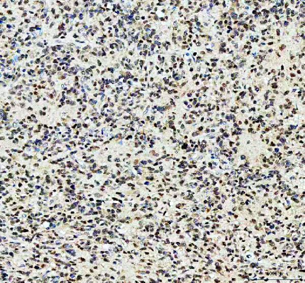
Real IHC data IHC analysis of TCF4/TCF7L2 using anti-TCF4/TCF7L2 antibody (A00431-2). TCF4/TCF7L2 was detected in a paraffin-embedded section of human glioma tissue. Heat mediated antigen retrieval was performed in EDTA buffer (pH 8.0, epitope retrieval solution). The tissue section was blocked with 10% goat serum. The tissue section was then incubated with 1:200 rabbit anti-TCF4/TCF7L2 Antibody (A00431-2) overnight at 4°C. Peroxidase Conjugated Goat Anti-rabbit IgG was used as secondary antibody and incubated for 30 minutes at 37°C. The tissue section was developed using HRP Conjugated Rabbit IgG Super Vision Assay Kit (Catalog # SV0002) with DAB as the chromogen.
Anti-TCF4/TCF7L2 Antibody
Cat # A00431-2
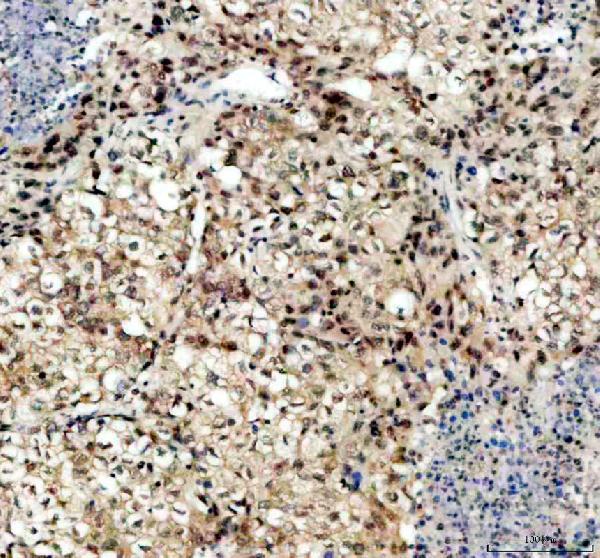
Real IHC data Immunohistochemical analysis of paraffin-embedded human colonic cancer, using TCF7L2 Antibody.
Anti-TCF7L2 Rabbit Monoclonal Antibody
Cat # M00431
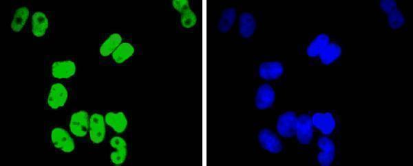
Real IF data ICC staining TCF7L2 in Hela cells (green). The nuclear counter stain is DAPI (blue). Cells were fixed in paraformaldehyde, permeabilised with 0.25% Triton X100/PBS.
Anti-Transcription factor 7-like 2 TCF7L2 Antibody
Cat # A00431-1

A00431-2 has paraffin-section IHC images from human glioma, human lung cancer and rat brain (A00431-2 image captions); M00431 has a paraffin-section IHC image from human colonic cancer (M00431 image caption). A00431-1 has an ICC image of paraformaldehyde-fixed, permeabilised HeLa cells (A00431-1 image caption), and its applications include IF and IHC (catalog).

Which to pick: For tissue IHC, choose A00431-2 for its human and rat paraffin-section examples with EDTA retrieval (A00431-2 image captions), or M00431 if a monoclonal antibody is preferred for human colonic cancer paraffin sections (catalog: clone FIB-20; M00431 image caption); the tissue captions do not report the fixative. For IF/ICC, A00431-1 has a HeLa ICC image showing paraformaldehyde fixation and Triton X100 permeabilisation (A00431-1 image caption). For cross-species tissue work, A00431-2 has pictured human and rat IHC examples (A00431-2 image captions); mouse reactivity is listed for all three SKUs (catalog).

Each figure is that product's own IHC / IF validation image from its datasheet.

References

  1. UniProt Consortium. UniProt entry Q9NQB0 (TF7L2_HUMAN, Transcription factor 7-like 2).
  2. Human Protein Atlas. TCF7L2 tissue IHC expression (reliability: Supported).
  3. Human Protein Atlas. TCF7L2 subcellular location (ICC-IF): Mainly localized to the nucleoplasm. In addition localized to the nuclear bodies and cytosol..
  4. Human Protein Atlas. TCF7L2 antibody validation summary (2 antibodies).
  5. Limited TCF7L2 expression in MS lesions. PloS one 2013 — PMC3748032.
  6. The type 2 diabetes associated rs7903146 T allele within TCF7L2 is significantly under-represented in Hereditary Multiple Exostoses: insights into pathogenesis. Bone 2015 — PMC4300120.
  7. Tcf7L2 is essential for neurogenesis in the developing mouse neocortex. Neural development 2018 — PMC5946422.
  8. Pancreatic alpha cell-selective deletion of Tcf7l2 impairs glucagon secretion and counter-regulatory responses to hypoglycaemia in mice. Diabetologia 2017 — PMC5423960.
  9. PubMed PMID:9065401 — UniProt-cited evidence.
  10. PubMed PMID:10919662 — UniProt-cited evidence.
  11. PubMed PMID:19602480 — UniProt-cited evidence.