THBS2 / Thrombospondin-2 · IHC design guide

Design Immunohistochemistry for THBS2

Plan THBS2 paraffin-section IHC around the granular cytoplasmic pattern reported in several tissues (HPA tissue IHC). Compare staining with the tissue profile while allowing for differences between RNA and protein locations for this secreted protein (HPA tissue IHC).

Evidence assembled Oct 2026 · For research use; verify linked source records and product datasheet before use
Immunohistochemistry protocol sheet for THBS2 (IHC for THBS2): expected localisation Cytoplasm in tissue sections (HPA tissue IHC), antibody A03253, validated IHC image, and IHC protocol steps
Printable THBS2 IHC protocol sheet — expected localisation Cytoplasm in tissue sections (HPA tissue IHC), antibody A03253, controls and protocol steps. Open the full THBS2 IHC guide →

THBS2 Immunohistochemistry Experimental Design Guide

Expected localisation, validated protocols, controls and antibodies — the at-a-glance facts below, then the full design guide.

Must know before staining
Expected localisation Cytoplasm in tissue sections (HPA tissue IHC)
Staining pattern Granular cytoplasmic staining in several tissues (HPA tissue IHC)
Antigen retrieval Citrate pH 6.0 HIER, 95–98 °C, 20 min (rule: cytoplasmic / membrane antigen)
Positive control ⓘ Adipose tissue+4 more · see all
Negative control ⓘ None in HPA (detected in all 45 tissues); use no-primary + isotype controls
Important caveats
Reasons your staining may differ from the expected pattern.
Fixation Keep fixation consistent across paraffin sections (standard IHC practice; not target-specific)
Caveat Secreted protein: RNA and protein locations may differ (HPA tissue IHC)
Regulation Blood vessel-enhanced RNA (HPA tissue RNA)
Isoform / epitope 0 isoforms listed; signal peptide 1–18 precedes chain 19–1172 (UniProt)
Section 1

Recommended THBS2 IHC & IF Protocols

The catalog antibody’s IHC-P protocol (datasheet) is accompanied by four published THBS2 IHC protocols (PMC11178913; PMC8853287; PMC5727893; PMC4189190).

Recommended immunohistochemistry (IHC-P) protocol parameters
SampleParaffin-embedded rat testis tissue; fixative not specified (datasheet A03253)
FixationImage fixative and duration unreported (datasheet A03253); verify before use.
Sectioning4–5 µm sections on charged slides (standard)
DeparaffinisationXylene, graded ethanol series to water (standard)
Antigen retrievalHeat-induced epitope retrieval in citrate buffer, pH 6.0, 20 min at 95–98 °C (standard rule: cytoplasmic / membrane antigen)
Peroxidase block3% H2O2, 10 min, room temperature (standard)
Blocking10% normal serum of the secondary host, 30 min, room temperature (standard)
Primary antibodyRabbit anti-THBS2, 1:100 (datasheet A03253)
Primary incubationOvernight at 4 °C (standard)
DetectionHRP-polymer secondary, DAB chromogen 5–10 min (standard)
CounterstainHematoxylin, blue, dehydrate and mount (standard)
Expected resultTHBS2-positive staining in adipocytes of adipose tissue (HPA tissue IHC: Medium). HPA tissue profile: Granular cytoplasmic expression in several tissues. No signal in the no-primary control.
💡Decision noteStart with citrate pH 6.0 HIER at 95–98 °C for 20 min (page retrieval rule); two published protocols also use citrate retrieval (PMC5727893; PMC4189190).
Section 2

What Is the Expected THBS2 Staining Pattern?

THBS2 IHC shows granular cytoplasmic staining in several tissues, including adipocytes and glandular cells (HPA: Approved tissue IHC). Interpret this pattern with its medium RNA–protein consistency in mind: HPA describes THBS2 as secreted, and UniProt records a signal peptide and no transmembrane segment (HPA tissue IHC; UniProt P35442 topology).

What am I looking at on my slide?
Granular cytoplasmic stain in adipocytes or glandular cellsFits the observed tissue pattern; these cells show medium staining (HPA tissue IHC).
Nuclear-only stainDoes not fit the granular cytoplasmic tissue pattern; assess artefact (HPA tissue IHC).
Strong stain in oral mucosa squamous cellsHPA reports low staining there; assess cross-reactivity or endogenous detection activity (HPA tissue IHC).
Diffuse stain across cells and surrounding tissueObscures the reported granular pattern; assess nonspecific background (HPA tissue IHC).
No stain in adipocytes on an otherwise valid sectionUnexpected against medium staining reported for adipocytes; assess assay sensitivity (HPA tissue IHC).
💡Expected THBS2 appearanceCall a positive result when granular cytoplasmic staining is visible at medium intensity in an HPA-listed cell type such as adipocytes; nuclear-only staining is discordant (HPA tissue IHC).
How each factor affects the staining
Tissue interpretationRNA and protein locations may differ for secreted THBS2; IHC–RNA consistency is medium (HPA tissue IHC).
Processing and topologySignal peptide is residues 1–18; no transmembrane segment is recorded (UniProt P35442).
IF/ICC: where is THBS2 seen?Mainly plasma membrane; cytosol and centrosome also reported (HPA ICC-IF).
Why is my staining missing, weak or wrong?
SituationLikely causeNext action
No stain in adipocytesAssay sensitivity may be insufficient; adipocytes show medium staining (HPA tissue IHC).General IHC practice: check positive control, retrieval and antibody dilution.
Diffuse background obscures granulesThe reported granular cytoplasmic pattern cannot be resolved (HPA tissue IHC).General IHC practice: check blocking, washes and detection controls.
Nuclear-only stainingDiscordant with the reported tissue pattern (HPA tissue IHC).General IHC practice: review controls and antibody specificity.
Strong oral mucosa squamous-cell stainUnexpected against HPA's low staining; cross-reactivity is possible (HPA tissue IHC).General IHC practice: compare with a negative control and another validated antibody.
Staining without primary antibodyEndogenous detection activity or reagent background is possible (general IHC practice).General IHC practice: check peroxidase blocking and detection reagents.

Sample controls for THBS2 IHC & IF

🧪Run adipose tissue first and assess staining in adipocytes (HPA: Medium in adipocytes). HPA detects THBS2 in all 45 scored tissues, so use no-primary and isotype controls for the negative comparison; cells without visible signal on the positive slide provide background reference, but should not be assumed THBS2-negative (HPA: no negative tissue listed).
Positive control tissue: Adipose tissue (Adipocytes, HPA Medium)
Negative control tissue: None in HPA: THBS2 is detected in all 45 scored tissues. Use a no-primary (secondary-only) and an isotype control instead.
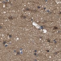
ICC-IF cell lines (HPA subcellular resource): HPA ICC-IF images show THBS2 in A-431, U-251MG, U2OS, ASC52telo, with annotated localisation: Plasma membrane (approved) (HPA subcellular).
Technical controls: Include a no-primary, secondary-only control, an isotype control matched to the primary antibody’s host species, clonality and concentration, and THBS2-knockout tissue or a validated peptide-block control (standard IHC practice). For chromogenic detection in adipose tissue, block endogenous peroxidase and assess staining against the no-primary control (standard IHC practice).
⚠️Feasibility: A target-specific fixation window and retrieval dependency are unreported in the supplied evidence; the selected A03253 paraffin-section caption also leaves the fixative unreported (A03253 tissue-IHC caption). The evidence does not establish whether frozen sections or IF are easier for THBS2 (HPA: ICC-IF images available). In adipose sections, lipid extraction can leave empty adipocyte spaces that should not be scored as negative staining (standard IHC practice).

HPA tissue IHC evidence for THBS2

Comprehensive Human Protein Atlas IHC scoring per tissue (reliability: Approved — Medium consistency between antibody staining and RNA expression data. Secreted protein, tissue location of RNA and protein is expected to differ.). Rows are taken directly from the HPA tissue chart — click any row's HPA link to view the source.

Positive expression · recommended positive controls

TissueCell typeLevelEvidenceSource
Adipose tissue Adipocytes Medium Protein (IHC) HPA →
Adrenal gland Glandular cells Medium Protein (IHC) HPA →
Appendix Glandular cells Medium Protein (IHC) HPA →
Breast Glandular cells Medium Protein (IHC) HPA →
Bronchus Respiratory epithelial cells Medium Protein (IHC) HPA →

Undetected expression · recommended negative controls

TissueCell typeLevelEvidenceSource
None in HPA: THBS2 is detected in all 45 scored tissues. Use a no-primary (secondary-only) and an isotype control instead.
Section 3

Advanced THBS2 IHC Tips

Use the catalog antibody’s paraffin-section image and the supplied localization evidence to plan THBS2 staining, controls, and scoring.

What retrieval should I start with for THBS2 in paraffin sections?
Start with citrate buffer at pH 6.0, heated to 95–98 °C for 20 min (page retrieval rule). Cool the sections before blocking, and keep heating and cooling conditions consistent across slides so differences in staining can be compared (standard IHC practice). The catalog image documents THBS2 staining in paraffin-embedded rat testis at 1:100, but its caption does not report a retrieval method (catalog image: A03253). If signal remains weak, compare a second retrieval condition on adjacent sections with the same detection settings; assess tissue damage and background alongside signal (standard IHC practice).
How should I troubleshoot weak staining when fixation details are missing?
The THBS2-specific effect of fixation on staining is unknown from the supplied evidence; the rat-testis caption identifies paraffin embedding but leaves the fixative unreported (catalog image: A03253). Record each specimen’s fixative, fixation time, processing history, and section age before comparing staining intensity across blocks (standard IHC practice). For a new specimen series, compare routinely processed sections from known fixation conditions using identical pH 6.0 retrieval and detection settings (page retrieval rule; standard IHC practice). Include a previously staining control section in each run, and interpret a weak study section only after that control performs as expected (standard IHC practice).
Should THBS2 appear in cytoplasm, at cell borders, or in extracellular material?
In tissue IHC, the supplied profile describes granular cytoplasmic THBS2 staining across several tissues (HPA tissue IHC). THBS2 has a signal peptide at residues 1–18, lacks a transmembrane segment, and can bind fibrinogen, fibronectin, laminin, and type V collagen (UniProt P35442). The cell-image dataset additionally reports approved plasma-membrane localization, with cytosol and centrosome as additional locations (HPA subcellular). Score cellular granules, border-associated signal, and extracellular staining separately; inspect adjacent morphology and a no-primary control before assigning diffuse matrix color to THBS2 (standard IHC practice). Do not require identical cellular and extracellular distributions when comparing sections, because secreted protein and its source cells may occupy different locations (HPA tissue IHC).
Could epitope placement explain different THBS2 staining patterns?
Check the antibody’s stated immunogen or epitope before interpreting a compartment-specific IHC result; an epitope is not supplied for the catalog image (catalog image: A03253). The record lists 0 isoforms, seven glycosylation sites, and a mature chain spanning residues 19–1172 (UniProt P35442). Its annotated regions include TSP type-1 repeats and a C-terminal domain, so document which region an antibody recognizes when comparing reagents (UniProt P35442). If two antibodies give discordant patterns, test them on adjacent sections with matched retrieval and detection, then compare their stated epitope regions and control staining before attributing the difference to THBS2 processing (standard IHC practice).
How can IF help investigate an ambiguous THBS2 IHC pattern?
Plan IF as a separate validation experiment: the supplied catalog image documents paraffin-section chromogenic IHC, while the subcellular dataset reports ICC/IF images in A-431, U-251MG, U2OS, and ASC52telo (catalog image: A03253; HPA subcellular). Multiplex THBS2 with a validated marker for the expected cell population, such as the glandular cells identified in tissue IHC, and include single-label controls (HPA tissue IHC; standard IF practice). Choose a red or far-red THBS2 fluorophore after checking tissue autofluorescence in each channel (standard IF practice). Because the antibody epitope is unspecified, compare nonpermeabilized and gently permeabilized preparations when testing surface-accessible versus intracellular signal; evaluate whether either pattern agrees with the approved plasma-membrane, cytosol, or centrosome locations (catalog image: A03253; HPA subcellular; standard IF practice).
How do I distinguish THBS2 staining from chromogenic background?
Run a no-primary control through the same secondary reagent and chromogen steps to reveal detection-related color (standard IHC practice). For peroxidase-based detection, apply a peroxidase block and inspect the control before interpreting brown DAB deposits; these are general workflow steps, not THBS2-specific evidence (standard IHC practice). Titrate the catalog antibody around its documented 1:100 paraffin-section image condition while keeping exposure and development times matched between sections (catalog image: A03253; standard IHC practice). Review tissue folds, section edges, damaged areas, and the counterstain under the same illumination, then prioritize reproducible cellular or matrix-associated signal that exceeds the matched control (standard IHC practice; UniProt P35442).
What should I quantify when THBS2 stains cells and extracellular areas? ⚠ ANSWER MARKED FOR VERIFICATION
Define cellular and extracellular regions before scoring, because THBS2 is an adhesive protein that can bind matrix components and its tissue IHC profile includes granular cytoplasmic staining (UniProt P35442; HPA tissue IHC). For cellular staining, record the percentage of positive cells and an H-score using fixed intensity categories across slides (standard IHC practice). For extracellular signal, measure positive area or optical density per mm² of eligible tissue, excluding folds, tears, and necrotic regions by a predefined rule (standard IHC practice). Normalize cellular counts to the number of eligible cells and extracellular measurements to analyzed tissue area; report both compartments separately with control-corrected thresholds (standard IHC practice).
When is a THBS2-positive IHC result convincing?
A convincing result is reproducible in intact tissue, exceeds the no-primary control, and has a pattern compatible with granular cytoplasmic staining or plausible matrix association (HPA tissue IHC; UniProt P35442; standard IHC practice). Check the stained cell population against local morphology: the supplied tissue profile reports medium staining in adipocytes and several glandular-cell populations, but does not establish every cell type as positive (HPA tissue IHC). Treat color confined to section edges, necrosis, or sites showing control-slide peroxidase activity as potential artefact (standard IHC practice). Interpret membrane-only signal cautiously in tissue sections, since the approved plasma-membrane location comes from a separate subcellular dataset rather than the supplied tissue-IHC profile (HPA subcellular; HPA tissue IHC).
Boster reagents

Best THBS2 / Thrombospondin-2 IHC Antibodies

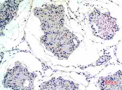
A03253 has a paraffin-section rat-testis IHC image (image caption) and lists IF use (catalog). Its listed reactivity covers human, mouse, and rat (catalog).

Real IHC data Immunohistochemical analysis of paraffin-embedded rat-testis, antibody was diluted at 1:100
Anti-Thrombospondin 2 Antibody
Cat # A03253

A03253 is the only card; its IHC image shows paraffin-embedded rat testis at 1:100 (A03253 image caption). A03253 lists IF with a 1:50 dilution and human, mouse, and rat reactivity, but supplies no IF image (A03253 catalog).

Which to pick: For tissue IHC, A03253 has a rat-testis paraffin-section image, although IHC is absent from its listed applications (A03253 image caption; A03253 catalog). For IF, A03253 is listed at 1:50; ICC and clonality are unspecified (A03253 catalog). For cross-species work, A03253 lists human, mouse, and rat reactivity, but its IHC image documents only rat testis; the fixative is unreported (A03253 catalog; A03253 image caption).

Each figure is that product's own IHC / IF validation image from its datasheet.

References

  1. UniProt Consortium. UniProt entry P35442 (TSP2_HUMAN, Thrombospondin-2).
  2. Human Protein Atlas. THBS2 tissue IHC expression (reliability: Approved).
  3. Human Protein Atlas. THBS2 subcellular location (ICC-IF): Mainly localized to the plasma membrane. In addition localized to the cytosol and centrosome..
  4. Human Protein Atlas. THBS2 antibody validation summary (3 antibodies).
  5. Thrombospondin 2, matrix Gla protein and digital analysis identified distinct fibroblast populations in fibrostenosing Crohn's disease. Scientific reports 2024 — PMC11178913.
  6. THBS2 is Closely Related to the Poor Prognosis and Immune Cell Infiltration of Gastric Cancer. Frontiers in genetics 2022 — PMC8853287.
  7. Detection of early pancreatic ductal adenocarcinoma with thrombospondin-2 and CA19-9 blood markers. Science translational medicine 2017 — PMC5727893.
  8. Down regulation of Thrombospondin2 predicts poor prognosis in patients with gastric cancer. Molecular cancer 2014 — PMC4189190.
  9. PubMed PMID:8406456 — UniProt-cited evidence.
  10. PubMed PMID:14702039 — UniProt-cited evidence.
  11. PubMed PMID:14574404 — UniProt-cited evidence.