This website uses cookies to ensure you get the best experience on our website.
- Table of Contents
2 Citations
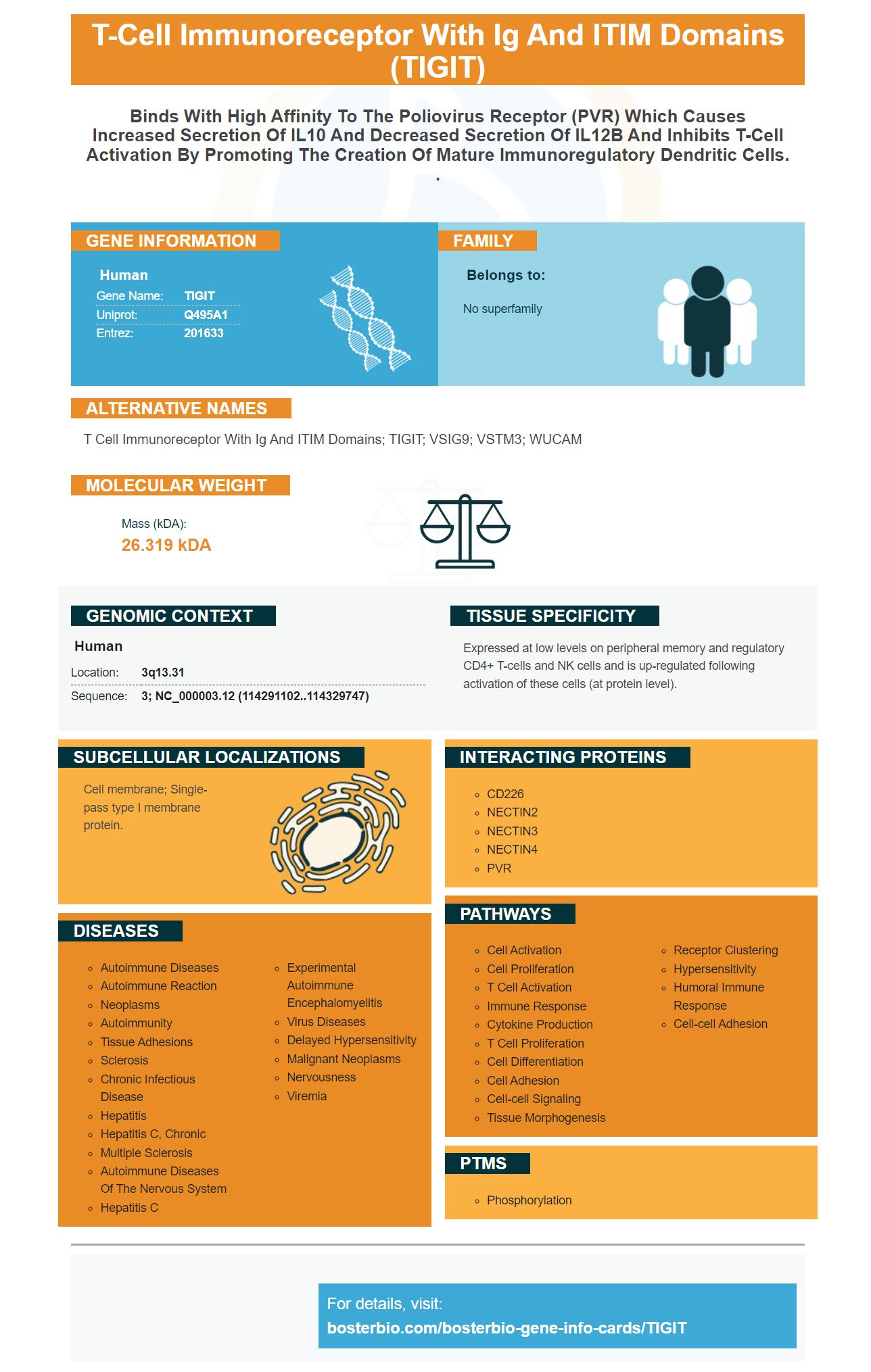
Facts about T-cell immunoreceptor with Ig and ITIM domains.

| Human | |
|---|---|
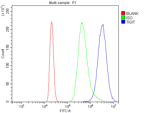
| Gene Name: | TIGIT |
| Uniprot: | Q495A1 |
| Entrez: | 201633 |

| Belongs to: |
|---|
| No superfamily |

T cell immunoreceptor with Ig and ITIM domains; TIGIT; VSIG9; VSTM3; WUCAM
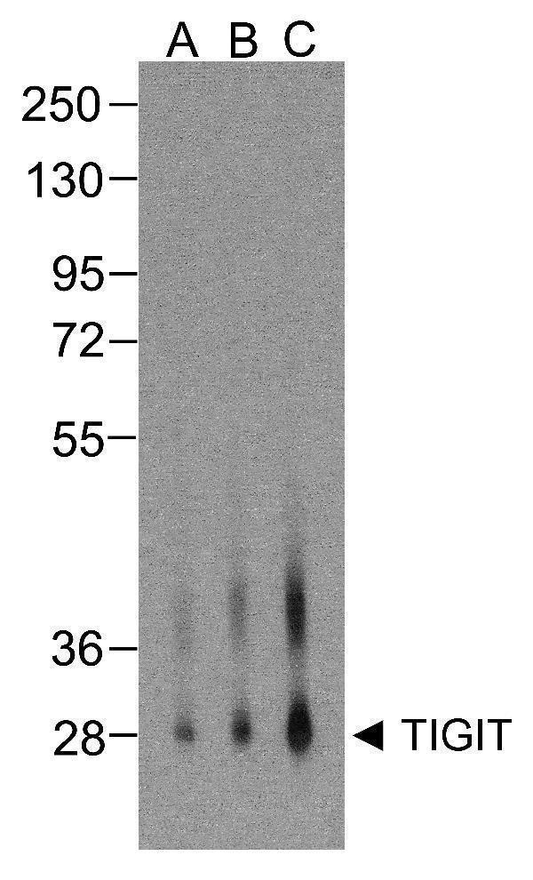
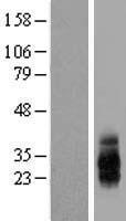
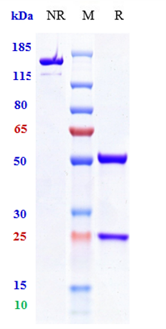
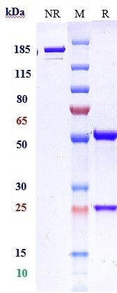
Mass (kDA):
26.319 kDA

| Human | |
|---|---|
| Location: | 3q13.31 |
| Sequence: | 3; NC_000003.12 (114291102..114329747) |
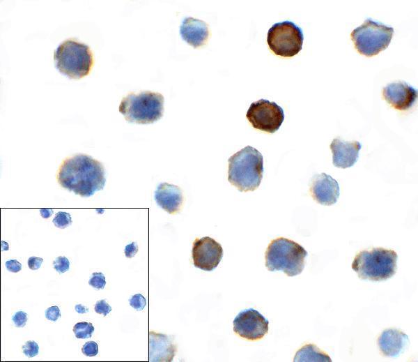
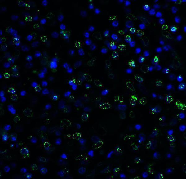
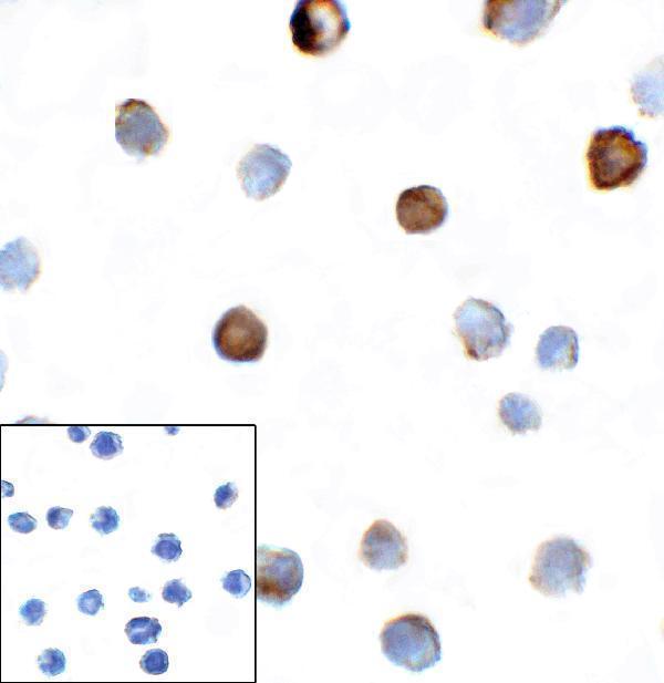
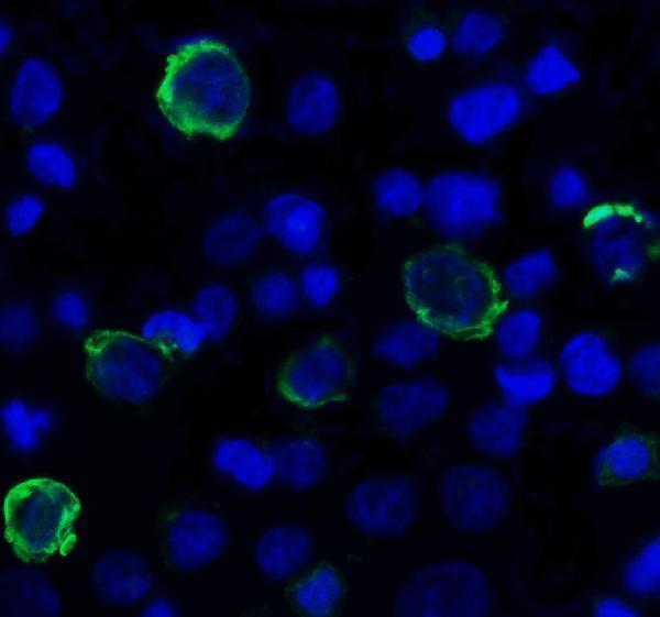
Expressed at low levels on peripheral memory and regulatory CD4+ T-cells and NK cells and is up-regulated following activation of these cells (at protein level).
Cell membrane; Single-pass type I membrane protein.


PMID: 19011627 by Yu X., et al. The surface protein TIGIT suppresses T cell activation by promoting the generation of mature immunoregulatory dendritic cells.
PMID: 22421438 by Stengel K.F., et al. Structure of TIGIT immunoreceptor bound to poliovirus receptor reveals a cell-cell adhesion and signaling mechanism that requires cis-trans receptor clustering.