This website uses cookies to ensure you get the best experience on our website.


- Table of Contents
For paraffin-section TIMM17A IHC, start with 0.5–1 μg/mL of the catalog antibody (datasheet A12168-1). Assess cytoplasmic staining using high-staining cardiomyocytes as a reference, while accounting for the medium consistency between antibody staining and RNA data (HPA tissue IHC).

Expected localisation, validated protocols, controls and antibodies — the at-a-glance facts below, then the full design guide.
| Expected localisation | Cytoplasmic tissue staining (HPA tissue IHC); mitochondrial inner membrane (UniProt) | |
| Staining pattern | Cytoplasmic staining; high in cardiomyocytes (HPA tissue IHC) | |
| Antigen retrieval | Citrate pH 6 HIER, heat-mediated (datasheet A12168-1) | |
| Positive control | Esophagus+4 more · see all | |
| Negative control | None in HPA (detected in all 45 tissues); use no-primary + isotype controls |
| Fixation | Keep fixation consistent across paraffin sections (standard IHC practice; not target-specific) | |
| Caveat | Antibody staining has medium consistency with RNA data (HPA tissue IHC) | |
| Regulation | No expression regulator annotated (UniProt) | |
| Isoform / epitope | No isoforms or cleavage annotated; 3 transmembrane segments (UniProt) |
The catalog antibody’s IHC-P protocol is accompanied by one published TIMM17A IHC protocol using rat tissue slides (PMC6257833: Immunohistochemical detection).
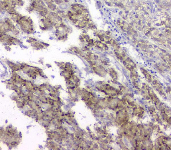
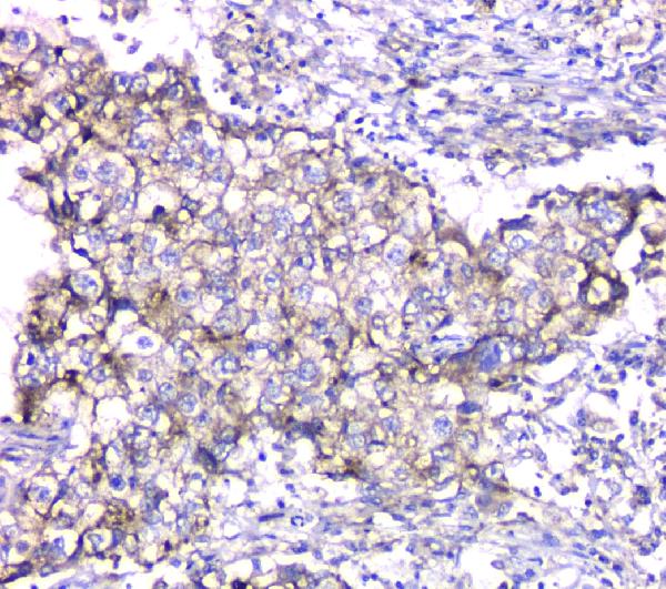
| Sample | Paraffin-embedded human liver cancer tissue; fixative not specified (datasheet A12168-1) |
| Fixation | Image fixative and duration unreported (datasheet A12168-1); verify before use. |
| Sectioning | 4–5 µm sections on charged slides (standard) |
| Deparaffinisation | Xylene, graded ethanol series to water (standard) |
| Antigen retrieval | Heat retrieval: Citrate pH 6, 20 min (datasheet A12168-1) |
| Peroxidase block | 3% H2O2, 10 min, room temperature (standard) |
| Blocking | 10% goat serum (datasheet A12168-1) |
| Primary antibody | Rabbit anti-TIMM17A, 0.5-1μg/ml (datasheet A12168-1) |
| Primary incubation | Overnight at 4 °C (datasheet A12168-1) |
| Detection | Streptavidin-biotin complex (SABC), DAB chromogen (datasheet A12168-1) |
| Counterstain | Hematoxylin, blue, dehydrate and mount (standard) |
| Expected result | TIMM17A-positive staining in squamous epithelial cells of esophagus (HPA tissue IHC: High). HPA tissue profile: General cytoplasmic expression. No signal in the no-primary control. |
TIMM17A resides in the mitochondrial inner membrane and has 3 transmembrane segments (UniProt Q99595 topology). In paraffin section IHC, expect cytoplasmic staining consistent with that location; HPA describes general cytoplasmic expression and rates its tissue IHC evidence Approved, with medium consistency between staining and RNA data (HPA tissue IHC). High staining is reported in squamous epithelial cells, cardiomyocytes, and selected glandular cells (HPA tissue IHC).
| Cytoplasmic stain in esophageal or oral squamous epithelium, cardiomyocytes, or rectal or thyroid glandular cells. | These cells have High HPA tissue IHC staining. A cytoplasmic pattern fits mitochondrial localization; chromogenic IHC need not resolve individual mitochondria (HPA tissue IHC; UniProt Q99595 localization; standard IHC practice). |
| Predominantly nuclear or extracellular stain, with little cytoplasmic signal in an HPA High cell population. | This poorly fits the mitochondrial inner membrane assignment (UniProt Q99595 localization). Review controls before calling it specific; HPA also lists nucleoplasm as an uncertain IF location, so nuclear signal alone cannot settle specificity (HPA subcellular ICC-IF; standard IHC practice). |
| Strong stain in a cell type that lacks an established positive pattern in this payload. | Possible cross-reactivity or endogenous detection activity warrants checking (standard IHC practice). Absence from HPA's positive list is not evidence that the cell type must be negative; HPA reports low tissue specificity (HPA tissue IHC). |
| Widespread, even stain over cells and surrounding tissue, obscuring cell boundaries. | Diffuse background limits interpretation of the expected cytoplasmic pattern (HPA tissue IHC; standard IHC practice). Assess a no-primary control and the detection and blocking steps before assigning TIMM17A expression (standard IHC practice). |
| No stain in a well-preserved HPA High population on the same run. | A technical failure becomes plausible when a documented positive is absent, though one negative section cannot establish its cause (HPA tissue IHC; standard IHC practice). Check controls, retrieval, antibody dilution, and detection (standard IHC practice). |
| Compartment and membrane topology | TIMM17A is an inner mitochondrial membrane protein with 3 |
| Tissue and cell choice | HPA reports High staining in esophageal and oral squamous epithelium, cardiomyocytes, and rectal and thyroid glandular cells; pancreatic exocrine cells and skeletal myocytes are Low (HPA tissue IHC). Compare like cell types when judging intensity (standard IHC practice). |
| Strength of IHC evidence | HPA calls tissue IHC Approved, with medium staining-to-RNA consistency; antibody HPA010083 is IHC Approved (HPA tissue IHC; HPA antibodies). These labels support use of the observed pattern but do not prove that every stained cell is specific (standard IHC practice). |
| Processing and modification record | The UniProt record lists one 171-aa chain, no signal peptide or propeptide, and no annotated glycosylation or modified residues (UniProt Q99595). It supplies no basis here for predicting a distinct processed staining compartment (UniProt Q99595). |
| Q: What does IF/ICC add? | A: HPA reports enhanced mitochondrial and uncertain nucleoplasmic localization; its IF images include A-549, SiHa, and U2OS (HPA subcellular ICC-IF). This context helps assess compartment claims, while IF/ICC methods belong to the separate guide (HPA subcellular ICC-IF). |
| Situation | Likely cause | Next action |
|---|---|---|
| No signal in esophageal squamous cells or cardiomyocytes. | Both are HPA High populations, so absent stain raises concern about the IHC run or specimen quality (HPA tissue IHC; standard IHC practice). | Confirm tissue preservation and a positive control; review retrieval, antibody dilution, and detection against the validated IHC procedure (standard IHC practice). |
| Weak signal in pancreatic exocrine cells or skeletal myocytes. | Both are listed Low in HPA tissue IHC; weak staining there can fit the reported pattern (HPA tissue IHC). | Judge run performance in an HPA High population on the same run before changing conditions (HPA tissue IHC; standard IHC practice). |
| Nuclear stain dominates an HPA High cell population. | Mitochondria are the enhanced IF location, while nucleoplasm is uncertain; nuclear dominance has ambiguous specificity (HPA subcellular ICC-IF; UniProt Q99595 localization). | Compare cytoplasmic staining and a no-primary control, then review the IHC-validated antibody's pattern; do not score nuclear stain alone as a confirmed positive (HPA tissue IHC; standard IHC practice). |
| Broad stain persists in a no-primary control. | Signal without primary antibody points toward detection-system or endogenous activity rather than TIMM17A binding (standard IHC practice). | Review blocking and the chromogenic detection controls; interpret the primary-stained slide only after control background is resolved (standard IHC practice). |
| Unexpected cells stain strongly while expected cells do not. | Cross-reactivity or uneven assay performance is possible; HPA's low tissue specificity prevents treating every unlisted cell as a known negative (HPA tissue IHC; standard IHC practice). | Compare cell identity, compartment, and matched controls with an HPA High reference; avoid assigning specificity from intensity alone (HPA tissue IHC; standard IHC practice). |
| Cytoplasmic stain is too diffuse to score individual cells. | Excess background can obscure the general cytoplasmic profile reported by HPA (HPA tissue IHC; standard IHC practice). | Check no-primary background, blocking, antibody dilution, and detection conditions before scoring cell-level intensity (standard IHC practice). |


Comprehensive Human Protein Atlas IHC scoring per tissue (reliability: Approved — Medium consistency between antibody staining and RNA expression data.). Rows are taken directly from the HPA tissue chart — click any row's HPA link to view the source.
| Tissue | Cell type | Level | Evidence | Source |
|---|---|---|---|---|
| Esophagus | Squamous epithelial cells | High | Protein (IHC) | HPA → |
| Heart muscle | Cardiomyocytes | High | Protein (IHC) | HPA → |
| Oral mucosa | Squamous epithelial cells | High | Protein (IHC) | HPA → |
| Rectum | Glandular cells | High | Protein (IHC) | HPA → |
| Thyroid gland | Glandular cells | High | Protein (IHC) | HPA → |
| Tissue | Cell type | Level | Evidence | Source |
|---|---|---|---|---|
| None in HPA: TIMM17A is detected in all 45 scored tissues. Use a no-primary (secondary-only) and an isotype control instead. | ||||
Troubleshooting guidance for TIMM17A chromogenic IHC in paraffin sections, with one note on adapting the experiment to IF.
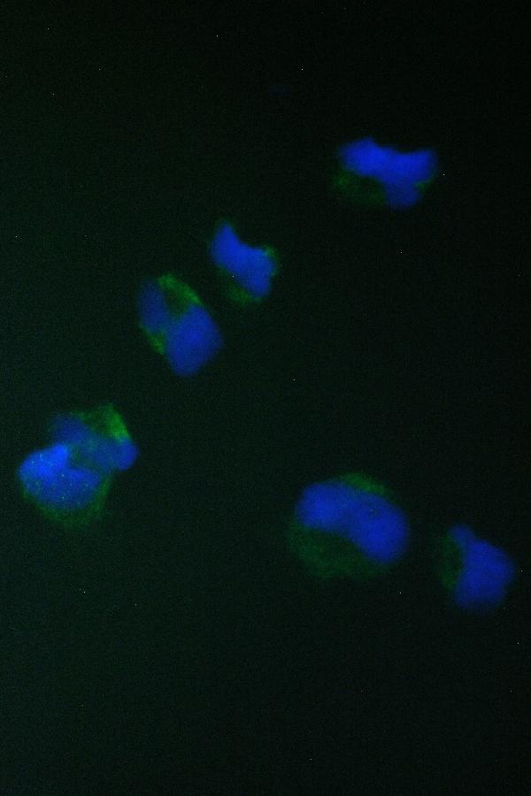
A12168-1 has real paraffin-section IHC images from human cancer tissues and rat lung, plus IF/ICC images from HeLa and A549 cells (catalog image captions).
A12168-1 is listed for IHC and IF/ICC in human, mouse, and rat (catalog applications and reactivity). Its IHC images show human liver, lung, and mammary cancer sections and rat lung; its IF images show HeLa and A549 cells (catalog image captions).
Which to pick: For chromogenic IHC on paraffin sections, choose A12168-1: its own caption documents citrate retrieval at pH 6 and primary antibody at 1 μg/ml; the fixative and clone are unreported (catalog IHC caption; catalog clone field). For IF/ICC, A12168-1 has images from HeLa and A549 cells (catalog IF captions). For cross-species work, A12168-1 lists human, mouse, and rat reactivity; rat tissue IHC is pictured, while mouse tissue IHC is unpictured (catalog reactivity; catalog IHC captions).