TIMP2 / Metalloproteinase inhibitor 2 · IHC design guide

Design Immunohistochemistry for TIMP2

Plan TIMP2 paraffin IHC around extracellular deposits and cytoplasmic staining (HPA tissue IHC). Use bronchial ciliated-cell tips as a high-staining reference (HPA tissue IHC), and titrate the catalog antibody at 0.5–1 µg/ml (datasheet).

Evidence assembled Oct 2026 · For research use; verify linked source records and product datasheet before use
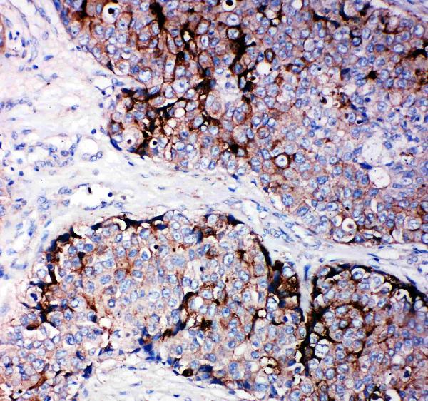
Immunohistochemistry protocol sheet for TIMP2 (IHC for TIMP2): expected localisation Extracellular deposits and cytoplasm (HPA tissue IHC), antibody PA1076, validated IHC image, and IHC protocol steps
Printable TIMP2 IHC protocol sheet — expected localisation Extracellular deposits and cytoplasm (HPA tissue IHC), antibody PA1076, controls and protocol steps. Open the full TIMP2 IHC guide →

TIMP2 Immunohistochemistry Experimental Design Guide

Expected localisation, validated protocols, controls and antibodies — the at-a-glance facts below, then the full design guide.

Must know before staining
Expected localisation Extracellular deposits and cytoplasm (HPA tissue IHC)
Staining pattern Extracellular and cytoplasmic staining; bronchial ciliary tips high (HPA tissue IHC)
Antigen retrieval Citrate pH 6.0 HIER, 95–98 °C, 20 min (rule: cytoplasmic / membrane antigen)
Positive control ⓘ Bronchus+3 more · see all
Negative control ⓘ Adipose tissue+4 more · see all
Important caveats
Reasons your staining may differ from the expected pattern.
Fixation Matched tissue-IHC evidence does not establish the fixation claim. Validate the specimen-specific method before use. (selected-SKU IHC image PA1076)
Caveat Secreted TIMP2 may deposit away from producing cells (UniProt; HPA tissue IHC)
Regulation Specific regulators of staining intensity are unreported (UniProt; HPA tissue IHC)
Isoform / epitope No annotated isoforms or transmembrane split; mature chain 27–220 (UniProt)
Section 1

Recommended TIMP2 IHC & IF Protocols

Compare the catalog antibody’s IHC-P protocol with four published TIMP2 chromogenic IHC methods (PMC8487594; PMC4466407; PMC8787141; PMC5472630).

Recommended immunohistochemistry (IHC-P) protocol parameters
SampleTissue sections; selected-image fixative not specified (standard IHC workflow)
FixationImage fixative and duration unreported (datasheet PA1076); verify before use.
Sectioning4–5 µm sections on charged slides (standard)
DeparaffinisationXylene, graded ethanol series to water (standard)
Antigen retrievalHeat-induced epitope retrieval in citrate buffer, pH 6.0, 20 min at 95–98 °C (standard rule: cytoplasmic / membrane antigen)
Peroxidase block3% H2O2, 10 min, room temperature (standard)
Blocking10% normal serum of the secondary host, 30 min, room temperature (standard)
Primary antibodyRabbit anti-TIMP2, 0.5-1μg/ml (datasheet PA1076)
Primary incubationOvernight at 4 °C (standard)
DetectionHRP-polymer secondary, DAB chromogen 5–10 min (standard)
CounterstainHematoxylin, blue, dehydrate and mount (standard)
Expected resultTIMP2-positive staining in ciliated cells (tip of cilia) of bronchus (HPA tissue IHC: High). HPA tissue profile: Extracellular deposits and cytoplasmic expression in all tissues. No signal in the no-primary control.
💡Decision noteStart with citrate pH 6.0 retrieval at 95–98 °C for 20 min (page antigen retrieval rule); use the published tissue-specific treatments when matching those samples.
Section 2

What Is the Expected TIMP2 Staining Pattern?

TIMP2 is secreted, with no transmembrane segment (UniProt P16035); deposits, cytoplasm and bronchial ciliary tips stain (HPA: Approved).

What am I looking at on my slide?
Bronchial ciliary tips stain strongly (HPA: High).Fits the reported pattern; deposits and cytoplasmic stain may coexist (HPA: Approved).
Staining is confined to nuclei (HPA: secreted profile).Discordant with the reported pattern; assess for artefact (HPA; UniProt P16035).
Adipocytes stain strongly (HPA: not detected).Unexpected; assess cross-reactivity and endogenous detection activity (HPA; IHC practice).
Uniform haze covers the section (IHC practice).Suggests background; review blocking and detection controls (IHC practice).
Bronchial ciliary tips lack signal (HPA: High).Possible assay failure; check the positive control and detection steps (IHC practice).
💡Expected TIMP2 appearanceExpect High bronchial ciliary-tip staining (HPA); nuclear-only stain suggests artefact (IHC practice).
How each factor affects the staining
Secretion (UniProt P16035)Extracellular deposits and cytoplasmic staining are reported (HPA: Approved).
Processing (UniProt P16035)Signal peptide 1–26; mature chain 27–220 (UniProt P16035).
Tissue contrast (HPA)Bronchial ciliary tips: High; adipocytes: not detected (HPA).
Antibody validation (HPA)CAB010203 is IHC Approved; ICC validation is unlisted (HPA antibodies).
Why is my staining missing, weak or wrong?
SituationLikely causeNext action
No stain in bronchial ciliary tips (HPA: High).Possible retrieval or detection failure (IHC practice).Check a known-positive section and reagent sequence (IHC practice).
Nuclear-only staining (HPA: secreted profile).Possible nonspecific staining (IHC practice).Compare with a negative control and assess antibody dilution (IHC practice).
Strong adipocyte staining (HPA: not detected).Possible cross-reactivity or endogenous activity (IHC practice).Check antibody and detection controls (IHC practice).
Diffuse section-wide haze (IHC practice).Possible inadequate blocking or excess antibody (IHC practice).Review blocking and titrate the primary antibody (IHC practice).
Ciliary tips are hard to assess (HPA: bronchial tip stain).Section orientation may obscure cilia (IHC practice).Inspect intact ciliated epithelium with a counterstain (IHC practice).
What should IF/ICC show? (HPA: secreted summary)No ICC-IF images or main location are available (HPA subcellular).Use the dedicated IF/ICC guide; treat precise IF localisation as unverified here (HPA).

Sample controls for TIMP2 IHC & IF

🧪Run bronchus first and look for staining at the tips of cilia in ciliated cells (HPA: High in bronchus ciliated cells, tip of cilia). Use adipose tissue as a negative comparator, focusing on adipocytes (HPA: Not detected in adipocytes); on the bronchus slide, treat areas outside the ciliary tips as an internal background check against the no-primary control (HPA: ciliary-tip staining; standard IHC practice).
Positive control tissue: Bronchus (Ciliated cells (tip of cilia), HPA High)
Negative control tissue: Adipose tissue (HPA Not detected)
ICC-IF cell lines (HPA subcellular resource): HPA carries no ICC-IF cell line for TIMP2; derive a cell-line control from the positive tissue's cell type (Ciliated cells (tip of cilia)) and confirm it by RNA or western blot first.
Technical controls: Include no-primary (secondary-only), a primary-antibody host-species- and clonality-matched isotype control, and TIMP2 knockout tissue if available (standard IHC practice). For chromogenic detection in bronchus, quench endogenous peroxidase and block endogenous biotin if using biotin-based detection (standard IHC practice).
⚠️Feasibility: The supplied evidence reports no TIMP2-specific fixation window, fixation effect, or antigen-retrieval requirement; the PA1076 tissue-IHC caption does not state a fixative (PA1076 caption). Whether frozen sections or IF are easier cannot be established from the supplied evidence (HPA: no ICC-IF images). Bronchial ciliary-tip staining occupies a narrow area (HPA: High in ciliated cells at the tip of cilia), so check ciliary morphology and compare luminal-edge deposits with the no-primary control when interpreting chromogen (standard IHC practice).

HPA tissue IHC evidence for TIMP2

Comprehensive Human Protein Atlas IHC scoring per tissue (reliability: Approved — At least one protein variant secreted, tissue location of RNA and protein might differ and correlation is complex.). Rows are taken directly from the HPA tissue chart — click any row's HPA link to view the source.

Positive expression · recommended positive controls

TissueCell typeLevelEvidenceSource
Bronchus Ciliated cells (tip of cilia) High Protein (IHC) HPA →
Fallopian tube Ciliated cells (ciliary rootlets) Medium Protein (IHC) HPA →
Nasopharynx Basal cells Medium Protein (IHC) HPA →
Testis Peritubular cells Medium Protein (IHC) HPA →

Undetected expression · recommended negative controls

TissueCell typeLevelEvidenceSource
Adipose tissue Adipocytes Not detected Protein (IHC) HPA →
Adrenal gland Glandular cells Not detected Protein (IHC) HPA →
Appendix Glandular cells Not detected Protein (IHC) HPA →
Bone marrow Hematopoietic cells Not detected Protein (IHC) HPA →
Breast Adipocytes Not detected Protein (IHC) HPA →
Section 3

Advanced TIMP2 IHC Tips

Troubleshoot TIMP2 staining in paraffin sections by checking retrieval, compartment, controls, and scoring against its secreted expression pattern (UniProt P16035; HPA tissue IHC).

What retrieval should I start with if TIMP2 staining is weak?
Start with citrate buffer at pH 6.0 for heat-induced retrieval at 95–98 °C for 20 min (page retrieval specification). Cool sections consistently before antibody incubation, and compare any retrieval changes on adjacent sections from the same block (standard IHC practice). Include a known positive section, such as bronchus with ciliated-cell staining, so a weak result can be distinguished from a failed run (HPA tissue IHC). If staining remains weak, test another retrieval buffer only as a documented fallback while holding detection conditions constant (standard IHC practice). Excessive retrieval can damage morphology or increase diffuse staining, so judge signal alongside tissue preservation (standard IHC practice).
How can I assess whether fixation caused weak or uneven TIMP2 staining?
Target-specific sensitivity of TIMP2 to fixative choice or fixation time is unknown from the supplied evidence; the PA1076 tissue caption does not state a fixative (catalog caption PA1076). Record the available fixation history and compare sections with similar processing before attributing a difference to the antibody (standard IHC practice). On adjacent sections, check morphology and staining distribution, especially whether signal is confined to exposed edges or lost in poorly preserved regions (standard IHC practice). Run a positive control through the same retrieval and detection sequence, then repeat the stain with one processing variable changed at a time (standard IHC practice). Do not infer fixation sensitivity from TIMP2 topology or tissue staining patterns (UniProt P16035; HPA tissue IHC).
Where should TIMP2 staining appear, and when is cytoplasmic signal credible?
Expect extracellular deposits and cytoplasmic expression across tissues in the reported IHC profile (HPA tissue IHC). TIMP2 is secreted, has a 1–26 signal peptide, and has no transmembrane segment, so a uniformly sharp membrane outline warrants scrutiny (UniProt P16035). Compare the pattern with a matched negative control and with tissue architecture before assigning extracellular signal to a particular cell (standard IHC practice). Bronchial ciliated-cell tips show high staining, while fallopian-tube ciliary rootlets and testicular peritubular cells show medium staining in the supplied profile (HPA tissue IHC). Cytoplasmic staining can be credible, but report its cell type and location separately from extracellular deposits (HPA tissue IHC; standard IHC practice).
Can epitope placement explain a change in TIMP2 staining after retrieval?
The supplied record lists no TIMP2 isoforms and places the mature chain at residues 27–220 after cleavage of the 1–26 signal peptide (UniProt P16035). It identifies an NTR domain at residues 27–152 and a C-terminal interaction with MMP2, but gives no epitope for the catalog antibody (UniProt P16035; supplied product evidence). Therefore, do not assign a staining difference to signal-peptide cleavage, domain masking, or an isoform without antibody-specific epitope evidence (standard IHC interpretation). Compare retrieval conditions on adjacent sections while keeping antibody dilution and detection constant, and record changes in both intensity and location (standard IHC practice). The record reports 0 glycosylation sites and 0 modified residues; it does not establish retrieval sensitivity (UniProt P16035).
How should I check TIMP2 localisation by multiplex immunofluorescence?
For the separate IF/ICC workflow, pair TIMP2 with a validated marker of the expected cell type, such as a ciliated-cell marker when examining bronchus (HPA tissue IHC; standard IF practice). Select spectrally separated fluorophores and consider a far-red TIMP2 channel if tissue autofluorescence obscures shorter-wavelength signal (standard IF practice). For extracellular deposits, assess staining without permeabilisation first; for cytoplasmic signal, compare with a gently permeabilised section (UniProt P16035; HPA tissue IHC; standard IF practice). TIMP2 is secreted and has no transmembrane segment, so no membrane-facing epitope side can be assumed for this antibody (UniProt P16035; supplied product evidence). Include single-label and no-primary controls when judging colocalisation or background (standard IF practice).
What should I check when TIMP2 DAB staining looks diffuse?
First distinguish expected extracellular deposits from diffuse chromogen or staining that ignores tissue structure (HPA tissue IHC; standard IHC practice). Inspect the no-primary control and confirm that endogenous peroxidase blocking and wash steps were performed before DAB development (standard chromogenic IHC practice). Compare a lower primary-antibody concentration or shorter DAB development on adjacent sections, changing one variable at a time (standard IHC practice). Examine tissue edges, folds, damaged areas, and necrosis separately because local background can mimic widespread TIMP2 deposition (standard IHC practice). A positive control with the expected bronchial ciliated-cell pattern helps determine whether reducing background also removes credible signal (HPA tissue IHC; standard IHC practice).
How should I score TIMP2 when cells and extracellular deposits both stain? ⚠ ANSWER MARKED FOR VERIFICATION
Define cell compartments and extracellular regions before scoring, because the reported TIMP2 pattern includes cytoplasmic expression and extracellular deposits (HPA tissue IHC). For cells, record the percentage positive and staining intensity, or calculate an H-score with a stated intensity scale (standard IHC quantification practice). For extracellular signal, measure positive area or optical density per tissue area in mm², using the same threshold across comparable sections (standard IHC quantification practice). Normalise cell scores to the number of evaluable cells in the specified compartment and extracellular measurements to evaluable tissue area (standard IHC quantification practice). Exclude folds, necrosis, and blank space consistently, and report cell and deposit measurements separately (standard IHC quantification practice).
How can I distinguish credible TIMP2 staining from artefact?
A credible result follows tissue architecture and includes plausible cytoplasmic or extracellular signal rather than requiring one exclusively cellular pattern (HPA tissue IHC). Compare the stained cell type with the supplied profile: bronchial ciliated-cell tips are high, whereas adipocytes in adipose tissue are reported as not detected (HPA tissue IHC). Treat staining limited to section edges, folds, or necrotic areas as suspect and inspect an adjacent section (standard IHC practice). Signal in a no-primary control suggests detection background, including possible endogenous enzyme activity in a peroxidase-based assay (standard chromogenic IHC practice). Because TIMP2 is secreted, extracellular protein need not identify the cell that produced it (UniProt P16035; HPA reliability description).
Boster reagents

Best TIMP2 / Metalloproteinase inhibitor 2 IHC Antibodies

PA1076 has real paraffin-section IHC data from human mammary cancer tissue (IHC image caption); the catalog lists Human, Mouse, and Rat reactivity (catalog: reactivity).

Real IHC data Anti-TIMP2 antibody, PA1076, IHC(P) IHC(P): Human Mammary Cancer Tissue
Anti-Metalloproteinase inhibitor 2 TIMP2 Antibody
Cat # PA1076

PA1076 will render with an IHC(P) image from human mammary cancer tissue (IHC image caption). Its listed applications are ELISA and IHC, and its listed reactivity is Human, Mouse, and Rat (catalog: applications and reactivity).

Which to pick: Choose PA1076 for paraffin-section tissue IHC: its own image caption documents IHC(P) in human mammary cancer tissue, and its IHC dilution is 0.5–1 μg/ml (IHC image caption; datasheet: 0.5–1 μg/ml). For cross-species IHC, PA1076 lists Human, Mouse, and Rat reactivity (catalog: reactivity); its clone is unreported, so clonality cannot guide selection. No IF/ICC application or image is listed for PA1076, so this payload does not support an IF/ICC pick; the IHC caption does not report the fixative (catalog: applications and IF images; IHC image caption).

Each figure is that product's own IHC / IF validation image from its datasheet.

References

  1. UniProt Consortium. UniProt entry P16035 (TIMP2_HUMAN, Metalloproteinase inhibitor 2).
  2. Human Protein Atlas. TIMP2 tissue IHC expression (reliability: Approved).
  3. Human Protein Atlas. TIMP2 subcellular location (ICC-IF): Secreted.
  4. Human Protein Atlas. TIMP2 antibody validation summary (1 antibodies).
  5. Verbascoside alleviates renal fibrosis in unilateral ureteral obstruction rats by inhibiting macrophage infiltration. Iranian journal of basic medical sciences 2021 — PMC8487594.
  6. Differential Expression of ADAM23, CDKN2A (P16), MMP14 and VIM Associated with Giant Cell Tumor of Bone. Journal of Cancer 2015 — PMC4466407.
  7. Genetic Variants and Protein Alterations of Selenium- and T-2 Toxin-Responsive Genes Are Associated With Chondrocytic Damage in Endemic Osteoarthropathy. Frontiers in genetics 2021 — PMC8787141.
  8. EZH2-mediated epigenetic silencing of TIMP2 promotes ovarian cancer migration and invasion. Scientific reports 2017 — PMC5472630.
  9. PubMed PMID:2380196 — UniProt-cited evidence.
  10. PubMed PMID:2157214 — UniProt-cited evidence.
  11. PubMed PMID:8810321 — UniProt-cited evidence.